prevalence of toxigenic bacteria in some egyptian food abstract ...
prevalence of toxigenic bacteria in some egyptian food abstract ...
prevalence of toxigenic bacteria in some egyptian food abstract ...

Create successful ePaper yourself
Turn your PDF publications into a flip-book with our unique Google optimized e-Paper software.
PROCEEDING OF FIFTH SCIENTIFIC ENVIRONMENTAL CONFERENCE, 2010, ZAGAZIG UNI., 107 - 124PREVALENCE OF TOXIGENIC BACTERIA IN SOME EGYPTIANFOODNadia Mohamed Awny, Azza A. M. Abou Zeid and Mohamed Ahmed AbdoBotany Department, Faculty <strong>of</strong> Science, Zagazig University.ABSTRACTThirty three <strong>food</strong> samples represent<strong>in</strong>g seven different <strong>food</strong> products were collectedfrom the market <strong>in</strong> Sharkia Governorate. The <strong>food</strong> samples were <strong>in</strong>vestigated for their<strong>bacteria</strong>l burden <strong>in</strong>clud<strong>in</strong>g total mesophilic <strong>bacteria</strong>, spore formers, Staphylococcus aureusand Bacillus cereus us<strong>in</strong>g specific and selective nutrient media. Total mesophilic countsranged between 2.2x10 6 to1x10 9 cfu/g, spore form<strong>in</strong>g <strong>bacteria</strong> were 5.6x10 2 to 2x10 4 cfu/g.Bacilli related to cereus group were detected <strong>in</strong> all tested <strong>food</strong> samples (100%) and S. aureus<strong>in</strong> 4 <strong>food</strong> samples (12.12%). Thirty six spore form<strong>in</strong>g stra<strong>in</strong>s were chosen from MYP andBaird Parker’s agar plates and differentiated <strong>in</strong>to different species us<strong>in</strong>g sta<strong>in</strong><strong>in</strong>g andbiochemical tests accord<strong>in</strong>g to key <strong>of</strong> Bergey's manual. B. cereus was the most frequentspecies (19 isolates) represent<strong>in</strong>g 53 % <strong>of</strong> total isolated <strong>bacteria</strong>. Four stra<strong>in</strong>s isolated fromBaird Parker's agar medium were coagulase positive and confirmed as S. aureus.The identified stra<strong>in</strong>s were screened for their virulence factors us<strong>in</strong>g agar diffusionmethod. B. cereus stra<strong>in</strong>s CH, GT1, LB3 and G8 were found to be the most potent isolates.Also, the four S. aureus showed nearly equal potency concern<strong>in</strong>g the <strong>in</strong>vestigated virulencefactors.The cellular prote<strong>in</strong> pr<strong>of</strong>ile <strong>of</strong> the eight potent <strong>food</strong> borne pathogenic <strong>bacteria</strong> showed<strong>some</strong> similarities and differences between the exam<strong>in</strong>ed stra<strong>in</strong>s. B. cereus stra<strong>in</strong>s CH and GT1showed about 90% similarity while GT1 and LB3 showed highest difference (80%) <strong>in</strong> theoccurr<strong>in</strong>g prote<strong>in</strong> bands. Meanwhile, S. aureus S, S1 &S2 were more related as they showedabout 80% similar prote<strong>in</strong> bands.Separation <strong>of</strong> extracellular prote<strong>in</strong>s <strong>of</strong> the four exam<strong>in</strong>ed B. cereus stra<strong>in</strong>s us<strong>in</strong>g SDS-PAGE revealed the presence <strong>of</strong> prote<strong>in</strong> bands with molecular weights between 30 and 53 kDasuspected as hemolytic enterotox<strong>in</strong>s. Also, prote<strong>in</strong> bands hav<strong>in</strong>g molecular weights between22 and 33 kDa were observed <strong>in</strong> three stra<strong>in</strong>s (S1, S2 &S3) <strong>of</strong> exam<strong>in</strong>ed S. aureus stra<strong>in</strong>s.Us<strong>in</strong>g multiplex PCR technique, two pairs <strong>of</strong> primers (FHblC and RHblC) and (FCytK andR2Cytk) were used to detect the tox<strong>in</strong> genes (hblC and cytK) <strong>in</strong> suspected toxic B. cereusstra<strong>in</strong>s and five pairs namely:( SEA-3 & SEA-4);(SEB-1SEB-2.); (SEC-5SEC-6); (SED-1SED-2) and (SEE-1 SEE-2) were used to detect the five enterotox<strong>in</strong>s <strong>in</strong> S. aureus stra<strong>in</strong>s .The multiplex PCR amplification allowed rapid detection and identification <strong>of</strong> enterotox<strong>in</strong>genes <strong>in</strong> <strong>food</strong> borne <strong>bacteria</strong>.Key words: Food borne pathogens, S. aureus, Bacillus cereus, virulence factors, SDS-PAGE, prote<strong>in</strong> pr<strong>of</strong>ile,PCR, enterotox<strong>in</strong> genes, agarose gel electrophoresis.INTRODUCTIONExtracellular prote<strong>in</strong>s <strong>of</strong> pathogenic <strong>bacteria</strong> are ma<strong>in</strong> contributors <strong>of</strong> pathogenesisand are <strong>in</strong>disputably <strong>in</strong>volved <strong>in</strong> <strong>bacteria</strong>l virulence. These prote<strong>in</strong>s have a range <strong>of</strong> biologicalfunctions rang<strong>in</strong>g from host cell toxicity to more suitable alterations <strong>of</strong> the host cell for thebenefit <strong>of</strong> the <strong>in</strong>vader (Wooldridge, 2009).The virulence factors <strong>of</strong> pathogenic <strong>bacteria</strong> are divided <strong>in</strong>to several groups on thebasis <strong>of</strong> the mechanism <strong>of</strong> virulence and function. Of the important ones are secretaryprote<strong>in</strong>s such as tox<strong>in</strong>s and enzymes (Wu et al., 2008).
108PREVALENCE OF TOXIGENIC BACTERIA IN SOME EGYPTIAN FOODAmong these pathogenic <strong>bacteria</strong> are genus Bacillus and Staphylococcus which causea wide variety <strong>of</strong> diseases through production <strong>of</strong> their tox<strong>in</strong>s on several substrates, amongthese substrates "<strong>food</strong>" and this result<strong>in</strong>g <strong>in</strong> "<strong>food</strong> poison<strong>in</strong>g".Bacillus cereus, as spore former and gram positive bacilli, is able to survive andproliferate <strong>in</strong> a wide range <strong>of</strong> environment, <strong>in</strong>clud<strong>in</strong>g soil, water and many types <strong>of</strong> processed<strong>food</strong> such as herbs, spices, milk, meat, raw and cooked vegetables, boiled or fried rice, vanillasauce, custards, soups, ice cream and cereals (Mckillip, 2000; Granum 2001; Kotiranta etal., 2000 and L<strong>in</strong>dback et al., 2004). Numerous worldwide studies have reported on theimportance <strong>of</strong> B. cereus as a cause <strong>of</strong> <strong>food</strong> poison<strong>in</strong>g outbreaks but less causative agent <strong>of</strong>diarrhea (Kotiranta et al., 2000). Bacillus cereus causes diarrheal and emetic <strong>food</strong> poison<strong>in</strong>gand a variety <strong>of</strong> typically necrotic non gastro-<strong>in</strong>test<strong>in</strong>al <strong>in</strong>fections (Beecher and Macmillan,1990; Drobniewski, 1993; Beecher and Wong, 2000b; Granum and Baird-Parker, 2000;Mckillip, 2000; Callegan et al., 2002& Schoeni and Wong, 2005). Among the manypotential virulence factors <strong>of</strong> B. cereus, Haemolys<strong>in</strong> BL (HBL) is a unique and potent threecomponentpore-form<strong>in</strong>g tox<strong>in</strong> consist<strong>in</strong>g <strong>of</strong> three dist<strong>in</strong>ct prote<strong>in</strong>s, namely b<strong>in</strong>d<strong>in</strong>gcomponent (B), lytic component (L 1 ) and lytic component (L 2 ) (Beecher and Macmillan,1991 & Beecher and Wong, 1994b).Staphylococcus aureus is Gram +ve facultative anaerobic, non-spore former,coagulase positive cocci. Staphylococcal <strong>food</strong> poison<strong>in</strong>g is caused by the <strong>in</strong>gestion <strong>of</strong> <strong>food</strong>conta<strong>in</strong><strong>in</strong>g pre-formed tox<strong>in</strong>s secreted by the <strong>bacteria</strong>. These are known as Staphylococcalenterotox<strong>in</strong>s, and 8 serologically dist<strong>in</strong>ct types (A, B, C1, C2, C3, D, E, and F) have so farbeen recognized. Enterotox<strong>in</strong> F has now been shown to be identical biochemical to toxicshock syndrome tox<strong>in</strong> 1 (TSST-1) which produces toxic shock syndrome commonlyassociated with the use <strong>of</strong> tampons dur<strong>in</strong>g menstruation (Doyle, 1989). The staphylococcalenterotox<strong>in</strong>s are responsible for the symptoms associated with staphylococcal <strong>food</strong> poison<strong>in</strong>g(Kenny et al., 1993; Matsunaga et al., 1993 and Llewelyn and Cohen, 2002). The disease ischaracterized by symptoms <strong>in</strong>clud<strong>in</strong>g nausea, vomit<strong>in</strong>g, abdom<strong>in</strong>al cramps and diarrhealast<strong>in</strong>g from 24 to 48 h and the complete recovery usually occurs with<strong>in</strong> 1–3 days. Theenterotox<strong>in</strong> genes however are not uniformly distributed among S. aureus. D<strong>in</strong>ges et al.(2000) and Boerema et al.( 2006) reported the potency <strong>of</strong> SEs.SEA is the most common enterotox<strong>in</strong> recovered from <strong>food</strong> poison<strong>in</strong>g outbreaksBalaban and Rasooly (2000) and it is known that 59% <strong>of</strong> staphylococcal <strong>food</strong> poison<strong>in</strong>goutbreaks are caused by SEA to SEE (Bergdoll, 1989).Food-borne diseases are ma<strong>in</strong>ly caused by pathogenic <strong>bacteria</strong> which are eithertransmitted to humans from the animal reservoir or which contam<strong>in</strong>ate the <strong>food</strong> process l<strong>in</strong>e.Detection and isolation <strong>of</strong> pathogenic <strong>bacteria</strong> from <strong>food</strong> are <strong>of</strong>ten difficult due to the highnumber <strong>of</strong> contam<strong>in</strong>at<strong>in</strong>g and <strong>in</strong>digenous <strong>bacteria</strong> and a low number <strong>of</strong> the pathogenic<strong>bacteria</strong> <strong>of</strong> concern. In order to obta<strong>in</strong> even a modest sensitivity, most traditional isolationmethods <strong>in</strong>clude a selective enrichment and <strong>some</strong>-times a pre-enrichment step, both <strong>of</strong> whichare labor and time consum<strong>in</strong>g.There is, consequently, scope for improvement <strong>of</strong> detection and isolation methods,especially with respect to the time needed to produce a diagnosis. The last 20 to 30 years haveseen many developments <strong>in</strong> techniques and also the dawn<strong>in</strong>g <strong>of</strong> technologies, which werepredicted to change ways <strong>of</strong> detect<strong>in</strong>g pathogenic <strong>bacteria</strong> <strong>in</strong> <strong>food</strong>. Several reviews have dealtwith the use <strong>of</strong> DNA- probes and the PCR-technique <strong>in</strong> <strong>food</strong> microbiology (Hill, 1996; Hilland Keasler, 1991; Olsen et al., 1995; Wolcott, 1991). Based on the wide use that thetechniques quickly were put to for research purposes, the reviews unanimously praise thepotential <strong>of</strong> these techniques to overcome <strong>some</strong> <strong>of</strong> the <strong>in</strong>herent problems <strong>in</strong> detection andisolation <strong>of</strong> <strong>bacteria</strong>l pathogens from <strong>food</strong> (Olsen, 2000).
Nadia Mohamed Awny; Azza A. M. Abou Zeid and Mohamed Ahmed Abdo 109PCR-based techniques are used <strong>in</strong>creas<strong>in</strong>gly <strong>in</strong> <strong>food</strong>-microbiology research as they arewell developed and when applied as culture confirmation tests, they are reliable, fast andsensitive. PCR methods <strong>of</strong>fer a sensitive and specific detection <strong>of</strong> pathogens and candiscrim<strong>in</strong>ate virulent <strong>bacteria</strong> from avirulent members <strong>of</strong> the same species as well (Olsen,2000). In the last 10 years, many authors have proposed the use <strong>of</strong> PCR for the detection <strong>of</strong><strong>food</strong>-borne pathogens to replace the time-consum<strong>in</strong>g culture-based classical techniques(Gravet et al., 1999 and Miethke et al., 1992). They are rapid, easy to handle, sensitive andspecific and therefore constitute very valuable tools for rout<strong>in</strong>e applications.Several pathogens can be detected simultaneously <strong>in</strong> one step by multiplex PCR. Suchmultiplex methods <strong>of</strong> relevance to <strong>food</strong> microbiology have been used to detect variants <strong>of</strong><strong>food</strong> borne pathogens with special concern to enterotoxic stra<strong>in</strong>s <strong>of</strong> S. aureus and B. cereus(Becker et al., 1998; P<strong>in</strong>to et al., 2005; Gu<strong>in</strong>ebretiere et al., 2006 and Ngamwongsatit etal., 2008). Such methods show the potential for practical every day use <strong>of</strong> PCR methods <strong>in</strong><strong>food</strong> microbiology.Aim <strong>of</strong> WorkThe aim <strong>of</strong> this study was to evaluate the proteolytic activity <strong>of</strong> certa<strong>in</strong> <strong>food</strong> borne<strong>bacteria</strong> contam<strong>in</strong>at<strong>in</strong>g different <strong>food</strong> products collected from the Egyptian market withrespect to their virulence factors <strong>in</strong>clud<strong>in</strong>g extracellular degrad<strong>in</strong>g enzymes and enterotox<strong>in</strong>s.Us<strong>in</strong>g quick, specific and reliable molecular techniques <strong>in</strong> characteriz<strong>in</strong>g and typ<strong>in</strong>g<strong>toxigenic</strong> <strong>food</strong> borne stra<strong>in</strong>s was a ma<strong>in</strong> goal <strong>in</strong> this study.MATERIALS AND METHODSCollection <strong>of</strong> samplesReplicates <strong>of</strong> seven different <strong>food</strong> samples (Beef luncheon, defated Karish cheese,Koshary, Raw milk, Double cream cheese, Turkish cheese and raw meat) were collectedrandomly from different localities <strong>in</strong> Sharkia Governorate.The <strong>food</strong> samples were transformed <strong>in</strong> sterile conta<strong>in</strong>ers with<strong>in</strong> few hours to theBacteriology laboratory at Botany Department- Faculty <strong>of</strong> Science- Zagazig University underaseptic conditions.Microbiological analysis25gs <strong>of</strong> each <strong>food</strong> samples were homogenized <strong>in</strong> 225ml <strong>of</strong> sterile sal<strong>in</strong>e solution(0.85%NaCl). Decimal dilutions up to 10 -8 were prepared to enumerate total mesophilic<strong>bacteria</strong> (cfu/g) us<strong>in</strong>g pour plate technique and tryptone soya agar (TSA) medium, total sporeform<strong>in</strong>g <strong>bacteria</strong> (cfu/g) (<strong>food</strong> homogenates placed <strong>in</strong> water bath at 75 o C for 20 m<strong>in</strong>utes)us<strong>in</strong>g pour plate technique and TSA medium.B. cereus and S. aureus were enumerated and isolated by surface spread <strong>of</strong> 0.1mlaliquots on surface <strong>of</strong> mannitol yolk polymx<strong>in</strong> agar (MYP) and Baird Parker agar media,respectively. All the media used <strong>in</strong> the present <strong>in</strong>vestigation were prepared as described <strong>in</strong> themanuals <strong>of</strong> Difco (1994) and Oxoid (1990).Identification <strong>of</strong> <strong>bacteria</strong>Suspected colonies grow<strong>in</strong>g on specific media (MYP agar and Baird Parker agar) werepicked, purified and identified accord<strong>in</strong>g to procedures recommended by APHA (1992) andFDA (2001) us<strong>in</strong>g Bergey 's Manual <strong>of</strong> Systematic Bacteriology (Krieg and Holt, 1984);Rhodehamel and Harmon (2001) and Todar (2005).
110PREVALENCE OF TOXIGENIC BACTERIA IN SOME EGYPTIAN FOODDeterm<strong>in</strong>ation <strong>of</strong> virulence factorsProduced hemolys<strong>in</strong>, lecith<strong>in</strong>ase and protease enzymes were determ<strong>in</strong>ed us<strong>in</strong>g agarwell diffusion assay accord<strong>in</strong>g to Re<strong>in</strong>heimer et al. (1990) and Misra and Kuila (1992).Wells <strong>in</strong> blood agar, egg yolk agar and case<strong>in</strong> agar plates were filled with 40ul aliquots <strong>of</strong>filter-sterilized (0.45µm pore size) <strong>bacteria</strong>l cultures filtrates. Plates were <strong>in</strong>cubated at desiredtemperature for 24 h.Molecular Biology StudiesDeterm<strong>in</strong>ation <strong>of</strong> cellular and extracellular prote<strong>in</strong> patterns us<strong>in</strong>g SDS-PAGE techniqueaccord<strong>in</strong>g to Laemmli (1970) and LKB Application note (1977).The exam<strong>in</strong>ed <strong>bacteria</strong>l stra<strong>in</strong>s were grown <strong>in</strong> 50ml Tryptone Soya Broth (TSB) at30 o C for 24 hours. Bacterial cells were harvested by centrifugation and the filtrates wereseparated for further extraction <strong>of</strong> extracellular prote<strong>in</strong>s. Bacterial pellets were washed twiceus<strong>in</strong>g sterile bi-distilled water. The <strong>bacteria</strong>l pellets were sonicated, re-suspended <strong>in</strong> steriledistilled water and centrifuged. The precipitated cellular prote<strong>in</strong>s were then separated and resuspended<strong>in</strong> phosphate buffer pH 7.The supernatants separated by centrifugation were concentrated100 times us<strong>in</strong>g 70% saturated(NH 4 ) 2 . SO 4 . The precipitated extracellular prote<strong>in</strong>s were re-suspended <strong>in</strong> phosphate bufferpH 7. 100μl <strong>of</strong> each cellular and extracellular prote<strong>in</strong> preparations were mixed with 50μl <strong>of</strong>treatment buffer separately and boiled <strong>in</strong> a water bath for 5 m<strong>in</strong>utes then <strong>in</strong>jected <strong>in</strong>to thewells <strong>of</strong> the prepared polyacrylamide gel. The molecular weights <strong>of</strong> separated prote<strong>in</strong>s weredeterm<strong>in</strong>ed by electrophoresis compared with marker prote<strong>in</strong>s hav<strong>in</strong>g molecular weightsrang<strong>in</strong>g between 14 to 116 kDa after sta<strong>in</strong><strong>in</strong>g with commasie blue. The molecular weights <strong>of</strong>separated prote<strong>in</strong>s were determ<strong>in</strong>ed by electrophoresis compared with marker prote<strong>in</strong>s hav<strong>in</strong>gmolecular weights rang<strong>in</strong>g between 14 to 116 kDa after sta<strong>in</strong><strong>in</strong>g with commasie blue.DNA extraction for multiplex- PCRDNA templates <strong>of</strong> the tested <strong>bacteria</strong>l cultures were prepared from 4-hours cultures grown <strong>in</strong>TSB at 30 o C separately accord<strong>in</strong>g to the method described by Ngamwongsatit et al. (2008).Specific multiplex PCR amplification conditions for Staphylococcus aureus enterotox<strong>in</strong>sgenes sea, seb, sec, sed & see accord<strong>in</strong>g to P<strong>in</strong>to et al. (2005).PCR amplifications were conducted <strong>in</strong> a solution conta<strong>in</strong><strong>in</strong>g 1XPCR buffer (10mMTris–HCl, pH 8.8; 1.5mM MgCl 2 ,50 mM KCl,0.1% Triton X-100), 100 mM <strong>of</strong> each dNTP,1mM <strong>of</strong> each primer and 0.5U <strong>of</strong> thermostable DNA polymerase (DyNAzyme II DNApolymerase, F<strong>in</strong> enzymes Oy,F<strong>in</strong>land) and 5 μl <strong>of</strong> DNA template, <strong>in</strong> a f<strong>in</strong>al volume <strong>of</strong> 50μl.Amplification conditions were: 5 m<strong>in</strong> at 94 o C, 35 cycles <strong>of</strong> 30 s at 94 o C,45 s at thecorrespond<strong>in</strong>g anneal<strong>in</strong>g temperature and 45 s at 72 o C and a f<strong>in</strong>al extension <strong>of</strong> 10 m<strong>in</strong> at 72o C. PCR products (15 ml) were electrophoresed through 2% agarose gel <strong>in</strong> TAE buffer(40mM Tris–acetate,pH 8.0; 1mM Na 2 EDTA). Amplicons sizes were estimated us<strong>in</strong>g 100bpDNA ladder (Amersham, USA) run on the same gel.The oligonucleotides used <strong>in</strong> this work, their sequences, target positions and size <strong>of</strong>amplification fragments are summarized <strong>in</strong> the follow<strong>in</strong>g tabulation accord<strong>in</strong>g to (P<strong>in</strong>to et al.,2005) :
Nadia Mohamed Awny; Azza A. M. Abou Zeid and Mohamed Ahmed Abdo 111Oligonucleotides used for multiplex PCR amplification <strong>of</strong> S. aureus enterotox<strong>in</strong>s genesTargetgene Primer Size <strong>in</strong> bp Primer sequence(5'-3')T o CLocationwith<strong>in</strong>genesPoduct size<strong>in</strong> bpseasebsecsedseeSEA-3SEA-4SEB-1SEB-2SEC-5SEC-6SED-1SED-2SEE-1SEE-221222020212020202020CCTTTGGAAACGGTTAAAACGTCTGAACCTTCCCATCAAAAACTCGCATCAAACTGACAAACGGCAGGTACTCTATAAGTGCCGAACTAGACATAAAAGCTAGGCATTCTTTGTTGTAAGGTGGCTAGTTTGGTAATATCTCCTTAATGCTATATCTTATAGGGTAGATAAAGTTAAAAAACAAGCTAACTTACCGTGGACCCTTC60625860585654525460487–507592–613 127634–6531088–1110 478670–690913–894 244354–373652–671 317491–510640–659 170Specific multiplex PCR amplification conditions for B. cereus enterotox<strong>in</strong>s genes hblCand cytK accord<strong>in</strong>g to Nagamwongsatit et al. (2008):The multiplex PCR amplification was performed <strong>in</strong> a f<strong>in</strong>al volume <strong>of</strong> 20ul conta<strong>in</strong><strong>in</strong>g5μl <strong>of</strong> DNA templates with f<strong>in</strong>al concentration 1X PCR buffer (10mM Tris-HCl pH 8.3 and50 mM KCl), 1.5mM MgCl2, 200μM <strong>of</strong> each dNTP, 5U Taq DNA polymerase and 0.4μMhlbC primer & 0.2μM cytK primer. The oligonucleotides used <strong>in</strong> this work, theire sequences,target positions and size <strong>of</strong> amplification fragments are summarized <strong>in</strong> the follow<strong>in</strong>gtabulation accord<strong>in</strong>g to ( Nagamwongsatit et al., 2008) :Primers used for multiplex PCR amplification <strong>of</strong> B.cereus entrotox<strong>in</strong>s genesTargetgenehblCcytKPrimerFHblCRHblCFCytKR2CytKSize<strong>in</strong>bp19202020Primer sequence (5'—3') T o C Productsize <strong>in</strong> bpCCTATCAATACTCTCGCAATTTCCTTTGTTATACGCTGCCGACGTCACAAGTTGTAACACGTGTGTAAATACCCCAGTT54565858conc(uM)695 0.4565 0.2Reactions were carried out with the follow<strong>in</strong>g cycl<strong>in</strong>g conditions: <strong>in</strong>itial denaturationat 95 o C for 5 m<strong>in</strong>, followed by30 cycles <strong>of</strong> 94 o C for 45 sec, anneal<strong>in</strong>g at 54&56 o C for 1 m<strong>in</strong><strong>in</strong> case <strong>of</strong> hblC and at 58 o C <strong>in</strong> case <strong>of</strong> cytK, elongation at 72 o C for 2 m<strong>in</strong> and f<strong>in</strong>al extensionat72 o C for 5 m<strong>in</strong>.Amplicons were separated on 1.5% agarose gel and sizes were estimated us<strong>in</strong>g 100bpDNA ladder (Amersham, USA) run on the same gel.All multiplex PCR reactions were carried out <strong>in</strong> a GeneAmp PCR System 9700 (PEApplied Biosystems, Norwalk,CT, USA) thermal cycler.Primers used <strong>in</strong> this study were synthesized by Metabion International AG, Lena-Christ-Strasse 44/I, Deutschland.RESULTS AND DISCUSSIONContam<strong>in</strong>ated <strong>food</strong> is a real threat to human welfare. Food-borne diseases are ma<strong>in</strong>lycaused by pathogenic <strong>bacteria</strong> which are either transmitted to humans from the animalreservoir or which contam<strong>in</strong>ate the <strong>food</strong> process l<strong>in</strong>e. Bacillus cereus and Staphylococcusaureus currently attracted <strong>in</strong>creas<strong>in</strong>g attention due to their capability <strong>of</strong> produc<strong>in</strong>g a range <strong>of</strong>enterotox<strong>in</strong>s and tissue degrad<strong>in</strong>g enzymes (Lund and Granum, 1997, Klotz et al., 2003, DoCarmo et al. 2004 and Schoeni and Wong, 2005).
112PREVALENCE OF TOXIGENIC BACTERIA IN SOME EGYPTIAN FOODThe <strong>prevalence</strong> <strong>of</strong> <strong>toxigenic</strong> sta<strong>in</strong>s <strong>of</strong> B. cereus and S. aureus has been extensivelyreported <strong>in</strong> different starchy <strong>food</strong>s such as vegetables, pudd<strong>in</strong>gs, sauces, milk, dairy products,cereals, <strong>in</strong>fant cereal formulas fried and cooked rice and meat products, salads, salmon,meatball, pork entrails, pasteurized egg, ready to serve dishes and cakes (Agata et al., 2002;Schneider et al., 2004; Duc et al., 2005; P<strong>in</strong>to et al., 2005; Shaheen et al., 2006; K<strong>in</strong>g et al.,2007; Svensson et al., 2007 and US FDA/CFSAN, 2007). Thus it was important to evaluatethe <strong>food</strong> safety <strong>of</strong> different <strong>food</strong> products widely distributed and sold <strong>in</strong> Egypt.Table(1): Distribution <strong>of</strong> <strong>bacteria</strong>l load among <strong>food</strong> samplesFood product1-BeefluncheonNo. <strong>of</strong>samplesTotal mesophilic(37 O C)Cfu/gmTotal sporeformers (75 O C)Cfu/gmB.cereus Group(MYP)agarCfu/gmS. aureusBaird Parker agarCfu/gm1 2.4x10 8 1.7x10 3 1.4x10 2 -2 4x10 8 3x10 3 1.2x10 2 -3 6.5x10 8 2.8x10 3 1x10 2 -4 7x10 7 1.4x10 3 3x10 2 -2-Karish1 2.3x10 8 1.1x10 3 1.6x10 2 -cheese2 9x10 8 6.4x10 3 1x10 2 -(defated3 6.8x10 8 2.1x10 3 1.3x10 2 -cheese)4 1x10 9 7.3x10 3 3.3x10 2 -5 6x10 8 3.9x10 3 2x10 2 -3-Koshary 1 2.6x10 6 3.2x10 3 5x10 -2 3.7x10 6 1.5x10 3 7x10 -3 2x10 7 4.7x10 3 3x10 2 -4 1.2x10 7 3.3x10 3 2.7x10 2 -5 1x10 8 1.4x10 4 1.4x10 2 -4-Raw milk 1 2.8x10 6 5.6x10 2 4x10 -2 4x10 8 2.8x10 3 6x10 2 -5-Doublecream cheese6-Turkishcheese3 5.7x10 7 1.6x10 3 4.2x10 2 -4 6.3x10 6 3.3x10 2 3x10 -5 2.2x10 6 1,3x10 2 5x10 -1 3.3x10 7 1.6x10 4 2.7x10 2 2x10 22 5x10 6 6.4x10 3 1.2x10 2 3x10 23 5.8x10 6 1.5x10 3 3x10 2 -4 3x10 7 2x10 3 1.1x10 2 -1 5.2x10 7 8.8x10 3 4x10 2 4x10 22 9.7x10 6 2.2x10 3 1.8x10 2 2x10 23 8.1x10 6 3x10 3 2x10 2 -4 3.9x10 7 1.7x10 3 1.1x10 2 -5 7.4x10 7 4x10 3 5x10 2 -7-Raw meat 1 2.8x10 8 2x10 4 6x10 2 -2 6.6x10 8 9.3x10 3 4.8x10 2 -Total%3 4.6x10 8 1.8x10 4 3.3x10 3 -4 5.8x10 8 5.9x10 3 2x10 2 -5 3.2x10 8 3.8x10 3 1.7x10 2 -33 100% 100% 100% 12.12%Data <strong>in</strong> table (1) shows the <strong>prevalence</strong> and frequency <strong>of</strong> B. cereus and S. aureusamong tested <strong>food</strong> samples. Suspected colonies were counted and isolated us<strong>in</strong>g manitol eggyolk polymx<strong>in</strong> agar (MYP) and Baird Parker agar media, respectively. Identification <strong>of</strong> 19isolates related to B. cereus group and 4 isolates to S. aureus were further on confirmedaccord<strong>in</strong>g to key <strong>of</strong> Bergey. Thereafter, the identified isolates were screened for theircapability <strong>of</strong> produc<strong>in</strong>g <strong>some</strong> virulence factors namely: hemolys<strong>in</strong>, licith<strong>in</strong>ase and proteaseenzymes(table 2) us<strong>in</strong>g well agar diffusion assay as zone diameters (mm) on blood agar, egg
Nadia Mohamed Awny; Azza A. M. Abou Zeid and Mohamed Ahmed Abdo 113yolk agar and case<strong>in</strong> agar plates, respectively. B. cereus stra<strong>in</strong>s 1, 2, 3&4 and S. aureus stra<strong>in</strong>s5,6,7&8 were chosen as the most potent stra<strong>in</strong>s concern<strong>in</strong>g their virulence factors.Table (2): Test for virulence factors <strong>of</strong> toxic <strong>bacteria</strong>l isolatesNo. CodeZone <strong>in</strong> (mm)Identification Lecith<strong>in</strong>ase Haemolys<strong>in</strong> (Beta) Prote<strong>in</strong>ase (Case<strong>in</strong>)1 G 8 B.cereus 29 40 312 LB 3 B.cereus 40 37 313 K 2 B.cereus 31 25 284 L 4 B.cereus 30 28 285 LB 5 B.cereus 22 19 186 LB 6 B.cereus 28 23 217 GT 1 B.cereus 31 27 428 GT 3 B.cereus 25 28 219 M 5 B.cereus 23 31 2810 G 4 B.cereus 26 32 2711 M 2 B.cereus 32 21 3212 M 6 B.cereus 27 23 3413 M 8 B.cereus 26 28 2614 L 3 B.cereus 12 31 1815 G 5 B.cereus 13 33 2116 CH B.cereus 36 31 3817 M 7 B.cereus 33 30 3318 L 6 B.cereus 30 25 2119 G 2 B.cereus 13 11 1320 S S. aureus 30 33 2821 S1 S. aureus 21 30 2322 S3 S. aureus 34 23 2423 S3 S. aureus 31 28 31Bacillus cereus stra<strong>in</strong>s were observed to produce an emetic and diarrhoealenterotox<strong>in</strong>s beside other virulence factors <strong>in</strong>clud<strong>in</strong>g phospholipase protease and hemolys<strong>in</strong>s,one <strong>of</strong> which cereolys<strong>in</strong> is a thiol activated hemolys<strong>in</strong>. These virulence factors may becontributed to enteric and non enteric diseases ( Drobniewski, 1993). B. cereus emetic tox<strong>in</strong>has been associated with life threaten<strong>in</strong>g acute conditions such as fulm<strong>in</strong>ant liver failure andrhabdomylosis (Mahler et al., 1997 and Yokoyama et al., 1999). This tox<strong>in</strong> is unique amongenterotox<strong>in</strong>s s<strong>in</strong>ce it is resistant to proteolytic degradation, pH extremes and elevatedtemperatures surviv<strong>in</strong>g 121 o C for 90 m<strong>in</strong>utes (Granum and Lund, 1997). Lund et al. (2000)reported for the first time that the cytotoxic gene cytK <strong>of</strong> B. cereus (a cl<strong>in</strong>ical isolate) was theonly cause <strong>of</strong> severe <strong>food</strong> poison<strong>in</strong>g out break that killed three people. They also reported thatCytK tox<strong>in</strong> had necrotic and hemolytic action and was completely different from other B.cereus enterotox<strong>in</strong>s.Staphylococcus aureus produces one or more tox<strong>in</strong>s simultaneously. Classically, SEs havebeen divided <strong>in</strong>to five major serological types (SEA, SEB, SEC, SED, and SEE) on the basis<strong>of</strong> their antigenic properties (Su and Wong, 1997), SEA is the most common enterotox<strong>in</strong>recovered from <strong>food</strong> poison<strong>in</strong>g outbreaks (Balaban and Rasooly, 2000) and it is known that59% <strong>of</strong> staphylococcal <strong>food</strong> poison<strong>in</strong>g outbreaks are caused by SEA to SEE (Bergdoll, 1989).Staphylococcal <strong>food</strong> poison<strong>in</strong>g (SFP), a form <strong>of</strong> enteritis, is <strong>in</strong>toxication rather than a diseaseresult<strong>in</strong>g from <strong>in</strong>gestion <strong>of</strong> <strong>food</strong> contam<strong>in</strong>ated with preformed staphylococcal enterotox<strong>in</strong>s(Bergdoll et al. 1974). Symptoms <strong>of</strong> SFP usually occur with<strong>in</strong> 1-6 hrs after the <strong>food</strong> <strong>in</strong>takeand are characterized by nausea, vomit<strong>in</strong>g, abdom<strong>in</strong>al cramps and diarrhoea. These symptomsusually subside <strong>in</strong> 1-3 days but the patient rema<strong>in</strong>s sick for 7-10 days due to the result <strong>of</strong> toxicshock (Jett et al. 1994; Do Carmo et al. 2004).SDS-PAGE which be<strong>in</strong>g a common technique used for analysis <strong>of</strong> complex mixtures<strong>of</strong> prote<strong>in</strong>s is considered a suitable tool for study<strong>in</strong>g gene expression and f<strong>in</strong>gerpr<strong>in</strong>t<strong>in</strong>g <strong>of</strong>
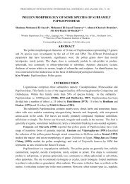
114PREVALENCE OF TOXIGENIC BACTERIA IN SOME EGYPTIAN FOODliv<strong>in</strong>g organisms (Smith, 1997 and Wong and Hancock, 2000). In this study, total cellularprote<strong>in</strong>s <strong>of</strong> selected virulent stra<strong>in</strong>s <strong>of</strong> B. cereus (GT1, CH, LB3 & G8) and S. aureus(S, S1,S2 & S3) were extracted and then fractionated by means <strong>of</strong> polyacrylamide gelelectrophoresis. The electrophoretic separation verify<strong>in</strong>g prote<strong>in</strong> patterns <strong>of</strong> selected stra<strong>in</strong>srevealed prote<strong>in</strong> bands with molecular weights rang<strong>in</strong>g between 33 to 108 kDa <strong>in</strong> case <strong>of</strong> B.cereus stra<strong>in</strong>s and between 17 to 220 kDa <strong>in</strong> case <strong>of</strong> S. aureus (Photo 1). Us<strong>in</strong>g gel proanalyzer the gel photo had been analyzed and revealed vary<strong>in</strong>g similarity percentages with<strong>in</strong>B. cereus stra<strong>in</strong>s between 20 to 90% and between 63.2 to 82.4% <strong>in</strong> S. aureus ). Also,difference <strong>in</strong> number <strong>of</strong> separated cellular prote<strong>in</strong> bands <strong>in</strong> the <strong>in</strong>vestigated stra<strong>in</strong>s wasobserved (fig1). B. cereus GT1 showed 9 prote<strong>in</strong> bands while B. cereus G8 showed only 7bands. Concern<strong>in</strong>g S. aureus stra<strong>in</strong>s S & S3 showed 19 bands and stra<strong>in</strong>s S1 & S2 17bands.Photo(1): Polyacrylamide gel electrophoresis show<strong>in</strong>g the cellular prote<strong>in</strong> pattern <strong>of</strong> virulent isolates.M: prote<strong>in</strong> marker (mixture <strong>of</strong> 7 purified prote<strong>in</strong>s with mol. wt. 116,97.4,66.2,37.6,28.5,18.4 and 14 kDa.Lanes (1,2,3 &4): B.cereus stra<strong>in</strong>sGH, GT1, LB3, G8. and lanes (5,6,7,&8) :S.aureus stra<strong>in</strong>s S, S1, S2, S3.Table(3): Similarity percentage betweenselected B. cereus stra<strong>in</strong>sTable(4):Similarity percentage betweenselected S. aureus stra<strong>in</strong>sStra<strong>in</strong>s S S1 S2 S3S 100 80 73.7 63.2S1 80 100 84.2 68.4S2 82.4 82.4 100 70.1S3 63.2 68.4 63.2 100Stra<strong>in</strong>s CH GT1 LB3 G8CH 100 89 33.3 44.4GT1 90 100 20 40LB3 30 20 100 20G8 57.1 57.1 28.6 100
Nadia Mohamed Awny; Azza A. M. Abou Zeid and Mohamed Ahmed Abdo 115MarkerB. cereus(CH)B. cereus(GT1)B. cereus(LB3)B. cereus(G8)S. aureus(S)S. aureus(S1)S. aureus(S2)S. aureus(S3)Fig (1): Pro-analysis <strong>of</strong> separated cellular prote<strong>in</strong> bands <strong>of</strong> the eight<strong>toxigenic</strong> stra<strong>in</strong>s on acrylamide gel show<strong>in</strong>g their <strong>in</strong>tensities andmolecular weights compared with marker prote<strong>in</strong> (6.8-214kDa).
116PREVALENCE OF TOXIGENIC BACTERIA IN SOME EGYPTIAN FOODPhoto(2): Polyacrylamide gel electrophoresis show<strong>in</strong>g the prote<strong>in</strong> pattern <strong>of</strong> cell free filtrates <strong>of</strong> the selected <strong>toxigenic</strong> isolates.M: prote<strong>in</strong> marker (mixture <strong>of</strong> 7 purified prote<strong>in</strong>s with mol. wt. 116,97.4,66.2,37.6,28.5,18.4 and 14 kDa.Lanes (1,2,3 &4): B.cereus stra<strong>in</strong>sCH, GT1,LB3 & G8 and lanes (5,6,7,&8) :S.aureus stra<strong>in</strong>s S, S1, S2, S3.Table(5): Similarity percentage betweenselected B. cereus stra<strong>in</strong>sStra<strong>in</strong>s CH GT1 LB3 G8CH 100 80 60 60GT1 80 100 80 60LB3 43 57 100 71.4G8 50 50 83.3 100Table(6): Similarity percentage betweenselected S. aureus stra<strong>in</strong>sStra<strong>in</strong>s S S1 S2 S3S 100 100 66.7 66.7S1 75 100 50 50S2 50 50 100 50S3 50 50 50 100Also, the pro-analysis <strong>of</strong> the extracellular prote<strong>in</strong> bands <strong>of</strong> the tested isolates revealedvary<strong>in</strong>g similarity between the virulent stra<strong>in</strong>s rang<strong>in</strong>g between50 to 83.3% <strong>in</strong> B. cereus andbetween 50 to 75% <strong>in</strong> S. aureus (tables5&6). Photo (2) showed obvious prote<strong>in</strong> bands <strong>in</strong> lanes1,2,3&4 hav<strong>in</strong>g molecular weights (19.26, 30.24, 33.98, 53.44 & 72.59 kDa) ; (19.6, 30. 12,33, 75, 35.53 & 53.19 kDa); (22.11, 30. 03, 33,67, 35.08, 43.24, 53.14 & 234.36 kDa)&(19.63,22.77, 30. 93, 33,53, 43.23 & 234.36 kDa); <strong>in</strong> case <strong>of</strong> B. cereus stra<strong>in</strong>s CH,GT1,LB3&G8, respectively. Meanwhile S. aureus stra<strong>in</strong>s S, S1, S2&S3 possessedextracellular prote<strong>in</strong>s with molecular weights (43.23, 93.486& 234.36kDa); (22.82, 43.54,53.676& 234.36kDa); (22.67, 33.21, 43.57 & 92.19kDa) & (30.39, 33.31, 43.74 &234.36kDa), respectively.Beecher and MacMillan (1990&1991) reported that molecular weights <strong>of</strong> B. cereusenterotox<strong>in</strong> prote<strong>in</strong>s were 35, 36 and 45 kDa for b<strong>in</strong>d<strong>in</strong>g prote<strong>in</strong> B, lytic prote<strong>in</strong> L1 (HBLD)and lytic prote<strong>in</strong> L2 (HBLC), respectively. Beecher and Wong (1994) also found that themolecular weight <strong>of</strong> B component was 37.8kDa, L1 was 38.5kDa and L2 was 43.2kDa.Schoeni and Wong (1999) reported that the three components isolated from prototype stra<strong>in</strong><strong>of</strong> B. cereus F837/76 have molecular weights <strong>of</strong> 37.5, 38.2 and 43.5 kDa, respectively. Theyalso added that an <strong>in</strong>dividual stra<strong>in</strong> could produce s<strong>in</strong>gle or multiple bands <strong>of</strong> eachcomponent. They observed two bands (38&42kDa) for B prote<strong>in</strong>, two L1prote<strong>in</strong>s(38&41kDa) and two L2 prote<strong>in</strong>s (both 43kDa) <strong>in</strong> a soil isolate encoded S1C stra<strong>in</strong>.
Nadia Mohamed Awny; Azza A. M. Abou Zeid and Mohamed Ahmed Abdo 117MarkerB. cereus(..)B. cereus(..)B. cereus(..)B. cereus(..)S. aureus(..)S. aureus(..)S. aureus(..)S. aureus(..)Fig(2): Pro-analysis <strong>of</strong> separated extra-cellular prote<strong>in</strong> bands <strong>of</strong> theeight <strong>toxigenic</strong> stra<strong>in</strong>s on acrylamide gel show<strong>in</strong>g their <strong>in</strong>tensitiesand molecular weights compared with marker prote<strong>in</strong> (6.8-214kDa).Concern<strong>in</strong>g S. aureus, the toxic shock caus<strong>in</strong>g SEs are s<strong>in</strong>gle cha<strong>in</strong> polypeptideshav<strong>in</strong>g a molecular weight rang<strong>in</strong>g from 27-29 kDa. SEs can withstand boil<strong>in</strong>g temperaturefor several m<strong>in</strong>utes, extremes <strong>of</strong> pH (3-11) and protease digestion by gastric enzymes
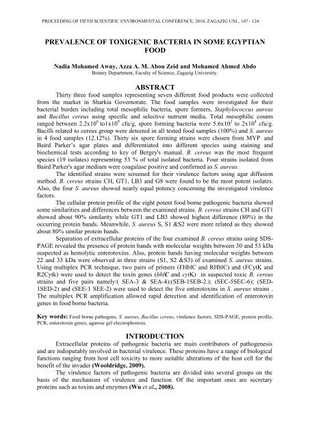
118PREVALENCE OF TOXIGENIC BACTERIA IN SOME EGYPTIAN FOOD(Soriano et al., 2002). Twenty different types <strong>of</strong> SEs, viz., SEA through SEE, SEG throughSER and SEU have already been discovered, however, only a few <strong>of</strong> the tox<strong>in</strong> serotypes arefrequently associated with <strong>food</strong> poison<strong>in</strong>g outbreaks (Mart<strong>in</strong> et al., 2004; Smyth et al., 2005and Fernández et al., 2006).M 1 2 3 4695bp565bpPhoto(3): Agarose gel show<strong>in</strong>g the PCR amplicons result<strong>in</strong>g from amplification <strong>of</strong> enterotox<strong>in</strong>s genes hblCand cytK us<strong>in</strong>g FHblC & RHblC and FCytk & R2Cytk primers. Lane M is 100bp DNA ladder marker;lanes 1,2,3&4 DNA amplicons <strong>of</strong> B. cereus G8,LB3 ,GT13&CH respectively. The gel reveals presence <strong>of</strong>both enterotox<strong>in</strong> genes (hblC &cytK) <strong>in</strong> stra<strong>in</strong>sGT1, LB3 & G8.Multiplex PCR technique has been recently used for rapid detection anddiscrim<strong>in</strong>ation <strong>of</strong> enterotox<strong>in</strong>s genes <strong>in</strong> B. cereus (Gu<strong>in</strong>ebretiere et al., 2006 andNagmwongsatit et al., 2008); and for direct detection <strong>of</strong> <strong>food</strong> contam<strong>in</strong>ation withentero<strong>toxigenic</strong> B. cereus as well (Ombui et al. 2008).Ngmwongsatit et al. (2008) have developed and evaluated group <strong>of</strong> new primerswhich were highly efficient <strong>in</strong> detect<strong>in</strong>g the tox<strong>in</strong> genes <strong>in</strong> 100% <strong>of</strong> their tested B. cereus andB. thur<strong>in</strong>gensis stra<strong>in</strong>s. Thus, it could be expected that the presence <strong>of</strong> either genes is an<strong>in</strong>dication for the presence <strong>of</strong> the whole operon.In this study the primers designed by Ngmwongsatit et al. (2008) were used underspecific multiplex PCR conditions, previously mentioned <strong>in</strong> materials and methods, to detectpresence <strong>of</strong> the enterotox<strong>in</strong> genes (hblC &cytK) <strong>in</strong> tested stra<strong>in</strong>s. Photo (3) revealed presence<strong>of</strong> amplified DNA fragments <strong>of</strong> the two tox<strong>in</strong> genes <strong>in</strong> three stra<strong>in</strong>s <strong>of</strong> B. cereus (CH, GT1 &G8) <strong>in</strong> one quick step. The tox<strong>in</strong> genes hblC & cytK predicted molecular sizes <strong>of</strong> 695 & 565bp, respectively.
Nadia Mohamed Awny; Azza A. M. Abou Zeid and Mohamed Ahmed Abdo 119478bp(seb)317bp(sed)244bp(sec)170bp(see)127bp(sea)Photo(4): Agarose gel show<strong>in</strong>g the PCR amplicons result<strong>in</strong>g from amplification <strong>of</strong> enterotox<strong>in</strong>s genes sea,seb, sec-1, sed and see us<strong>in</strong>g their specific primers. Lane M is 100bp DNA ladder marker; lanes 1,2,3&4DNA amplicons <strong>of</strong> S. aureus S,S1,S2&S3 respectively. The gel reveals presence <strong>of</strong> enterotox<strong>in</strong>s sea & sed<strong>in</strong> S. aureus S1 and sed <strong>in</strong> S. aureus S3.Regard<strong>in</strong>g the enterotox<strong>in</strong> genotype, previous studies on S. aureus proved that enterotox<strong>in</strong>PCR determ<strong>in</strong>ations are <strong>in</strong> a high agreement (97–100%) with the tox<strong>in</strong> production as def<strong>in</strong>ed byimmunoassays (Fueyo et al., 2001, Letertre et al., 2003 and McLauchl<strong>in</strong> et al., 2000).Enterotox<strong>in</strong> genotyp<strong>in</strong>g <strong>of</strong> tested stra<strong>in</strong>s revealed presence <strong>of</strong> sed gene <strong>in</strong> both stra<strong>in</strong>s S.aureus (S1 & S3) and sea gene <strong>in</strong> stra<strong>in</strong> (S1) only.P<strong>in</strong>to et al. (2005) found a total <strong>of</strong> 40 (30%) S. aureus <strong>food</strong> isolates positive for se genes.Among them, the sec genotype was the most frequent (22 stra<strong>in</strong>s,20% <strong>of</strong> total se positive stra<strong>in</strong>s) andsea the second more frequent (14 stra<strong>in</strong>s, 13%),which is <strong>in</strong> accordance with the results obta<strong>in</strong>ed by(Fueyo et al., 2001 ). Data on enterotox<strong>in</strong> genotype confirmed the group<strong>in</strong>g <strong>of</strong> stra<strong>in</strong>s nuc PCRpositive and se PCR positive <strong>in</strong> S. aureus clusters.On the basis <strong>of</strong> these results, we suggest amplification <strong>of</strong> enterotox<strong>in</strong> genes as targetgenes us<strong>in</strong>g multiplex PCR test as a rapid and valuable technique that can be applied directlyto s<strong>in</strong>gle colonies grow<strong>in</strong>g on selective plates for a rapid, accurate and unequivocalidentification <strong>of</strong> B. cereus and S. aureus. It could be implemented as an alternative tophenotypic and immunology-based tests <strong>in</strong> the rout<strong>in</strong>e <strong>food</strong> microbiological analysis.REFERENCESAgata, N.; Ohta, M. and Yokoyama, K. (2002). Production <strong>of</strong> Bacillus cereus emetic tox<strong>in</strong>(cerulide) <strong>in</strong> various <strong>food</strong>s. Int. J. Food Microbiol., 73: 23-27.APHA, (1992). Compendium <strong>of</strong> methods for the microbiological exam<strong>in</strong>ation ,3rd Edn.,Wash<strong>in</strong>gton, American Public Health Association.Balaban, N. and Rasooly, A. (2000). Staphylococcal enterotox<strong>in</strong>s, Int. J. Food Microbiol. 61: 1–10.Becker, K., R. Roth, and G. Peters. (1998). Rapid and specific detection <strong>of</strong> <strong>toxigenic</strong>Staphylococcus aureus: use <strong>of</strong> two multiplex PCR enzyme immunoassays for
120PREVALENCE OF TOXIGENIC BACTERIA IN SOME EGYPTIAN FOODamplification and hybridization <strong>of</strong> staphylococcal enterotox<strong>in</strong> genes, exfoliativetox<strong>in</strong> genes, and toxic shock syndrome tox<strong>in</strong> 1 gene. J. Cl<strong>in</strong>. Microbiol. 36:2548–2553.Beecher, D.J. and Macmillan, J.D. (1990). A novel bicomponent haemolys<strong>in</strong> from Bacilluscereus. Infect. Immun. 58, 2220–2227.Beecher, D.J. and Macmillan, J.D. (1991). Characterization <strong>of</strong> the components <strong>of</strong>haemolys<strong>in</strong> BL from Bacillus cereus. Infect. Immun. 59, 1778–1784.Beecher, D.J. and Wong, A.C.L. (1994). Identification <strong>of</strong> heamolys<strong>in</strong> Bl produc<strong>in</strong>g Bacilluscereus isolates by a discont<strong>in</strong>ous haemolytic pattern <strong>in</strong> blood agar. Appl. Environ.Microbial. 60:1646-1651. Bergdoll, M.S. (1989). Staphylococcus aureus, In :M.P. Doyle (Ed.), Foodborne <strong>bacteria</strong>l pathogens, Marcel Dekker, New York, pp.463–523.Beecher, D.J. and Wong, A.C.L. (2000b). Cooperative, synergistic and antagonistic haemolytic<strong>in</strong>teractions between haemolys<strong>in</strong> BL, phosphatidylchol<strong>in</strong>e phospholipase C andsph<strong>in</strong>gomyel<strong>in</strong>ase from Bacillus cereus. Microbiology 146, 3033–3039.Bergdoll, M.S. (1989). Staphylococcus aureus. In: Foodborne Bacterial Pathogens (Doyle, M.P.,ed.). Marcel Dekker, Inc., New York, NY, USA, pp. 463-523.Bergdoll, M. S.; Huang, I.Y. and Schantz, E. J. (1974). Chemistry <strong>of</strong> the staphylococcalenterotox<strong>in</strong>s. Journal <strong>of</strong> Agricultural and Food Chemistry, 22(1):9-13.Boerema J.A.; Clemens, R. and Brightwel, G. (2006). Evaluation <strong>of</strong> molecular methods todeterm<strong>in</strong>e entero<strong>toxigenic</strong> status and molecular genotype <strong>of</strong> bov<strong>in</strong>e, ov<strong>in</strong>e, humanand <strong>food</strong> isolates <strong>of</strong> Staphylococcus aureus. Int.J. Food Microbiol 107: 192-201.Callegan, M.C.; Cochran, D.C.; Kane, S.T.; Gilmore, M.S.; Gom<strong>in</strong>et, M. and Lereclus,D. (2002). Contribution <strong>of</strong> membrane-damag<strong>in</strong>g tox<strong>in</strong>s to Bacillusendophthalmitis pathogenesis. Infect. Immun. 70, 5381–5389.Difco, Manual (1994). (10 th ed). Dehydrated culture media reagents for microbial. DIFCOLaboratories. Detroit Michigan 48232 USA.D<strong>in</strong>ges, M. M.; Orw<strong>in</strong>, P. M., and Schlievert, P. M. (2000). Exotox<strong>in</strong>s <strong>of</strong> Staphylococcusaureus. Cl<strong>in</strong>ical Microbiology Review, 13, 16–34.DoCarmo, L. S.; Cumm<strong>in</strong>gs, C.; L<strong>in</strong>ardi, V. R.; Dias, R. S.; De Souza, J. M.; De Sena,M. J.; Dos Santos, D. A.; Shupp, J. W.; Peres Pereira, R.K. and Jett, M.(2004). A case study <strong>of</strong> a massive staphylococcal <strong>food</strong> poison<strong>in</strong>g <strong>in</strong>cident. FoodBorne Pathogens and Disease, December 2004, vol. 1, no. 4, p. 241-246.Doyle, M.P. (eds) (1989). Food borne <strong>bacteria</strong>l pathogens. Marcel Dekker, New York.Drobniewski, F. (1993). Bacillus cereus and related species. Cl<strong>in</strong> Microbiol Rev. (4): 324-338.Duc, L.H.; Dong, T.C.; Logan, N.A.; Sutherland, A.D.; Taylor, J. and Cutt<strong>in</strong>g, S.M.(2005). Cases <strong>of</strong> emesis associated with <strong>bacteria</strong>l contam<strong>in</strong>ation <strong>of</strong> an <strong>in</strong>fantbreakfast cereal product. Int. J. Food Microbiol., 102: 245-251.FDA, (2001). "Food and Drug Adm<strong>in</strong>istration", Bacteriological Analytical Manual. E.J.Rhodhehamel and S.M. Harmon, Chapter, 14.FDA/CFSAN, (2007). U.S. Food and Drug Adm<strong>in</strong>istration. Center for <strong>food</strong> safety andapplied nutrition. Bacillus cereus and other Bacillus spp. In: "Food BornePathogenic Microorganisms and Natural Tox<strong>in</strong>s Handbook". mow/las/dav/ear.Fernanez, M. M.; De Marzi, M. C.; Berguer, P; Burzyn, D; Langley, R. J.; Piazzon, I ;Mariuzza, R. A. and Malchiodi, E. L (2006). B<strong>in</strong>d<strong>in</strong>g <strong>of</strong> natural variants <strong>of</strong>staphylococcal superantigens SEG and SEI to TCR and MHC class II molecule.Molecular Immunology. 43( 7):927-938.Fueyo, J.M.; Mart<strong>in</strong>, M.C.; Gonzalez-Hevia, M.A. and Mendoza, M.C. (2001).Enterotox<strong>in</strong> production and DNA f<strong>in</strong>gerpr<strong>in</strong>t<strong>in</strong>g <strong>in</strong> Staphylococcus aureus isolated
Nadia Mohamed Awny; Azza A. M. Abou Zeid and Mohamed Ahmed Abdo 121from human and <strong>food</strong> samples. Relations between genetic types and enterotox<strong>in</strong>s.Int. J. Food Microbiol. 67, 139–145.Gravet, A.; Rondeau, M.; Harf-Monteil, C.; Grunenberger, F.; Monteil, H. ; Scheftel, J.M. and Prevost, G. (1999). Predom<strong>in</strong>ant Staphylococcus aureus isolated fromantibiotic-associated diarrhea is cl<strong>in</strong>ically relevant and produces enterotox<strong>in</strong> A andthe bicomponent tox<strong>in</strong> LukE-LukD. J. Cl<strong>in</strong>. Microbiol.37:4012–4019.Granum, P.E (2001). Bacillus cereus. In: " Food Microbiolog Fundamentals and Frontiers",Doyle, M.P. ( Ed.), pp. 373-381. Wash<strong>in</strong>gton, DC. USA: ASM press.Granum, P.E. and Baird-Parker, T.C. (2000). Bacillus species. In: Lund, B., Baird-Parker, T.,Gould, G. (Eds.), The microbiological safety and quality <strong>of</strong> <strong>food</strong>. AspenPublisher, Gaitherburg, MD, USA, pp. 1029–1039.Granum P.E., Lund T. (1997): Bacillus cereus and its <strong>food</strong> poison<strong>in</strong>g tox<strong>in</strong>s. FEMSMicrobiol. Lett. 157, 223–228.Gu<strong>in</strong>ebretiere, M.; Fagerlund, A.; Granum, P.E. and Nguyen-The, C. (2006). Rapiddiscrim<strong>in</strong>ation <strong>of</strong> cytK-1 and cytK-2 genes <strong>in</strong> Bacillus cereus stra<strong>in</strong>s by a novelPCR system. FEMS Microbiol. Lett., 59(1): 74-80.Hill, W.E. (1996). The polymerase cha<strong>in</strong> reaction: application for the detection <strong>of</strong> <strong>food</strong> bornepathogens. Critical reviews <strong>in</strong> <strong>food</strong> science and nutrition. 36:123-173.Hill, W.E.; and Keasler, S.P. (1991): identification <strong>of</strong> <strong>food</strong> borne pathogens by nucleic acidhybridization . International journal <strong>of</strong> <strong>food</strong> microbiology 12: 67- 76.Hilliard, N.J.; Schelonka, R.L. and Waites, K. (2003). Bacillus cereus bacteremia <strong>in</strong> apreterm neonate. J Cl<strong>in</strong> Microbiol 41:3441–3444.Jett, M.; Neill, R.; Welch, C.; Boyle, T.; Bernton, E.; Hoover, D.; Lowell, G.; Hunt, R.E.;Chatterjee, S. and Gemski, P. (1994). Identification <strong>of</strong> staphylococcalenterotox<strong>in</strong> B sequences important for <strong>in</strong>duction <strong>of</strong> lymphocyte proliferation byus<strong>in</strong>g synthetic peptide fragments <strong>of</strong> tox<strong>in</strong>. Infection and Immunity. 62(8):3408-3415.Kenny, K.; Reiser, R. F. ; Bastida-Corcucra, F. D. and Norcross, N. L. (1993).Production <strong>of</strong> enterotox<strong>in</strong>s and toxic shock syndrome tox<strong>in</strong> by bov<strong>in</strong>e mammaryisolates <strong>of</strong> Staphylococcus aureus. J. Cl<strong>in</strong>. Microbiol. 31:706–707.K<strong>in</strong>g, N.J.; Whyte, R. and Hudson, J.A. (2007). Presence and significance <strong>of</strong> Bacilluscereus <strong>in</strong> dehydrated potatoes. J.Food Prot., 70(2): 514-520.Klotz, M.; Opper, S.; Heeg, K. and Zimmermann, S. (2003). detection <strong>of</strong> Staphylococcusaureus Enterotox<strong>in</strong>s A to D by real-time Fluorescence PCR assay. Journal <strong>of</strong>cl<strong>in</strong>ical microbiology. 41(10): 4683-4687.Kotiranta, A.; Lounatmaa, K. and Haapasalo M. (2000). Epidemiology and pathogenesis<strong>of</strong> Bacillus cereus <strong>in</strong>fections. Microbiol Infect 2:189–198.Krieg, N.R and Holt, J.G. (1984): Bergey,s Manual <strong>of</strong> Systematic Bacteriology vol. 1 ,2 .9thed . Baltimore ,pp 1599.Laemmli, U.K. (1970).Cleavage <strong>of</strong> structural prote<strong>in</strong>s dur<strong>in</strong>g the assembly <strong>of</strong> the head <strong>of</strong>bacteriophage T4. Nature(London), 227: 680-685.Letertre, C.; Perelle, S.; Dilasser, F. and Fach, P. (2003).Identification <strong>of</strong> a new putativeenterotox<strong>in</strong> SEU encoded by the egc cluster <strong>of</strong> Staphylococcus aureus. J. Appl.Microbiol. 95:38–43.L<strong>in</strong>dback, T.; Fagerlund, A.; Rodland, M.S. and Granum, P.E (2004). Characterization <strong>of</strong>the Bacillus cereus Nhe enterotox<strong>in</strong>. Microbiology 150:3959–3967.LKB Application note (1997). SDS-PAGE.Llewelyn M, Cohen J (2002). Superantigens: Microbial agents that corrupt immunity. LancetInfect Dis 2: 156-162
122PREVALENCE OF TOXIGENIC BACTERIA IN SOME EGYPTIAN FOODLund, T. and Granum, P.E. (1997). Comparison <strong>of</strong> biological effect <strong>of</strong> the two differententerobtox<strong>in</strong> complexes isolated from three different stra<strong>in</strong>s <strong>of</strong> Bacilus cereus.Microbiology, 143:3329-3336.Mahler, H.; Pasi, A.; Kramer, J.M.; Schulte, P.; Scog<strong>in</strong>g, A.C.; Bar, W. andKrahenbuhl, S. (1997). Fulm<strong>in</strong>ant liver failure <strong>in</strong> association with emetic tox<strong>in</strong><strong>of</strong> Bacillus cerus. New England Journal <strong>of</strong> Medic<strong>in</strong>e, 336: 1142-1148.Mart<strong>in</strong>, M.C.; Fueyo, J.M.; Gonzalez-Hevia, M.A. and Mendoza, M.C. (2004). Geneticprocedures for identification <strong>of</strong> entero<strong>toxigenic</strong> stra<strong>in</strong>s <strong>of</strong> Staphylococcus aureusfrom three <strong>food</strong> poison<strong>in</strong>g outbreaks. International Journal <strong>of</strong> Food Microbiology.94( 3):279-286.Matsunaga, T., S. Kamata, N. Kakiichi, and A. Uccida. (1993). Characteristics <strong>of</strong>Staphylococcus aureus isolated from peracute, acute and chronic bov<strong>in</strong>e mastitis.J. Vet. Med. Sci. 55:297–300.McKillip, J.L. (2000): Prevalence and expression <strong>of</strong> enterotox<strong>in</strong>s <strong>in</strong> B. cereus and other B.spp., a literature review. Antonie van Leeuwenhoek 77, 393–399.McLauchl<strong>in</strong>, J.; Narayanan, G. L.; Mithani, V. and O’Neill, G. (2000). The detection <strong>of</strong>enterotox<strong>in</strong>s and toxic shock syndrome tox<strong>in</strong> genes <strong>in</strong> Staphylococcus aureus bypolymerase cha<strong>in</strong> reaction. J. Food Prot. 63(4):479–488.Miethke, T., C. Wahl, K. Heeg, B. Echtenacher, P. H. Krammer, and H. Wagner. (1992).T cell-mediated lethal shock triggered <strong>in</strong> mice by the superantigen staphylococcalenterotox<strong>in</strong> B: critical role <strong>of</strong> tumor necrosis factor. J. Exp. Med. 175:91–98.Misra, A.K. and Kuila, R.K. (1992). Use <strong>of</strong> Bifidobacterium bifidum <strong>in</strong> the manufacture <strong>of</strong>bifidus milk and its anti<strong>bacteria</strong>l activity. Lait,72(2): 213-220Ngamwongsatit, P.; Busari, W.; Pianariyanon, P.; Pulsrikarn, C.; Ohba, M.; Assavanig,A and Panbangred, W. (2008). Broad distribution <strong>of</strong> entertox<strong>in</strong> genes (hblCDA,nheABC, cytK, and entFM) among Bacillus thur<strong>in</strong>giensis and Bacillus cereus asshown by novel primers. International journal <strong>of</strong> <strong>food</strong> microbiology. 121:352-356.Olsen, J. E. (2000). DNA-based methods for detection <strong>of</strong> <strong>food</strong>-borne <strong>bacteria</strong>l pathogens.Food research <strong>in</strong>ternational. 33 (3-4):257-266.Olsen, J. E.; Aabo, S.; Rasmussen, O. F. and Rossen, L. (1995). Oligo-nucleotide probesspecific for the genus Salmonella and for S. typhimurium. Letters <strong>in</strong> AppliedMicrobiology. 20:160-166.Ombui, J.N.; Gitahi, N. and Gicheru, M. (2008). Direct detection <strong>of</strong> Bacillus cereusenterotox<strong>in</strong> genes <strong>in</strong> <strong>food</strong> by multiplex polymerase cha<strong>in</strong> reaction. InternationalJournal <strong>of</strong> Integrative Biology, 2(3):172.Oxoid Manual (1990). Culture media, <strong>in</strong>clud<strong>in</strong>g <strong>in</strong> gradients and other laboratory services.P<strong>in</strong>to, B; Chenoll, E. and Aznar, R. (2005). Identification and typ<strong>in</strong>g <strong>of</strong> <strong>food</strong> borneStaphylococcus aureus by PCR-based techniques. Syst. Appl.microbial. 37:4012–4019.Re<strong>in</strong>heimer, J.A.; Demkow, M.R.; Canditi, M.C. Austr, B. (1990). J.Dairy Technol., 45:5-9.Rhodehamel, E.J. and Harmon, S.M. (2001). Bacteriological Analytical Manual Onl<strong>in</strong>e.Chapter 14, Bacillus cereus. U.S. FDA, Center for Food Safety & AppliedNutrition.Schneider, K.R.; Parish, M.E.; Goodrich, R.M. and Cook<strong>in</strong>gham, T. (2004). Prevent<strong>in</strong>g<strong>food</strong> borne illness: Bacillus cereus and Bacillus anthracis. University <strong>of</strong> Florida,IFAS Extension. FSHNO04-05.http://edis.ifas.ufl.edu.Schoeni, J.L. and Wong, A.C.L. (2005). B. cereus <strong>food</strong> poison<strong>in</strong>g and its tox<strong>in</strong>s. J. FoodProt. 68, 636–648.
Nadia Mohamed Awny; Azza A. M. Abou Zeid and Mohamed Ahmed Abdo 123Shaheen, R.; Andersson, M.A.; Apetroaie, C.; Schulz, Ehl<strong>in</strong>g-Schulz, Ollila<strong>in</strong>en, V. andSalk<strong>in</strong>oja- Salonen, M.S. (2006). Potential <strong>of</strong> selected <strong>in</strong>fant <strong>food</strong> formulas forproduction <strong>of</strong> Bacillus cereus emetic tox<strong>in</strong>, cerulide. Int. J. Food Microbiol.,107(3): 287-294.Smyth, D. S.; Hartigan, P. J.; Meaney, W. J.; Fitzgerald, J. R.; Deobald, C. F.; Bohach,G. A. and Smyth, . J.(2005). Superantigen genes encoded by the egc cluster andSaPIbov are predom<strong>in</strong>ant among Staphylococcus aureus isolates from cows, goats,sheep, rabbits and poultry. Journal <strong>of</strong> Medical Microbiology. 54(4):401- 411.Soriano, J. M; Font, G; Molto, J. C. and Manes, J. (2002). Entero<strong>toxigenic</strong> staphylococciand their tox<strong>in</strong>s <strong>in</strong> restaurant <strong>food</strong>s. Trends <strong>in</strong> Food Science and Technology.13(2):60-67.Su, Y.C., Lee Wong, A. (1997): Current perspectives on detection <strong>of</strong> staphylococcalenterotox<strong>in</strong>s. J. Food. Protect. 60, 195–202.Svensson, B.; Monthan, A.; Gu<strong>in</strong>ebretiere, H.M.; Nguyen, C. and Christiansson, A.(2007). Tox<strong>in</strong> production potential and the detection <strong>of</strong> tox<strong>in</strong> genes among stra<strong>in</strong>s<strong>of</strong> Bacillus cereus group isolated along the dairy production cha<strong>in</strong>. InternationalDairy Journal 17:1201-1208.Todar, K. (2005). Textbook <strong>of</strong> Bacteriology. The Genus Bacillus. Onl<strong>in</strong>e.Wolcott, M.J. (1991). DNA-based rapid methods for the detection <strong>of</strong> <strong>food</strong> borne pathogens.Journal <strong>of</strong> <strong>food</strong> protection. 54:387-401.Wooldridge, K. (2009). Secretory mechanisms and role pathogenesis. In: <strong>bacteria</strong>l secretedprote<strong>in</strong>s. Published by Caister Academic Press.U.K.Wu, H. J.; Wang, A. H. J. and Jenn<strong>in</strong>gs, P. M. (2008). Discovery <strong>of</strong> virulence factors <strong>of</strong>pathogenic <strong>bacteria</strong>. Current opnion <strong>in</strong> chemical biology. 12:93-101.Yokoyama, K.; Ito, M.; Agata, N.; Isobe, M.; Shibayama, K.; Horii, T. and Ohta, M.(1999). Pathological effect <strong>of</strong> synthetic cerulide, an emetic tox<strong>in</strong> <strong>of</strong> Bacilluscereus, is reversible <strong>in</strong> mice. FEMS. Immunol. Med. Microbiol., 24: 115-120.
124PREVALENCE OF TOXIGENIC BACTERIA IN SOME EGYPTIAN FOODBaird Parker’sMYP agaragarG8LB3GT1CHLB3GT!GT1CHS1SS2kDakDaPCR