Scanning Near-Field Optical Microscopy and Spectroscopy as a ...
Scanning Near-Field Optical Microscopy and Spectroscopy as a ...
Scanning Near-Field Optical Microscopy and Spectroscopy as a ...
You also want an ePaper? Increase the reach of your titles
YUMPU automatically turns print PDFs into web optimized ePapers that Google loves.
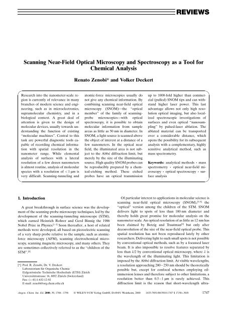
REVIEWS<strong>Scanning</strong> <strong>Near</strong>-<strong>Field</strong> <strong>Optical</strong> <strong>Microscopy</strong> <strong>and</strong> <strong>Spectroscopy</strong> <strong>as</strong> a Tool forChemical AnalysisRenato Zenobi* <strong>and</strong> Volker DeckertResearch into the nanometer-scale regionis currently of relevance in manybranches of modern science <strong>and</strong> engineering,such <strong>as</strong> in microelectronics,supramolecular chemistry, <strong>and</strong> in abiological context. A great deal ofattention is given to the design ofmolecular devices, usually towards underst<strong>and</strong>ingthe function of existingªmolecular machinesº. Central to thist<strong>as</strong>k are powerful diagnostic tools capableof recording chemical informationwith spatial resolution in thenanometer range. While elementalanalysis of surfaces with a lateralresolution of a few dozen nanometersis almost routine, analysis of molecularspecies with a resolution of < 1 mm isvery difficult. <strong>Scanning</strong>-tunneling <strong>and</strong>atomic-force microscopies usually donot give any chemical information. Bycombining scanning near-field opticalmicroscopy (SNOM)Ðthe ªopticalmemberº of the family of scanningprobemicroscopiesÐwith opticalspectroscopy, it is possible to obtainmolecular information from sampleare<strong>as</strong> <strong>as</strong> little <strong>as</strong> 50 nm in diameter. InSNOM, a light source is scanned abovethe object of interest at a distance of afew nanometers. In the optical nearfield, the illuminated area is not subjectto the Abbe diffraction limit, butmerely by the size of the illuminatingsource. High quality SNOM probes canbe reproducibly prepared by a chemical-etchingmethod. These etchedprobes have an optical transmissionup to 1000-fold higher than commercial(pulled) SNOM tips <strong>and</strong> can withst<strong>and</strong>higher l<strong>as</strong>er power. This l<strong>as</strong>tadvantage allows not only high resolutionoptical imaging, but also localizedspectroscopic investigations ofsurfaces <strong>and</strong> even optical ªnanosamplingºby pulsed-l<strong>as</strong>er ablation. Theablated material can be transportedover a considerable distance, whichopens the possibility for its subsequentanalysis with a complementary, highlysensitive analytical method, such <strong>as</strong>m<strong>as</strong>s spectrometry.Keywords: analytical methods ´ m<strong>as</strong>sspectrometry ´ optical near-field microscopy´ optical spectroscopy ´ surfaceanalysis1. IntroductionA great breakthrough in surface science w<strong>as</strong> the developmentof the scanning-probe microscopy techniques, led by thedevelopment of the scanning-tunneling microscope (STM),which earned Heinrich Rohrer <strong>and</strong> Gerd Binnig the 1986Nobel Prize in Physics. [1±3] Soon thereafter, a host of relatedmethods were developed, all b<strong>as</strong>ed on piezoelectric scanningof a very sharp probe relative to the sample, such <strong>as</strong> atomicforcemicroscopy (AFM), scanning electrochemical microscopy,scanning magnetic microscopy, <strong>and</strong> many others. Theyare sometimes collectively referred to <strong>as</strong> the ªchildren of theSTMº. [4][*] Prof. R. Zenobi, Dr. V. DeckertLaboratorium für Organische ChemieEidgenössische Technische Hochschule (ETH) ZürichUniversitätsstr<strong>as</strong>se 16, 8092 Zürich (Switzerl<strong>and</strong>)Fax: (‡ 41) 1-632-12-92E-mail: zenobi@org.chem.ethz.chOf particular interest to applications in molecular science isscanning near-field optical microscopy (SNOM), [5±8] theªopticalº version among the children of the STM. SNOMdelivers light to spots of less than 100 nm diameter <strong>and</strong>thereby holds great promise for molecular analysis on thenanometer scale. An optical resolution of <strong>as</strong> little <strong>as</strong> 12 nm h<strong>as</strong>been claimed by Betzig <strong>and</strong> Trautman [9] but only afterdeconvolution of the size of the near-field optical probe. Thisspatial resolution h<strong>as</strong> not been reproduced lately by otherresearchers. Delivering light to such small spots is not possibleby conventional optical methods, such <strong>as</strong> by a focussed l<strong>as</strong>erbeam. It is also impossible to resolve features separated byless than l/2 by conventional optical microscopy, where l isthe wavelength of the illuminating light. This limitation isimposed by the Abbe diffraction limit. At visible wavelengths,a resolution approaching 200 ± 250 nm should be theoreticallypossible but, except for confocal schemes employing oilimmersionlenses <strong>and</strong> therefore subject to other limitations, aresolution better than 0.5 ± 1 mm is rarely achieved. Thisdiffraction limit is the re<strong>as</strong>on that short-wavelength ultra-Angew. Chem. Int. Ed. 2000, 39, 1746 ± 1756 WILEY-VCHVerlag GmbH, D-69451 Weinheim, 2000 1433-7851/00/3910-1747 $ 17.50+.50/0 1747


