Scanning Near-Field Optical Microscopy and Spectroscopy as a ...
Scanning Near-Field Optical Microscopy and Spectroscopy as a ...
Scanning Near-Field Optical Microscopy and Spectroscopy as a ...

Create successful ePaper yourself
Turn your PDF publications into a flip-book with our unique Google optimized e-Paper software.
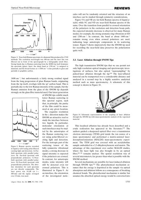
<strong>Optical</strong> <strong>Near</strong>-<strong>Field</strong> <strong>Microscopy</strong>REVIEWSscattering tensor of thefully symmetric vibrationalmodes, a strong decre<strong>as</strong>e insignal intensity is expectedif the polarizers are crossed.In contr<strong>as</strong>t, for anisotropicmodes, some intensity willstill be detected even forcrossed polarizers. This isillustrated in Figure 9. Atan interface, the orientationof the investigated moleculeswill not be r<strong>and</strong>omly oriented <strong>and</strong> the structure of aninterface can be studied through symmetry considerations.Figure 9 A <strong>and</strong> 9 B are far-field Raman spectra of liquid p-xylol, while 9 C <strong>and</strong> 9 D are near-field Raman spectra of thesame. Over the transition from parallel to crossed orientationof the polarizers in the excitation <strong>and</strong> detection beam path,the expected intensity decre<strong>as</strong>e is observed for many Ramanmodes, for example, the strong aromatic-ring vibrations at 820<strong>and</strong> 1200 cm 1 . In contr<strong>as</strong>t, the b<strong>and</strong> at about 1600 cm 1remains strong even when crossed polarizers are used,indicating large anisotropic components in its scatteringtensor. Figure 9 shows impressively that the SNOM tip usedfor recording the near-field data preserves the polarizationquite well.Figure 8. <strong>Near</strong>-field Raman spectrum of a diamond film produced by CVDmethods. The excitation wavelength w<strong>as</strong> 488 nm <strong>and</strong> the l<strong>as</strong>er line w<strong>as</strong>filtered out in front of the spectrograph by a holographic notch filter.Strong b<strong>and</strong>s due to Raman scattering from the SERS tip itself dominatethe spectrum (gl<strong>as</strong>s). Inset: the sharp b<strong>and</strong> at 1330 cm 1 is <strong>as</strong>signed todiamond, <strong>and</strong> the broad feature at 1600 cm 1 is due partially to gl<strong>as</strong>s <strong>and</strong>partially to graphitic carbon.1600 cm 1 , but unfortunately a fairly strong residual signalfrom the long progression of gl<strong>as</strong>s Raman b<strong>and</strong>s, originatingfrom the tip itself, overlaps with the sp 2 carbon b<strong>and</strong>. This ispartially due to the low Raman intensity of the sample, but theRaman emission from the gl<strong>as</strong>s of the SNOM tip dependsstrongly on the gl<strong>as</strong>s fiber material used. Our latest generationof SNOM tips exhibit muchlower Raman scattering inthis spectral region, suchthat, in principle, the purityof the film could be me<strong>as</strong>uredat any given location.The exquisite resolutionin the z direction makes theSNOM an attractive tool tostudy the interface betweentwo liquids. In particular,molecular orientation atsuch interfaces may be studiedby the anisotropies ofthe Raman scattering tensorusing polar-filtered excitinglight <strong>and</strong> detection.For the isotropic part of theFigure 9. Raman spectra recordedin liquid p-xylol using polar-filteredexciting light <strong>and</strong> detection. A) Farfieldspectrum with parallel polarizers;B) Far-field spectrum withperpendicular polarizers; C) <strong>Near</strong>fieldspectrum with parallel polarizers;D) <strong>Near</strong>-field spectrum withperpendicular polarizers. The absoluteintensities for each spectrumare not directly comparable. Thel<strong>as</strong>er power w<strong>as</strong> 10 mW (at l ˆ488 nm) <strong>and</strong> the signal, averagedover 10 min, w<strong>as</strong> used.3.3. L<strong>as</strong>er Ablation through SNOM TipsThe high transmission SNOM tips that we use permit notonly high resolution optical imaging <strong>and</strong> spectroscopic investigationsof surfaces, but also optical ªnanosamplingº bypulsed-l<strong>as</strong>er ablation through the tip. [66] The l<strong>as</strong>er-ablatedmaterial can be transported over a considerable distance <strong>and</strong>analyzed in a second step by a highly sensitive analyticalmethod such <strong>as</strong> m<strong>as</strong>s spectrometry. A schematic of thisconcept is shown in Figure 10.Figure 10. Schematic representation of the coupling of l<strong>as</strong>er ablationthrough the SNOM tip with m<strong>as</strong>s-spectrometric analysis of the vaporizedmaterial.This localized ablation h<strong>as</strong> already been described <strong>and</strong> acrude realization h<strong>as</strong> appeared in the literature. [67] Theauthors guided a sharpened optical fiber over a transmissionelectron microscopy (TEM) grid inside the ion source of am<strong>as</strong>s spectrometer <strong>and</strong> performed a matrix-<strong>as</strong>sisted l<strong>as</strong>erdesorption/ionization(MALDI) m<strong>as</strong>s-spectrometry experiment.The TEM grid w<strong>as</strong> covered with an acetylcholinesample embedded in a 2,5-dihydroxybenzoic acid matrix. Theadvantage of this experiment over earlier MALDI studies,where the l<strong>as</strong>er light w<strong>as</strong> also brought in by an optical[68, 69]fiber, remains unclear. The spatial resolution w<strong>as</strong> ofseveral micrometers, far greater than what is possible with theSNOM method.Several mechanisms are possible for l<strong>as</strong>er-induced ablationthrough SNOM tips. [70] The photochemical mechanism <strong>as</strong>sumesthat the energy is deposited in the sample via opticalabsorption <strong>and</strong> ablation follows <strong>as</strong> a consequence of breakingchemical bonds. The photothermal mechanism is similar but<strong>as</strong>sumes the absorbed optical energy would be converted intoAngew. Chem. Int. Ed. 2000, 39, 1746 ± 1756 1753