Chapter 4 SEGMENTATION
Chapter 4 SEGMENTATION
Chapter 4 SEGMENTATION

You also want an ePaper? Increase the reach of your titles
YUMPU automatically turns print PDFs into web optimized ePapers that Google loves.
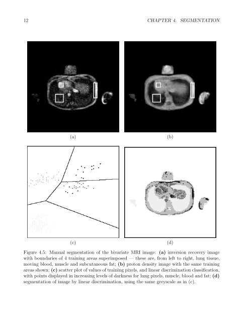
12 CHAPTER 4. <strong>SEGMENTATION</strong>(a)(b)(c)(d)Figure 4.5: Manual segmentation of the bivariate MRI image: (a) inversion recovery imagewith boundaries of 4 training areas superimposed — these are, from left to right, lung tissue,moving blood, muscle and subcutaneous fat; (b) proton density image with the same trainingareas shown; (c) scatter plot of values of training pixels, and linear discrimination classification,with points displayed in increasing levels of darkness for lung pixels, muscle, blood and fat; (d)segmentation of image by linear discrimination, using the same greyscale as in (c).