BD Pharmingen BrdU Flow Kits Instruction Manual - BD Biosciences
BD Pharmingen BrdU Flow Kits Instruction Manual - BD Biosciences
BD Pharmingen BrdU Flow Kits Instruction Manual - BD Biosciences

You also want an ePaper? Increase the reach of your titles
YUMPU automatically turns print PDFs into web optimized ePapers that Google loves.
<strong>BD</strong> <strong>Pharmingen</strong><br />
<strong>BrdU</strong> <strong>Flow</strong> <strong>Kits</strong><br />
<strong>Instruction</strong> <strong>Manual</strong><br />
FITC <strong>BrdU</strong> <strong>Flow</strong> Kit<br />
Catalog No. 559619 (50 tests)<br />
Catalog No. 557891 (4 x 50 tests)<br />
APC <strong>BrdU</strong> <strong>Flow</strong> Kit<br />
Catalog No. 552598 (50 tests)<br />
Catalog No. 557892 (4 x 50 tests)
ii<br />
<strong>BD</strong> <strong>Pharmingen</strong> <strong>BrdU</strong> <strong>Flow</strong> <strong>Kits</strong><br />
Copyrights<br />
© 2011, Becton, Dickinson and Company. All rights reserved. No part of this publication may<br />
be reproduced, transmitted, transcribed, stored in retrieval systems, or translated into any<br />
language or computer language, in any form or by any means: electronic, mechanical,<br />
magnetic, optical, chemical, manual, or otherwise, without prior written permission from<br />
<strong>BD</strong> <strong>Biosciences</strong>.<br />
The information in this guide is subject to change without notice. <strong>BD</strong> <strong>Biosciences</strong> reserves the<br />
right to change its products and services at any time to incorporate the latest technological<br />
developments. Although this guide has been prepared with every precaution to ensure<br />
accuracy, <strong>BD</strong> <strong>Biosciences</strong> assumes no liability for any errors or omissions, nor for any damages<br />
resulting from the application or use of this information. <strong>BD</strong> <strong>Biosciences</strong> welcomes customer<br />
input on corrections and suggestions for improvement.<br />
Trademarks<br />
<strong>BD</strong>, <strong>BD</strong> Logo and all other trademarks are property of Becton, Dickinson and Company.<br />
© 2011 <strong>BD</strong><br />
Regulatory information<br />
<strong>BD</strong> flow cytometers are Class 1 Laser Products.<br />
For Research Use Only. Not for use in diagnostic or therapeutic procedures.<br />
History<br />
Revision Date Change made<br />
23-12721-00 Rev. 01 11/2011 New document
Contents<br />
Chapter 1: Introduction . . . . . . . . . . . . . . . . . . . . . . . . . . . . . . . . . . . . . 5<br />
Purpose of the kit . . . . . . . . . . . . . . . . . . . . . . . . . . . . . . . . . . . . . . . 6<br />
Limitations . . . . . . . . . . . . . . . . . . . . . . . . . . . . . . . . . . . . . . . . . . . . 8<br />
Kit contents . . . . . . . . . . . . . . . . . . . . . . . . . . . . . . . . . . . . . . . . . . . . 9<br />
Storage and handling . . . . . . . . . . . . . . . . . . . . . . . . . . . . . . . . . . . . 12<br />
Chapter 2: Before you begin . . . . . . . . . . . . . . . . . . . . . . . . . . . . . . . . 15<br />
<strong>BrdU</strong> <strong>Flow</strong> Kit protocol overview . . . . . . . . . . . . . . . . . . . . . . . . . . 16<br />
Required materials . . . . . . . . . . . . . . . . . . . . . . . . . . . . . . . . . . . . . . 18<br />
Reagent preparation . . . . . . . . . . . . . . . . . . . . . . . . . . . . . . . . . . . . 18<br />
Chapter 3: Staining protocol . . . . . . . . . . . . . . . . . . . . . . . . . . . . . . . . 19<br />
In vitro labeling of cells with <strong>BrdU</strong> . . . . . . . . . . . . . . . . . . . . . . . . . 20<br />
In vivo labeling of mouse cells with <strong>BrdU</strong> . . . . . . . . . . . . . . . . . . . . 21<br />
<strong>BrdU</strong> <strong>Flow</strong> Kit staining protocol . . . . . . . . . . . . . . . . . . . . . . . . . . . 22<br />
Chapter 4: Instrument setup . . . . . . . . . . . . . . . . . . . . . . . . . . . . . . . . 27<br />
Instrument setup guidelines . . . . . . . . . . . . . . . . . . . . . . . . . . . . . . . 28<br />
FITC <strong>BrdU</strong> instrument setup example . . . . . . . . . . . . . . . . . . . . . . . 29<br />
APC <strong>BrdU</strong> instrument setup example . . . . . . . . . . . . . . . . . . . . . . . . 32<br />
Chapter 5: Analysis . . . . . . . . . . . . . . . . . . . . . . . . . . . . . . . . . . . . . . . 35<br />
Analysis of stained cell samples . . . . . . . . . . . . . . . . . . . . . . . . . . . . 36<br />
Chapter 6: Reference . . . . . . . . . . . . . . . . . . . . . . . . . . . . . . . . . . . . . . 41<br />
References . . . . . . . . . . . . . . . . . . . . . . . . . . . . . . . . . . . . . . . . . . . . 42<br />
For Research Use Only. Not for use in diagnostic or therapeutic procedures.
This section covers the following topics:<br />
� Purpose of the kit (page 6)<br />
� Limitations (page 8)<br />
� Kit contents (page 9)<br />
� Storage and handling (page 12)<br />
For Research Use Only. Not for use in diagnostic or therapeutic procedures.<br />
1<br />
Introduction
6<br />
<strong>BD</strong> <strong>Pharmingen</strong> <strong>BrdU</strong> <strong>Flow</strong> <strong>Kits</strong><br />
Purpose of the kit<br />
<strong>BrdU</strong> staining The immunofluorescent staining of incorporated<br />
bromodeoxyuridine (<strong>BrdU</strong>) and flow cytometric analysis<br />
provide a high-resolution technique to determine the<br />
frequency and nature of individual cells that have<br />
synthesized DNA. In this method, <strong>BrdU</strong> (an analog of<br />
the DNA precursor thymidine) is incorporated into<br />
newly synthesized DNA by cells entering and progressing<br />
through the S (DNA synthesis) phase of the cell cycle. 1-4<br />
The incorporated <strong>BrdU</strong> is stained with specific anti-<strong>BrdU</strong><br />
fluorescent antibodies. The levels of cell-associated <strong>BrdU</strong><br />
are then measured by flow cytometry. Often, staining<br />
with a dye that binds to total DNA such as 7-aminoactinomycin<br />
D (7-AAD) is coupled with<br />
immunofluorescent <strong>BrdU</strong> staining. With this<br />
combination, two-color flow cytometric analysis permits<br />
the enumeration and characterization of cells that are<br />
actively synthesizing DNA (<strong>BrdU</strong> incorporation) in terms<br />
of their cell cycle position (ie, G0/1, S, or G2/M phase<br />
defined by 7-AAD staining intensities). 5,6<br />
Prolonged exposure of cells to <strong>BrdU</strong> allows for the<br />
identification and analysis of actively cycling, as opposed<br />
to non-cycling, cell fractions. Pulse labeling of cells with<br />
<strong>BrdU</strong> at various time points, permits the determination<br />
of cell-cycle kinetics. <strong>BrdU</strong> incorporation studies have<br />
been used in a variety of experimental protocols. These<br />
include in vitro and in vivo (eg, intraperitoneal injection<br />
or administration via drinking water) labeling systems.<br />
For Research Use Only. Not for use in diagnostic or therapeutic procedures.
Other uses of the<br />
kit<br />
Chapter 1: Introduction<br />
An important feature of the <strong>BD</strong> <strong>Pharmingen</strong> <strong>BrdU</strong><br />
<strong>Flow</strong> Kit is that it provides reagents for<br />
immunofluorescent <strong>BrdU</strong> staining with a protocol that is<br />
compatible with the use of additional fluorescent<br />
antibodies specific for other cellular molecules. These<br />
molecules may include cell surface antigens or<br />
intracellular proteins (eg, cytokines, cyclins, and other<br />
proteins) the expression or activity of which may be<br />
related to the cell’s activation, entry, and progression<br />
through cell cycle or cell death. This is possible because<br />
the <strong>BrdU</strong> <strong>Flow</strong> Kit staining protocol avoids DNA<br />
denaturing agents such as acid, ethanol, and high<br />
temperatures that can result in altered cellular lightscattering<br />
characteristics and limit the recognition of<br />
cellular antigens by fluorescent antibodies. 7-11<br />
Fluorescent antibodies that are capable of recognizing<br />
cell surface antigens or proteins in cells that have been<br />
fixed with paraformaldehyde and permeabilized with<br />
saponin can be used with the <strong>BrdU</strong> <strong>Flow</strong> Kit. However,<br />
not all antibody clones and fluorochromes are<br />
compatible with paraformaldehyde fixation. With the<br />
combination of reagents, the expression levels of various<br />
surface or intracellular proteins can be measured by flow<br />
cytometry relative to the cell’s DNA synthetic activity<br />
(<strong>BrdU</strong> incorporation level). For example, the <strong>BrdU</strong> <strong>Flow</strong><br />
Kit can be used with fluorescent anti-cytokine antibodies<br />
in time-course analyses of cultured cells following in<br />
vitro mitogenic stimulation of quiescent lymphoid cell<br />
populations. In this way, the levels of a particular<br />
cytokine (eg, the T-cell growth and differentiation factor,<br />
IL-2) that are expressed prior to, at, and following the<br />
onset of DNA synthesis (during the first major round of<br />
cell-cycle activity) can be studied.<br />
For Research Use Only. Not for use in diagnostic or therapeutic procedures.<br />
7
8<br />
<strong>BD</strong> <strong>Pharmingen</strong> <strong>BrdU</strong> <strong>Flow</strong> <strong>Kits</strong><br />
Limitations<br />
Many high-resolution studies of this type are possible<br />
with the use of the <strong>BD</strong> <strong>Pharmingen</strong> <strong>BrdU</strong> <strong>Flow</strong> Kit and<br />
other selected flow cytometry reagents. The kit ensures<br />
consistent results by providing the critical reagents<br />
necessary to implement the staining protocol. These<br />
individual components have been rigorously tested for<br />
their suitability to perform multiparameter analyses of<br />
incorporated <strong>BrdU</strong> levels, cell surface antigen expression,<br />
and expression of intracellular antigens by individual<br />
cells. <strong>BrdU</strong> uptake can also be analyzed in frozen or<br />
paraffin embedded tissue sections. The <strong>BD</strong><br />
<strong>Pharmingen</strong> <strong>BrdU</strong> In-Situ <strong>Kits</strong> (Catalog Nos. 550803<br />
and 551321) provide the reagents that allow you to<br />
perform two-color staining in tissue sections.<br />
Assay limitations The <strong>BD</strong> <strong>Pharmingen</strong> <strong>BrdU</strong> <strong>Flow</strong> Kit staining procedure<br />
includes the fixative paraformaldehyde.<br />
Paraformaldehyde can alter epitopes on antigens and<br />
inhibit recognition by some antibodies after fixation. It is<br />
important that the antibody reagents used to stain<br />
proteins with this procedure be capable of binding to<br />
paraformaldehyde-fixed epitopes. Reagents that are<br />
compatible with other fixatives (eg, ethanol) may not<br />
work with the <strong>BD</strong> <strong>Pharmingen</strong> <strong>BrdU</strong> <strong>Flow</strong> Kit staining<br />
procedure.<br />
Reagent<br />
limitations<br />
Both the <strong>BD</strong> Perm/Wash Buffer (1X) and the <strong>BD</strong><br />
Cytoperm Permeabilization Buffer Plus should be used<br />
with fixed cell samples only. Use of these buffers on<br />
unfixed cells will cause cell damage.<br />
For Research Use Only. Not for use in diagnostic or therapeutic procedures.
Kit contents<br />
Chapter 1: Introduction<br />
Contents The kits (Catalog Nos. 559619 and 552598) contain the<br />
following components. Because some kit components are<br />
stored at 4°C and others are stored at –80°C, the kit<br />
components are shipped separately. See Storage and<br />
handling (page 12) for details on storage.<br />
Reagent Quantity<br />
Fluorochrome-conjugated anti-<strong>BrdU</strong> Antibody One 65-µL vial<br />
<strong>BD</strong> Cytofix/Cytoperm Buffer One 25-mL bottle<br />
<strong>BD</strong> Perm/Wash Buffer (10X) Two 25-mL bottles<br />
<strong>BD</strong> Cytoperm Permeabilization Buffer Plus One 10-mL bottle<br />
7-AAD One 1-mL vial<br />
The following items are shipped separately.<br />
Reagent Quantity<br />
<strong>BrdU</strong> (10 mg/mL) Five 0.5-mL vials<br />
DNase Five 300-µL vials<br />
Reagents Some kit reagents are supplied as concentrated stock<br />
solutions and need to be diluted either with deionized<br />
water, 1X Dulbecco’s PBS (DPBS), or with <strong>BD</strong> Perm/<br />
Wash Buffer. See Reagent preparation (page 18) for<br />
information. The concentrations of the kit components<br />
follow.<br />
For Research Use Only. Not for use in diagnostic or therapeutic procedures.<br />
9
10<br />
<strong>BD</strong> <strong>Pharmingen</strong> <strong>BrdU</strong> <strong>Flow</strong> <strong>Kits</strong><br />
Fluorochrome-conjugated anti-<strong>BrdU</strong> Antibody. A single<br />
vial contains 65 µL of fluorochrome-conjugated anti-<br />
<strong>BrdU</strong> antibody stock solution and is sufficient for<br />
staining 50 samples (10 6 cells/sample). The FITC <strong>BrdU</strong><br />
<strong>Flow</strong> Kit (Catalog No. 559619) comes with 65 µL of<br />
FITC-conjugated anti-<strong>BrdU</strong> antibody. The APC <strong>BrdU</strong><br />
<strong>Flow</strong> Kit (Catalog No. 552598) comes with 65 µL of<br />
APC-conjugated anti-<strong>BrdU</strong> antibody.<br />
Note: If you run out of anti-<strong>BrdU</strong> antibody, an<br />
additional <strong>BrdU</strong> flow kit must be purchased. We do not<br />
recommended using any other anti-<strong>BrdU</strong> antibody clone<br />
or formulation from the <strong>BD</strong> <strong>Biosciences</strong> catalog in<br />
conjunction with this kit.<br />
<strong>BD</strong> Cytofix/Cytoperm Buffer. A 25-mL bottle of<br />
<strong>BD</strong> Cytofix/Cytoperm Buffer is provided in a ready-touse<br />
formulation. <strong>BD</strong> Cytofix/Cytoperm Buffer<br />
constitutes a single-step fixation and permeabilization<br />
reagent, designed for use in intracellular staining. It<br />
contains a mixture of the fixative paraformaldehyde and<br />
the detergent saponin. This reagent serves to preserve<br />
cell morphology, fix cellular proteins, and permeabilize<br />
cells for the subsequent immunofluorescent staining of<br />
intracellular proteins.<br />
<strong>BD</strong> Perm/Wash Buffer. Two 25-mL bottles contain a<br />
concentrated (10X) stock solution of <strong>BD</strong> Perm/Wash<br />
Buffer. The <strong>BD</strong> Perm/Wash Buffer mixture contains fetal<br />
bovine serum and the reversible permeabilization<br />
detergent reagent saponin.<br />
For Research Use Only. Not for use in diagnostic or therapeutic procedures.
Chapter 1: Introduction<br />
<strong>BD</strong> Cytoperm Permeabilization Buffer Plus. One 10-mL<br />
bottle of buffer is provided. It is specially formulated for<br />
the <strong>BrdU</strong> <strong>Flow</strong> Kit and is used as a staining enhancer and<br />
secondary permeabilization reagent. It contains fetal<br />
bovine serum. One hundred microliters of this buffer is<br />
used for each sample. <strong>BD</strong> Cytoperm Permeabilization<br />
Buffer Plus can be purchased separately from <strong>BD</strong><br />
(Catalog No. 561651, 10 mL).<br />
7-AAD. One 1-mL vial of 7-AAD is provided. 7-aminoactinomycin<br />
D (7-AAD) is a fluorescent dye for labeling<br />
DNA for flow cytometric analysis. It contains fetal<br />
bovine serum. Twenty microliters of 7-AAD is used for<br />
staining each sample (10 6 cells/sample).<br />
Note: Source of all serum proteins is from USDAinspected<br />
abattoirs located in the United States.<br />
<strong>BrdU</strong> and DNase <strong>BrdU</strong>. Five vials of <strong>BrdU</strong> Solution are provided. Each<br />
vial contains 0.5 mL of a 10-mg/mL <strong>BrdU</strong> (32.5-mM)<br />
solution diluted in 1X DPBS. The <strong>BrdU</strong> solution<br />
provided is prepared aseptically (0.22-µm filtered), and<br />
contains no preservative; therefore we recommend that<br />
the solution be handled under aseptic conditions. This<br />
stock solution can be injected intraperitoneally (IP) into<br />
animals or diluted to a 1-mM solution for in vitro<br />
labeling. For in vivo labeling by IP injection, see In vivo<br />
labeling of mouse cells with <strong>BrdU</strong> (page 21).<br />
DNase. Five vials of DNase are provided. Each vial<br />
contains 300 µL of a 1-mg/mL solution of DNase in 1X<br />
DPBS. DNase can be purchased separately from Sigma<br />
(Catalog No. D-4513).<br />
For Research Use Only. Not for use in diagnostic or therapeutic procedures.<br />
11
12<br />
<strong>BD</strong> <strong>Pharmingen</strong> <strong>BrdU</strong> <strong>Flow</strong> <strong>Kits</strong><br />
Storage and handling<br />
Storage Antibody, buffers, and 7-AAD. Store the fluorochromeconjugated<br />
anti-<strong>BrdU</strong> antibody, <strong>BD</strong> Cytofix/Cytoperm<br />
Buffer, <strong>BD</strong> Perm/Wash Buffer, <strong>BD</strong> Cytoperm<br />
Permeabilization Buffer Plus, and 7-AAD at 2 to 8°C.<br />
Keep the fluorochrome-conjugated anti-<strong>BrdU</strong> antibody<br />
and 7-AAD protected from light.<br />
Warnings and<br />
precautions<br />
Unused portions of 1X <strong>BD</strong> Cytofix/Cytoperm Buffer can<br />
be stored at 2 to 8°C.<br />
<strong>BrdU</strong> and DNase. Store the <strong>BrdU</strong> and DNase at –80°C.<br />
The <strong>BrdU</strong> solution has been shown to be stable for up to<br />
4 months at 2 to 8°C, or it can be refrozen. Avoid<br />
multiple freeze-thaw cycles.<br />
DNase stock solution (1 mg/mL) may be refrozen once<br />
before it loses activity.<br />
<strong>BD</strong> Cytofix/Cytoperm Buffer contains 4%<br />
paraformaldehyde and is classified as harmful and a<br />
suspected carcinogen. The following risk and safety<br />
precautions should be followed when handling this<br />
product:<br />
Limited evidence of a carcinogenic effect (R40). May<br />
cause sensitization by skin contact (R43). Keep out<br />
of the reach of children (S2). Keep away from food,<br />
drink, and animal feed or feeding materials (S13).<br />
Wear suitable protective clothing (S36). Wear<br />
suitable gloves (S37). If swallowed, seek medical<br />
attention immediately and show this container or<br />
label (S46). Not recommended for interior use on<br />
large surface areas (S52).<br />
For Research Use Only. Not for use in diagnostic or therapeutic procedures.
Chapter 1: Introduction<br />
The <strong>BD</strong> Cytoperm Permeabilization Buffer Plus contains<br />
10% dimethyl sulfoxide and is harmful. Follow these<br />
precautions:<br />
Harmful by inhalation, if in contact with skin and if<br />
swallowed (R20/21/22). Avoid contact with eyes<br />
(S25). In case of contact with eyes, rinse immediately<br />
with plenty of water and seek medical advice (S26).<br />
After contact with skin, wash immediately with<br />
plenty of water (S28). Wear suitable protective<br />
clothing (S36). This material and its container must<br />
be disposed of as hazardous waste (S60).<br />
<strong>BrdU</strong> contains 1% Broxuridin and is harmful. Follow<br />
these precautions:<br />
Harmful by inhalation, if in contact with skin and if<br />
swallowed (R20/21/22). Keep container in a wellventilated<br />
place (S9). Do not breathe gas/fumes/<br />
vapour/spray (S23). Wear suitable protective<br />
clothing and gloves (S36/37). This material and its<br />
container must be disposed of as hazardous waste<br />
(S60).<br />
7-AAD contains 0.6% dimethyl sulfoxide and is an<br />
irritant and a potential carcinogen. Follow these<br />
precautions:<br />
Irritating to eyes, respiratory system, and skin (R36/<br />
37/38). Avoid contact with skin and eyes (S24/25).<br />
In case of contact with eyes, rinse immediately with<br />
plenty of water and seek medical advice (S26). Wear<br />
suitable protective clothing, gloves, and eye/face<br />
protection (S36/37/39). Keep out of reach of<br />
children (S2). Do not breathe gas/fumes/vapour/<br />
spray (S23). If swallowed, rinse mouth with water<br />
(only if the person is conscious), seek medical advice<br />
immediately, and show this container or label (S46/<br />
64). This material and its container must be disposed<br />
of as hazardous waste (S56/60).<br />
For Research Use Only. Not for use in diagnostic or therapeutic procedures.<br />
13
14<br />
<strong>BD</strong> <strong>Pharmingen</strong> <strong>BrdU</strong> <strong>Flow</strong> <strong>Kits</strong><br />
The anti-<strong>BrdU</strong> antibody and 7-AAD contain sodium<br />
azide. Sodium azide yields highly toxic hydrazoic acid<br />
under acidic conditions. Dilute azide compounds in<br />
running water before discarding to avoid accumulation<br />
of potentially explosive deposits in plumbing.<br />
For Research Use Only. Not for use in diagnostic or therapeutic procedures.
This section covers the following topics:<br />
� <strong>BrdU</strong> <strong>Flow</strong> Kit protocol overview (page 16)<br />
� Required materials (page 18)<br />
� Reagent preparation (page 18)<br />
For Research Use Only. Not for use in diagnostic or therapeutic procedures.<br />
2<br />
Before you begin
16<br />
<strong>BD</strong> <strong>Pharmingen</strong> <strong>BrdU</strong> <strong>Flow</strong> <strong>Kits</strong><br />
<strong>BrdU</strong> <strong>Flow</strong> Kit protocol overview<br />
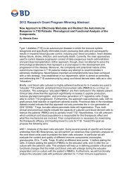
Options for<br />
storing cells<br />
prior to staining<br />
The <strong>BD</strong> <strong>Pharmingen</strong> <strong>BrdU</strong> <strong>Flow</strong> Kit staining procedure<br />
offers several time-saving options for sample handling.<br />
With this staining protocol, it is possible to stain and<br />
analyze samples in a single day. The entire staining<br />
procedure requires approximately 3 hours.<br />
Alternatively, samples may be fixed and stored for<br />
various lengths of time prior to staining. Due to the time<br />
intervals required for cell activation, <strong>BrdU</strong> incubation,<br />
and other factors that are necessary to prepare cells prior<br />
to staining, you may wish to store samples and complete<br />
the staining protocol at a later time.<br />
� If short-term sample storage prior to staining is<br />
desired, Option 1 in the following flow chart allows<br />
you to store cells overnight after the initial fixation<br />
step.<br />
� If longer sample storage is desired, Option 2 allows<br />
you to store frozen samples indefinitely, following<br />
the initial fixation step.<br />
For Research Use Only. Not for use in diagnostic or therapeutic procedures.
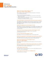
Chapter 2: Before you begin<br />
In vitro or in vivo labeling<br />
of cells with <strong>BrdU</strong><br />
Option 1 Option 2<br />
Overnight cell storage<br />
prior to intracellular staining<br />
Washing fixed cells and store at<br />
4°C overnight in staining buffer<br />
Single Day Staining Procedure<br />
Immunofluorescent staining<br />
of cell surface antigens<br />
Fixing and permeabilizing cells<br />
with <strong>BD</strong> Cytofix/Cytoperm Buffer<br />
Permeabilizing cells with<br />
<strong>BD</strong> Cytoperm Permeabilization<br />
Buffer Plus<br />
Repeating incubation of cells with<br />
<strong>BD</strong> Cytofix/Cytoperm Buffer<br />
Treating cells with DNase to<br />
expose <strong>BrdU</strong> epitopes<br />
Immunofluorescent staining with fluorochromeconjugated<br />
anti-<strong>BrdU</strong> and appropriate<br />
intracellular antigen-specific antibodies (2)<br />
Staining of DNA for cell cycle<br />
analysis using 7-AAD (3)<br />
Resuspending cells in staining buffer<br />
and analyzing on a flow cytometer<br />
Long-term cell storage<br />
prior to intracellular staining<br />
Washing cells in staining buffer<br />
and store indefinitely at –80°C<br />
in freezing medium (1)<br />
Wash freshly thawed cells<br />
with staining buffer<br />
The following notes pertain to the numbers in<br />
parentheses found in the flow chart.<br />
1. Recipe for Freezing Medium: 10% dimethyl<br />
sulfoxide (DMSO) + 90% heat-inactivated fetal<br />
bovine serum (FBS).<br />
2. The immunofluorescent staining of cell surface<br />
antigens can be done at the same time as staining<br />
intracellular antigens provided the antibodies<br />
recognize paraformaldehyde-fixed epitopes.<br />
3. If you do not wish to stain for total DNA content,<br />
then the 7-AAD staining step can be omitted and<br />
fluorescent data for another parameter can be<br />
measured in that channel.<br />
For Research Use Only. Not for use in diagnostic or therapeutic procedures.<br />
17
18<br />
<strong>BD</strong> <strong>Pharmingen</strong> <strong>BrdU</strong> <strong>Flow</strong> <strong>Kits</strong><br />
Required materials<br />
Materials<br />
required but not<br />
provided<br />
Reagent preparation<br />
In addition to the reagents provided in the<br />
<strong>BD</strong> <strong>Pharmingen</strong> <strong>BrdU</strong> <strong>Flow</strong> Kit, the following items are<br />
also required.<br />
� For the FITC kit, you will need a flow cytometer<br />
equipped with a 488-nm laser capable of detecting<br />
FITC and 7-AAD. For the APC kit, you will need a<br />
flow cytometer with a 488-nm laser capable of<br />
detecting 7-AAD and a 633- to 640-nm laser capable<br />
of detecting APC.<br />
� <strong>BD</strong> Falcon 12 x 75-mm sample acquisition tubes<br />
for a flow cytometer (Catalog No. 352008)<br />
� Staining buffer, for example, <strong>BD</strong> <strong>Pharmingen</strong> Stain<br />
Buffer (FBS) [Catalog No. 554656] or 1X DPBS +<br />
3% heat-inactivated FBS + 0.09% sodium azide<br />
Introduction Dilute the following reagents prior to use.<br />
Procedures Fluorochrome-conjugated anti-<strong>BrdU</strong> Antibody. Dilute<br />
an appropriate amount of the antibody stock solution<br />
1:50 with 1X <strong>BD</strong> Perm/Wash Buffer. Fifty microliters of<br />
the diluted antibody is used to stain each sample.<br />
<strong>BD</strong> Perm/Wash Buffer. Dilute the concentrated stock<br />
buffer 1:10 with deionized water. Store unused portions<br />
of 1X <strong>BD</strong> Perm/Wash Buffer at 4°C.<br />
For Research Use Only. Not for use in diagnostic or therapeutic procedures.
This section covers the following topics:<br />
� In vitro labeling of cells with <strong>BrdU</strong> (page 20)<br />
� In vivo labeling of mouse cells with <strong>BrdU</strong> (page 21)<br />
� <strong>BrdU</strong> <strong>Flow</strong> Kit staining protocol (page 22)<br />
For Research Use Only. Not for use in diagnostic or therapeutic procedures.<br />
3<br />
Staining protocol
20<br />
<strong>BD</strong> <strong>Pharmingen</strong> <strong>BrdU</strong> <strong>Flow</strong> <strong>Kits</strong><br />
In vitro labeling of cells with <strong>BrdU</strong><br />
Introduction Many different protocols for in vitro <strong>BrdU</strong> labeling of<br />
cells have been reported. 12-15 We have found that<br />
incubating cells with <strong>BrdU</strong> at a final concentration of<br />
10 µM in cell culture medium (ie, 10 µL of 1-mM <strong>BrdU</strong><br />
per mL of culture medium) was effective for labeling a<br />
wide variety of human and mouse cell lines and normal<br />
cell populations. 15,16 Prolonged exposure of cells to<br />
<strong>BrdU</strong> allows for the identification of actively cycling cell<br />
populations. Pulse labeling of cells by brief <strong>BrdU</strong><br />
exposures at various time points permits the<br />
determination of cell-cycle kinetics.<br />
Use cells from the same population that are not <strong>BrdU</strong>labeled<br />
as a negative staining control for this assay. This<br />
allows you to determine background staining levels for<br />
the anti-<strong>BrdU</strong> monoclonal antibody.<br />
Before you begin For pulse-labeling experiments, the choice of time points<br />
and lengths of time for pulsing depends on the test cell<br />
population’s rate of cell cycle entry and progression. For<br />
example, an effective length of time for pulsing an<br />
actively proliferating cell line (eg, CTLL-2 cells) is 30 to<br />
45 minutes (ie, when the cells are in the logarithmic<br />
phase of cell proliferation).<br />
Determine time points and pulse-labeling time intervals<br />
that are optimal for each different cell line or cell<br />
population within a particular experimental system.<br />
Dilute the <strong>BrdU</strong> stock (10-mg/mL <strong>BrdU</strong> solution) to a<br />
1-mM solution by adding 31 µL to 1 mL of either 1X<br />
DPBS or culture medium (this is a dilution of 32X). Add<br />
10 µL of the 1-mM solution to each mL of culture<br />
medium to obtain a final concentration of 10 µM. The<br />
molecular weight of <strong>BrdU</strong> is 307.1.<br />
For Research Use Only. Not for use in diagnostic or therapeutic procedures.
Chapter 3: Staining protocol<br />
Procedure To label cells in vitro:<br />
1. Carefully add 10 µL of <strong>BrdU</strong> solution (1 mM <strong>BrdU</strong><br />
in 1X DPBS) directly to each mL of tissue culture<br />
medium.<br />
Avoid disturbing the cells in any way (eg,<br />
centrifugation steps or temperature changes) that<br />
may disrupt the normal cell cycling patterns. The cell<br />
culture density should not exceed 2 x 10 6 cells/mL.<br />
2. Incubate the treated cells for the desired length of<br />
time.<br />
In vivo labeling of mouse cells with <strong>BrdU</strong><br />
Introduction Two common methods reported for in vivo <strong>BrdU</strong><br />
labeling of cells include the intraperitoneal (IP) injection<br />
of a <strong>BrdU</strong>-containing solution into mice and the feeding<br />
of mice with <strong>BrdU</strong> that is added to their drinking<br />
water. 16-22 However, these methods have not been<br />
routinely tested at <strong>BD</strong> <strong>Biosciences</strong>.<br />
Injecting <strong>BrdU</strong><br />
via the<br />
intraperitoneal<br />
route<br />
A 10 mg/mL solution of <strong>BrdU</strong> in sterile 1X DPBS is<br />
provided for in vivo use. Inject mice IP with 100 to 200<br />
µL (1–2 mg) of <strong>BrdU</strong> solution. 17,19,21 Incorporation of<br />
<strong>BrdU</strong> can be readily detected in thymus and bone<br />
marrow in as little as 1 hour post injection.<br />
For Research Use Only. Not for use in diagnostic or therapeutic procedures.<br />
21
22<br />
<strong>BD</strong> <strong>Pharmingen</strong> <strong>BrdU</strong> <strong>Flow</strong> <strong>Kits</strong><br />
Introduction of<br />
<strong>BrdU</strong> through<br />
drinking water<br />
Dilute <strong>BrdU</strong> to 0.8 mg/mL in the drinking water. The<br />
<strong>BrdU</strong> mixture should be made up fresh and changed<br />
daily. 18,23 Prolonged feeding of <strong>BrdU</strong> can have toxic<br />
effects for the animal. Some researchers have reported<br />
lethal effects associated with 14 days of continuous <strong>BrdU</strong><br />
feeding. 21 For longer term studies, some researchers have<br />
reported that feeding mice with <strong>BrdU</strong> for 9 consecutive<br />
days followed by a changeover to normal water has<br />
worked effectively. 18 <strong>BrdU</strong> incorporation by cells from<br />
these animals has been detected past 70 days. 18<br />
<strong>BrdU</strong> <strong>Flow</strong> Kit staining protocol<br />
Before you begin When treating 10 or more samples, thaw the entire vial<br />
of DNase solution and add 700 µL of 1X DPBS to make<br />
a working stock solution of 300 µg/mL. If treating fewer<br />
than 10 samples, take a 30-µL aliquot (1 mg/mL) of<br />
DNase solution per sample and refreeze the remaining 1<br />
mg/mL DNase at –80°C.<br />
Procedure In addition to the labeled cells, stain an aliquot of<br />
unlabeled cells for use as a negative control.<br />
To stain the cells:<br />
1. (Optional) Stain cell surface antigens.<br />
a. Add <strong>BrdU</strong>-pulsed cells (10 6 cells in 50 µL of<br />
staining buffer) to 12 x 75-mm tubes.<br />
b. Add fluorescent antibodies specific for cellsurface<br />
markers in 50 µL of staining buffer (eg,<br />
<strong>BD</strong> <strong>Pharmingen</strong> Stain Buffer [FBS] Catalog No.<br />
554656) per tube and mix well.<br />
For Research Use Only. Not for use in diagnostic or therapeutic procedures.
Chapter 3: Staining protocol<br />
c. Incubate cells with antibodies for 15 minutes on<br />
ice.<br />
d. Wash cells by adding 1 mL of staining buffer per<br />
tube, centrifuge for 5 minutes at 200 to 300g,<br />
and discard the supernatant.<br />
2. Fix and permeabilize the cells with <strong>BD</strong> Cytofix/<br />
Cytoperm Buffer.<br />
a. Resuspend the cells in 100 µL of <strong>BD</strong> Cytofix/<br />
Cytoperm Buffer per tube.<br />
b. Incubate the cells for 15 to 30 minutes at room<br />
temperature or on ice.<br />
c. Wash the cells with 1 mL of 1X <strong>BD</strong> Perm/Wash<br />
Buffer. Centrifuge for 5 minutes at 200 to 300g,<br />
and discard the supernatant.<br />
Note: The presence of some precipitate in the<br />
10X <strong>BD</strong> Perm/Wash stock buffer is common. The<br />
precipitate will not affect the performance of the<br />
buffer. If desired, you can remove the precipitate<br />
prior to use by filtering the diluted 1X <strong>BD</strong> Perm/<br />
Wash Buffer through a 0.45-µm–pore filter.<br />
3. Incubate the cells with <strong>BD</strong> Cytoperm<br />
Permeabilization Buffer Plus.<br />
a. Resuspend the cells in 100 µL of <strong>BD</strong> Cytoperm<br />
Permeabilization Buffer Plus per tube.<br />
b. Incubate the cells for 10 minutes on ice.<br />
c. Wash the cells in 1 mL of 1X <strong>BD</strong> Perm/Wash<br />
Buffer (as in step 2c).<br />
For Research Use Only. Not for use in diagnostic or therapeutic procedures.<br />
23
24<br />
<strong>BD</strong> <strong>Pharmingen</strong> <strong>BrdU</strong> <strong>Flow</strong> <strong>Kits</strong><br />
4. Re-fix cells.<br />
a. Resuspend the cells in 100 µL of <strong>BD</strong> Cytofix/<br />
Cytoperm Buffer per tube.<br />
b. Incubate the cells for 5 minutes at room<br />
temperature or on ice.<br />
c. Wash the in 1 mL of 1X <strong>BD</strong> Perm/Wash Buffer<br />
(as in step 2c).<br />
5. Treat cells with DNase to expose incorporated<br />
<strong>BrdU</strong>. 24,25<br />
a. Resuspend the cells in 100 µL of diluted DNase<br />
(diluted to 300 µg/mL in DPBS) per tube, (ie,<br />
30 µg of DNase/10 6 cells).<br />
b. Incubate cells for 1 hour at 37°C.<br />
c. Wash the cells in 1 mL of 1X <strong>BD</strong> Perm/Wash<br />
Buffer (as in step 2c).<br />
6. Stain <strong>BrdU</strong> and intracellular antigens with<br />
fluorescent antibodies.<br />
a. Resuspend the cells in 50 µL of <strong>BD</strong> Perm/Wash<br />
Buffer containing diluted fluorescent anti-<strong>BrdU</strong><br />
and/or antibodies specific for intracellular<br />
antigens.<br />
b. Incubate the cells for 20 minutes at room<br />
temperature.<br />
c. Wash the cells in 1 mL of 1X <strong>BD</strong> Perm/Wash<br />
Buffer (as in step 2c).<br />
Note: Proceed to step 8 if you do not wish to stain for<br />
total DNA levels.<br />
7. Stain total DNA for cell cycle analysis. Resuspend<br />
the cells in 20 µL of the 7-AAD solution.<br />
8. Resuspend the cells in 1 mL of staining buffer.<br />
For Research Use Only. Not for use in diagnostic or therapeutic procedures.
Chapter 3: Staining protocol<br />
9. Acquire stained cells on a flow cytometer. For<br />
optimal resolution, acquire using a low flow rate.<br />
Run at a rate no greater than 400 events per second.<br />
Samples may be stored overnight at 4°C, protected from<br />
light, prior to analysis by flow cytometry.<br />
For Research Use Only. Not for use in diagnostic or therapeutic procedures.<br />
25
This section covers the following topics:<br />
� Instrument setup guidelines (page 28)<br />
� FITC <strong>BrdU</strong> instrument setup example (page 29)<br />
� APC <strong>BrdU</strong> instrument setup example (page 32)<br />
For Research Use Only. Not for use in diagnostic or therapeutic procedures.<br />
4<br />
Instrument setup
28<br />
<strong>BD</strong> <strong>Pharmingen</strong> <strong>BrdU</strong> <strong>Flow</strong> <strong>Kits</strong><br />
Instrument setup guidelines<br />
Introduction The information in the following instrument setup<br />
sections is intended as an example of the type of setup<br />
necessary for samples stained using the <strong>BrdU</strong> staining<br />
procedure. FITC <strong>BrdU</strong> instrument setup example<br />
(page 29) is specific to the <strong>BD</strong> <strong>Pharmingen</strong> FITC <strong>BrdU</strong><br />
<strong>Flow</strong> Kit (Catalog No. 559619) and APC <strong>BrdU</strong><br />
instrument setup example (page 32) applies to the <strong>BD</strong><br />
<strong>Pharmingen</strong> APC <strong>BrdU</strong> <strong>Flow</strong> Kit (Catalog No. 552598).<br />
Cytometer setup<br />
flow chart<br />
The instrument adjustments required might vary<br />
between instruments and between individual samples in<br />
a given experiment. It is often necessary to make further<br />
adjustments for different combinations of fluorescentconjugated<br />
antibodies. We recommend that you refer to<br />
a textbook on flow cytometry or on cell-cycle analysis by<br />
flow cytometry for more information. 26,27<br />
The following flow chart shows you the general steps<br />
involved in instrument setup.<br />
Adjust light scatter profiles and PMT settings<br />
Adjust 7-AAD – %PE<br />
Adjust PE – %7-AAD<br />
Adjust PE – %FITC or APC* (depending on the kit)<br />
Adjust FITC or APC* (depending on the kit) – %PE<br />
Adjust 7-AAD – %FITC or APC, if necessary (depending on the kit)<br />
Further compensation of PE – %7-AAD for bright fluorochromes<br />
*Typically little or no compensation is necessary between PE and APC.<br />
For Research Use Only. Not for use in diagnostic or therapeutic procedures.
FITC <strong>BrdU</strong> instrument setup example<br />
Experiment<br />
details<br />
Adjusting scatter<br />
and PMTs<br />
Adjusting<br />
compensation<br />
for 7-AAD and PE<br />
Chapter 4: Instrument setup<br />
Mouse S-phase T cells were labeled with 10 µM of <strong>BrdU</strong><br />
for 1 hour. The cells were processed according to the<br />
<strong>BrdU</strong> <strong>Flow</strong> Kit protocol. The samples were stained with<br />
FITC anti-<strong>BrdU</strong>, 7-AAD, and a PE-labeled surface<br />
marker.<br />
Note: PE was included to illustrate the amount of<br />
compensation necessary when including PE in the<br />
experiment. If you do not use PE, proceed directly to<br />
Adjusting the <strong>BrdU</strong>/7-AAD profile (page 32).<br />
To adjust scatter and PMTs:<br />
1. Adjust the FSC vs SSC parameters so that the cell<br />
populations are on scale.<br />
2. Adjust the PMTs so that the negative populations<br />
fall between channels 10 2 and 10 3 .<br />
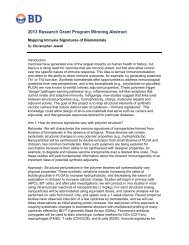
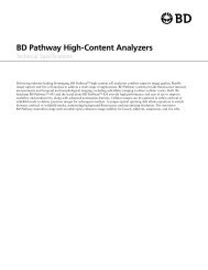
To adjust compensation for 7-AAD and PE:<br />
1. Adjust 7-AAD–%PE to bring the PE signal to the left<br />
(setting is ~209).<br />
uncompensated 7-AAD–%PE compensated 7-AAD–%PE<br />
For Research Use Only. Not for use in diagnostic or therapeutic procedures.<br />
29
30<br />
<strong>BD</strong> <strong>Pharmingen</strong> <strong>BrdU</strong> <strong>Flow</strong> <strong>Kits</strong><br />
2. Adjust PE–%7-AAD to bring the PE signal down<br />
(setting is ~3.0).<br />
uncompensated PE–%7-AAD compensated PE–%7-AAD<br />
The <strong>BrdU</strong> plot gets adjusted from the 7-AAD–%PE<br />
and PE–%7-AAD compensation adjustments. The<br />
following uncompensated plot is an example of how<br />
the data appears when PE is included.<br />
uncompensated 7-AAD vs FITC compensated 7-AAD vs FITC<br />
For Research Use Only. Not for use in diagnostic or therapeutic procedures.
Adjusting<br />
compensation<br />
for PE and FITC<br />
Chapter 4: Instrument setup<br />
To adjust compensation for PE and FITC:<br />
1. Adjust PE–%FITC to bring the FITC signal down<br />
(setting is ~65.4).<br />
uncompensated PE–%FITC compensated PE–%FITC<br />
2. Adjust FITC–%PE to bring the PE signal slightly to<br />
the left (setting is ~0.4).<br />
uncompensated FITC–%PE compensated FITC–%PE<br />
For Research Use Only. Not for use in diagnostic or therapeutic procedures.<br />
31
32<br />
<strong>BD</strong> <strong>Pharmingen</strong> <strong>BrdU</strong> <strong>Flow</strong> <strong>Kits</strong><br />
Adjusting the<br />
<strong>BrdU</strong>/7-AAD<br />
profile<br />
Start here if you stained using only the contents of the<br />
FITC <strong>BrdU</strong> <strong>Flow</strong> Kit with no additional drop-in<br />
antibodies.<br />
To adjust compensation for 7-AAD and FITC:<br />
1. Adjust 7-AAD–%FITC to bring the FITC signal to<br />
the left (setting is ~88).<br />
APC <strong>BrdU</strong> instrument setup example<br />
Initial<br />
instrument<br />
settings<br />
uncompensated 7-AAD–%FITC compensated 7-AAD–%FITC<br />
When staining cells with anti-<strong>BrdU</strong> APC, the initial setup<br />
is similar to the FITC anti-<strong>BrdU</strong> conjugate shown in the<br />
previous section. Shown here are cells stained with APC<br />
anti-<strong>BrdU</strong>, 7-AAD, and a PE-labeled surface marker. The<br />
important difference here is that APC anti-<strong>BrdU</strong> is<br />
detected with a separate laser.<br />
Note: PE was included to illustrate the amount of<br />
compensation necessary when including PE in the<br />
experiment. If you do not use PE, proceed directly to<br />
Adjusting the <strong>BrdU</strong>/7-AAD profile (page 34).<br />
For Research Use Only. Not for use in diagnostic or therapeutic procedures.
Adjusting scatter<br />
and PMTs<br />
Adjusting<br />
compensation<br />
for PE and 7-AAD<br />
Chapter 4: Instrument setup<br />
To adjust scatter and PMTs:<br />
1. Adjust the FSC vs SSC parameters so that the cell<br />
populations are on scale.<br />
2. Adjust the PMTs so that the negative populations<br />
fall between channels 10 2 and 10 3 .<br />
To adjust compensation for PE and 7-AAD:<br />
1. Adjust 7-AAD–%PE to bring the PE signal to the left<br />
(setting is ~308).<br />
uncompensated 7-AAD–%PE compensated 7-AAD–%PE<br />
2. Adjust PE–%7-AAD to bring the PE signal down<br />
(setting is ~0.5).<br />
uncompensated PE–%7-AAD compensated PE–%7-AAD<br />
For Research Use Only. Not for use in diagnostic or therapeutic procedures.<br />
33
34<br />
<strong>BD</strong> <strong>Pharmingen</strong> <strong>BrdU</strong> <strong>Flow</strong> <strong>Kits</strong><br />
Adjusting the<br />
<strong>BrdU</strong>/7-AAD<br />
profile<br />
The <strong>BrdU</strong> plot gets adjusted from the 7-AAD–%PE<br />
and PE–%7-AAD compensation adjustments. The<br />
following uncompensated plot is an example of how<br />
the data appears when PE is included.<br />
uncompensated 7-AAD vs APC compensated 7-AAD vs APC<br />
Start here if you stained using only the contents of the<br />
APC <strong>BrdU</strong> <strong>Flow</strong> Kit with no additional drop-ins.<br />
To adjust compensation for 7-AAD and APC:<br />
1. If the APC is very bright, adjust compensation<br />
between 7-AAD and APC. The following plot looks<br />
correct without compensation. The compensation<br />
settings for 7-AAD–%APC and APC–%7-AAD are<br />
both 0.<br />
uncompensated 7-AAD vs APC<br />
For Research Use Only. Not for use in diagnostic or therapeutic procedures.
This section covers the following topics:<br />
� Analysis of stained cell samples (page 36)<br />
For Research Use Only. Not for use in diagnostic or therapeutic procedures.<br />
5<br />
Analysis
36<br />
<strong>BD</strong> <strong>Pharmingen</strong> <strong>BrdU</strong> <strong>Flow</strong> <strong>Kits</strong><br />
Analysis of stained cell samples<br />
Introduction Some of the data presented in the following examples<br />
was acquired using a flow cytometer equipped with a<br />
488-nm laser, which excites FITC, PE, and 7-AAD.<br />
When staining with fluorochromes such as APC, flow<br />
cytometers with an additional laser light source were<br />
used. With the addition of each different fluorochrome<br />
used for multicolor staining, it becomes more critical to<br />
properly compensate overlaps in detection of emitted<br />
fluorescence signals. Fluorescence signals from 7-AAD<br />
are typically acquired in the linear signal amplification<br />
mode, whereas fluorescence signals generated by other<br />
fluorochromes are acquired in a logarithmic mode.<br />
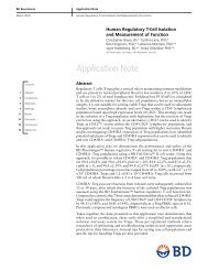
<strong>BrdU</strong> and total<br />
DNA staining<br />
The cell cycle positions and DNA synthetic activities of<br />
cells can be determined by analyzing the correlated<br />
expression of total DNA and incorporated <strong>BrdU</strong> levels.<br />
For Research Use Only. Not for use in diagnostic or therapeutic procedures.
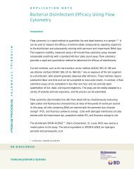
Chapter 5: Analysis<br />
Regions for the quantitative cell cycle analysis of<br />
populations that have been stained for incorporated<br />
<strong>BrdU</strong> and total DNA levels.<br />
Region Cell population<br />
R3 G0/G1<br />
R4 S phase<br />
R5 G2 + M<br />
R6 apoptotic<br />
Plot A: The measurement of cell-incorporated <strong>BrdU</strong><br />
(with anti-<strong>BrdU</strong> FITC) and total DNA content (with<br />
7-AAD) in D10.G4.1 cells. The D10.G4.1 cells were<br />
cultured with 10 µM of <strong>BrdU</strong> for 30 minutes. As shown<br />
by the regions applied to the 7-AAD vs <strong>BrdU</strong> dot plot,<br />
flow cytometric analysis of cells stained with the<br />
reagents provided in the <strong>BrdU</strong> <strong>Flow</strong> Kit allowed for the<br />
discrimination of cell subsets that were<br />
apoptotic—defined as sub-G0/G1 (R6, 5.6% of cells) or<br />
resided in G0/G1 (R3, 38.6%), S (R4, 38.6%), or G2 +<br />
M (R5, 14.4%) phases of the cell cycle and had recently<br />
synthesized DNA. 5,6 The 7-AAD signal data is displayed<br />
on a linear scale, as shown on the x-axis.<br />
Plot B: The measurement of cell-incorporated <strong>BrdU</strong><br />
(with anti-<strong>BrdU</strong> APC) and total DNA content (with<br />
7-AAD). Human PBMCs were stimulated with<br />
immobilized anti-human CD3 antibody, clone HIT3a,<br />
10 µg/mL for plate coating (Catalog No. 555336),<br />
soluble anti-human CD28 antibody, clone CD28.2 at<br />
2 µg/mL (Catalog No. 555725), recombinant human<br />
IL-2 at 10 ng/mL (Catalog No. 554603), and<br />
recombinant human IL-4 at 20 ng/mL (Catalog No.<br />
554605) for 2 days. The cells were then washed and<br />
subsequently expanded for 3 days in culture with<br />
medium containing recombinant IL-2 and IL-4. Finally<br />
the cells were harvested and restimulated for 4 hours<br />
with PMA (Sigma, Catalog No. P-8139, 5 ng/mL) and<br />
For Research Use Only. Not for use in diagnostic or therapeutic procedures.<br />
37
38<br />
<strong>BD</strong> <strong>Pharmingen</strong> <strong>BrdU</strong> <strong>Flow</strong> <strong>Kits</strong><br />
Sample data<br />
using the FITC<br />
<strong>BrdU</strong> <strong>Flow</strong> Kit<br />
protocol<br />
ionomycin (Sigma, Catalog No. I-0634, 500 ng/mL).<br />
Twenty micromoles of <strong>BrdU</strong> was added for the final<br />
hour. Regions are as in figure A with R6 being apoptotic<br />
(3.31%), R4 S phase (23.5%), R3 G0/G1 (64.3%), and<br />
R5 G2 + M (6.1%).<br />
Spleen cells from a BALB/c mouse were primed in vitro<br />
and restimulated with PMA, ionomycin, and Brefeldin A<br />
(a protein transport inhibitor to promote intracellular<br />
cytokine accumulation). During the final 1 hour of<br />
culture, the cells were pulsed with 50 µM of <strong>BrdU</strong>. The<br />
cells were then harvested and stained with anti-<strong>BrdU</strong><br />
FITC, 7-AAD, anti-mouse IL-4 PE, and anti-mouse CD4<br />
APC. The plots depict two-color staining patterns<br />
generated from the reanalyzed flow cytometric data for<br />
these cells.<br />
Multicolor flow cytometric analysis of stimulated mouse<br />
spleen cells that synthesized DNA and/or produced IL-4.<br />
For Research Use Only. Not for use in diagnostic or therapeutic procedures.
<strong>BrdU</strong> timecourse<br />
study<br />
Chapter 5: Analysis<br />
C57BL/6 mice were intraperitoneally injected with 1 mg<br />
of <strong>BrdU</strong> for various time intervals. The animals were<br />
sacrificed at 40 minutes, 2 hours, or 4 hours post<br />
injection. Thymus and bone marrow cells were removed<br />
and stained for <strong>BrdU</strong> and 7-AAD.<br />
Time-course study of in vivo <strong>BrdU</strong> pulsing in mice.<br />
Plots A show bone marrow and thymus cells obtained<br />
from mice that were pulsed with <strong>BrdU</strong> for 40 minutes.<br />
Notice the characteristic <strong>BrdU</strong>/7-AAD “horseshoe”<br />
fluorescence staining profile. Plots B show staining<br />
patterns for bone marrow and thymus cells from mice<br />
pulsed for 2 hours. The characteristic horseshoe pattern<br />
is present. In addition, a population of cells that have<br />
incorporated <strong>BrdU</strong> but reside in the G0/G1 compartment<br />
is discernible (ie, <strong>BrdU</strong>-positive cells without increased<br />
7-AAD content). Plots C show bone marrow and thymus<br />
cells from mice pulsed for 4 hours. The profile now<br />
shows a large population of <strong>BrdU</strong>-positive cells that are<br />
For Research Use Only. Not for use in diagnostic or therapeutic procedures.<br />
39
40<br />
<strong>BD</strong> <strong>Pharmingen</strong> <strong>BrdU</strong> <strong>Flow</strong> <strong>Kits</strong><br />
in G0/G1. The characteristic <strong>BrdU</strong>/7-AAD horseshoe<br />
pattern is much less discernible.<br />
For Research Use Only. Not for use in diagnostic or therapeutic procedures.
This section covers the following topic:<br />
� References (page 42)<br />
For Research Use Only. Not for use in diagnostic or therapeutic procedures.<br />
6<br />
Reference
42<br />
<strong>BD</strong> <strong>Pharmingen</strong> <strong>BrdU</strong> <strong>Flow</strong> <strong>Kits</strong><br />
References<br />
Cited<br />
publications<br />
1. Sasaki K, Murakami T, Takahashi M. <strong>Flow</strong><br />
cytometric analysis of cell proliferation kinetics<br />
using the anti-<strong>BrdU</strong>rd antibody. Gan To Kagaku<br />
Ryoho. 1989;16:2338-2344.<br />
2. Miltenburger HG, Sachse G, Schliermann M.<br />
S-phase cell detection with a monoclonal antibody.<br />
Dev Biol Stand. 1987;66:91-99.<br />
3. Vanderlaan M, Thomas CB. Characterization of<br />
monoclonal antibodies to bromodeoxyuridine.<br />
Cytometry. 1985;6:501-505.<br />
4. Gratzner HG, Leif RC. An immunofluorescence<br />
method for monitoring DNA synthesis by flow<br />
cytometry. Cytometry. 1981;1:385-393.<br />
5. Lacombe F, Belloc F, Bernard P, Boisseau MR.<br />
Evaluation of four methods of DNA distribution<br />
data analysis based on bromodeoxyuridine/DNA<br />
bivariate data. Cytometry. 1988;9:245-253.<br />
6. Dean PN, Dolbeare F, Gratzner H, Rice GC, Gray<br />
JW. Cell-cycle analysis using a monoclonal antibody<br />
to <strong>BrdU</strong>rd. Cell Tissue Kinet. 1984;17:427-436.<br />
7. Toba K, Winton EF, Bray RA. Improved staining<br />
method for the simultaneous flow cytofluorometric<br />
analysis of DNA content, S-phase fraction, and<br />
surface phenotype using single laser<br />
instrumentation. Cytometry. 1992;13:60-67.<br />
8. Sasaki K, Adachi S, Yamamoto T, Murakami T,<br />
Tanaka K, Takahashi M. Effects of denaturation<br />
with HCl on the immunological staining of<br />
bromodeoxyuridine incorporated into DNA.<br />
Cytometry. 1988;9:93-96.<br />
For Research Use Only. Not for use in diagnostic or therapeutic procedures.
Chapter 6: Reference<br />
9. Lakhanpal S, Gonchoroff NJ, Katzmann JA,<br />
Handwerger BS. A flow cytofluorometric double<br />
staining technique for simultaneous determination<br />
of human mononuclear cell surface phenotype and<br />
cell cycle phase. J Immunol Meth. 1987;96:35-40.<br />
10. Houck DW, Loken MR. Simultaneous analysis of<br />
cell surface antigens, bromodeoxyuridine<br />
incorporation and DNA content. Cytometry.<br />
1985;6:531-538.<br />
11. Moran R, Darzynkiewicz Z, Staiano-Coico L,<br />
Melamed MR. Detection of 5-bromodeoxyuridine<br />
(<strong>BrdU</strong>rd) incorporation by monoclonal antibodies:<br />
role of the DNA denaturation step. J Histochem<br />
Cytochem. 1985;33:821-827.<br />
12. Holm M, Thomsen M, Høyer M, Hokland P.<br />
Optimization of a flow cytometric method for the<br />
simultaneous measurement of cell surface antigen,<br />
DNA content, and in vitro <strong>BrdU</strong>rd incorporation<br />
into normal and malignant hematopoietic cells.<br />
Cytometry. 1998;32:28-36.<br />
13. Mehta BA, Maino VC. Simultaneous detection of<br />
DNA synthesis and cytokine production in<br />
staphylococcal enterotoxin B activated CD4+ T<br />
lymphocytes by flow cytometry. J Immunol Meth.<br />
1997;208:49-59.<br />
14. Endl E, Steinbach P, Knüchel R, Hofstädter F.<br />
Analysis of cell cycle-related Ki-67 and p120<br />
expression by flow cytometric <strong>BrdU</strong>rd-Hoechst/<br />
7AAD and immunolabeling technique. Cytometry.<br />
1997;29:233-241.<br />
15. Dolbeare F, Gratzner H, Pallavicini MG, Gray JW.<br />
<strong>Flow</strong> cytometric measurement of total DNA content<br />
and incorporated bromodeoxyuridine. Proc Natl<br />
Acad Sci USA. 1983;80:5573-5577.<br />
For Research Use Only. Not for use in diagnostic or therapeutic procedures.<br />
43
44<br />
<strong>BD</strong> <strong>Pharmingen</strong> <strong>BrdU</strong> <strong>Flow</strong> <strong>Kits</strong><br />
16. Penit C. In vivo thymocyte maturation. <strong>BrdU</strong><br />
labeling of cycling thymocytes and phenotypic<br />
analysis of their progeny support the single lineage<br />
model. J Immunol. 1986;137:2115-2121.<br />
17. Thoman ML. Early steps in T cell development are<br />
affected by aging. Cell Immunol. 1997;178:117-<br />
123.<br />
18. Tough DF, Sprent J. Turnover of naive- and memoryphenotype<br />
T cells. J Exp Med. 1994;179:1127-1135.<br />
19. von Boehmer H, Hafen K. The life span of naive<br />
alpha/beta T cells in secondary lymphoid organs.<br />
J Exp Med. 1993;177:891-896.<br />
20. Schittek B, Rajewsky K, Forster I. Dividing cells in<br />
bone marrow and spleen incorporate<br />
bromodeoxyuridine with high efficiency. Eur J<br />
Immunol. 1991;21:235-238.<br />
21. Rocha B, Penit C, Baron C, Vasseur F, Dautigny N,<br />
Freitas AA. Accumulation of bromodeoxyuridinelabeled<br />
cells in central and peripheral lymphoid<br />
organs: minimal estimates of production and<br />
turnover rates of mature lymphocytes. Eur J<br />
Immunol. 1990;20:1697-1708.<br />
22. Westermann J, Ronneberg S, Fritz FJ, Pabst R.<br />
Proliferation of lymphocyte subsets in the adult rat:<br />
a comparison of different lymphoid organs. Eur J<br />
Immunol. 1989;19:1087-1093.<br />
23. Robey E, Chang D, Itano A, Cado D, Alexander H,<br />
Lans D, Weinmaster G, Salmon P. An activated form<br />
of Notch influences the choice between CD4 and<br />
CD8 T cell lineages. Cell. 1996;87:483-492.<br />
24. Carayon P, Bord A. Identification of DNAreplicating<br />
lymphocyte subsets using a new method<br />
to label the bromo-deoxyuridine incorporated into<br />
the DNA. J Immunol Meth. 1992;147:225-230.<br />
For Research Use Only. Not for use in diagnostic or therapeutic procedures.
Chapter 6: Reference<br />
25. Gonchoroff NJ, Katzmann JA, Currie RM, Evans<br />
EL, Houck DW, Kline BC, Greipp PR, Loken MR.<br />
S-phase detection with an antibody to<br />
bromodeoxyuridine. Role of DNase pretreatment.<br />
J Immunol Meth. 1986;93:97-101.<br />
26. Shapiro H M, Practical <strong>Flow</strong> Cytometry, 3rd<br />
Edition, Wiley-Liss, New York.<br />
27. Gray JW, Darzynkiewicz Z, Eds, Techniques in cell<br />
cycle analysis, Humana Press, Clifton, New Jersey.<br />
For Research Use Only. Not for use in diagnostic or therapeutic procedures.<br />
45
23-12721-00 Rev. 01<br />
United States<br />
877.232.8995<br />
Canada<br />
800.268.5430<br />
Europe<br />
32.2.400.98.95<br />
Japan<br />
0120.8555.90<br />
Asia/Pacific<br />
65.6861.0633<br />
Latin America/Caribbean<br />
55.11.5185.9995<br />
Becton, Dickinson and Company<br />
<strong>BD</strong> <strong>Biosciences</strong><br />
2350 Qume Dr.<br />
San Jose, CA 95131 USA<br />
(US) Ordering 855.236.2772<br />
Technical Service 877.232.8995<br />
Fax 800.325.9637<br />
answers@bd.com<br />
bdbiosciences.com