BD BioCoatâ„¢ Matrigelâ„¢ Invasion Chambers ... - BD Biosciences
BD BioCoatâ„¢ Matrigelâ„¢ Invasion Chambers ... - BD Biosciences
BD BioCoatâ„¢ Matrigelâ„¢ Invasion Chambers ... - BD Biosciences

You also want an ePaper? Increase the reach of your titles
YUMPU automatically turns print PDFs into web optimized ePapers that Google loves.
<strong>BD</strong> <strong>Biosciences</strong><br />
<strong>BD</strong> BioCoat Matrigel <strong>Invasion</strong> <strong>Chambers</strong><br />
Frequently Asked Questions<br />
The <strong>BD</strong> BioCoat Matrigel <strong>Invasion</strong><br />
Chamber is a useful tool to study<br />
the invasion of malignant cells. For<br />
cells to metastasize, they must be<br />
able to secrete proteases that break<br />
down the basement membrane as<br />
well as migrate. <strong>Invasion</strong> through a<br />
<strong>BD</strong> Matrigel matrix-coated cell culture<br />
insert has become the gold standard<br />
for low throughput quantitative and<br />
qualitative measurement of cellular<br />
metastatic potential, including<br />
investigation of the mechanisms of<br />
cell invasion and inhibition of cell<br />
invasion 1-10 .<br />
<strong>BD</strong> <strong>Biosciences</strong><br />
bdbiosciences.com<br />
How do I prepare <strong>BD</strong> BioCoat Matrigel invasion chambers for use?<br />
Remove the package from the -20°C and allow it to come to room temperature.<br />
Confirm that the inserts are in the notches of the companion plates. Add warm<br />
bicarbonate based media to the interior of the inserts. See <strong>BD</strong> BioCoat Matrigel<br />
invasion chamber guidelines for use for exact volumes. Allow to rehydrate for two<br />
hours in a 5% CO 2 incubator.<br />
NOTE: If you do not wish to use all of the inserts in the same experiment, do not<br />
allow them to thaw, as repeated freeze-thaws will damage the <strong>BD</strong> Matrigel barrier.<br />
Open the package under aseptic conditions and using sterile forceps, transfer the<br />
inserts to a separate companion plate to thaw. Wrap up the unused inserts in the<br />
original packaging, and quickly return to the -20°C freezer.<br />
Should I add chemoattractant into the upper or lower chambers?<br />
The chemoattractant should be placed in the lower, or basolateral, chamber. It<br />
slowly diffuses into the upper, or apical, chamber setting up a chemotactic gradient<br />
for the cells to follow. See <strong>BD</strong> BioCoat Matrigel invasion chamber guidelines for<br />
use for detailed instructions.<br />
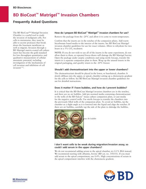
Does it matter if I have bubbles, and how do I prevent bubbles?<br />
It is critical that the <strong>BD</strong> BioCoat Matrigel invasion chambers are in the notches,<br />
and there are no air bubbles. Add pre-warmed media containing chemoattractant<br />
to the wells of the <strong>BD</strong> Falcon tissue culture companion plate, or just media<br />
for the negative control wells. Use sterile forceps to transfer the chambers into<br />
the previously filled wells of the companion plate. To avoid air bubbles, tip the<br />
chamber at a slight angle as it is lowered into the liquid and align the notches. If<br />
there are air bubbles, carefully tap the side of the plate to dislodge the bubbles.<br />
Air bubble<br />
I don't want cells to be weak during migration/invasion assay, so<br />
could I add serum in the upper chambers?<br />
We do not recommend adding serum to the apical chamber, try 0.1% BSA instead.<br />
We recommend starting with albumin, bovine (<strong>BD</strong> cat. no. 354331). If you must<br />
add serum to the apical compartment, use 0.4%. High concentrations of serum in<br />
the apical compartment interfere with the chemotactic gradient.
When I used the <strong>BD</strong> BioCoat Matrigel invasion chambers and the<br />
<strong>BD</strong> BioCoat control inserts, the cells were uneven on the bottom of<br />
the insert?<br />
There will be some normal variability, and you should count multiple fields. All<br />
conditions should be run in triplicate. Always check for air bubbles when setting<br />
up your assay, since cells will not migrate through the dry patches. Always use<br />
<strong>BD</strong> Falcon 24-well cell culture insert companion plates that come with the system<br />
(cat. no. 353504) and <strong>BD</strong> Falcon 6-well cell culture insert companion plate<br />
(cat. no. 353502) – do not use standard multiwell plates. Make sure the inserts are<br />
in the positioning notches of the companion plates, see below.<br />
Feeding Position<br />
Slide the insert into the side<br />
of the well for easy pipet<br />
access<br />
Ensure that insert sits<br />
properly in plate<br />
Positioning<br />
Notches<br />
What will the cells look like under the microscope?<br />
Cells will look slightly different depending on cell type. The cells will have an<br />
irregular shape. The pores will be round or ovoid and be refractile.<br />
Pore<br />
If incubation times are long, do cells proliferate after migration/<br />
invasion?<br />
We recommend doing a migration control on uncoated inserts and calculating<br />
the percent invasion by dividing the number of cells that invaded through the<br />
<strong>BD</strong> Matrigel invasion chamber by the number of cells that migrated through the<br />
control insert and multiplying by 100. This controls for proliferation. See the<br />
<strong>BD</strong> BioCoat Matrigel invasion chamber guidelines for use for full details.<br />
What reagent do you recommend for staining the cells on the<br />
membrane?<br />
We recommend any colorimetric stain that differentially stains the cytoplasm<br />
and the nucleus such as hematoxylin and eosin (H&E) or Wrights stain. Two<br />
examples of these types of stains are Diff-Quik from VWR (cat. no. 47733-150)<br />
or Hemacolor ® Stain Set from Merck KGaA, Darmstadt, Germany (cat. no.<br />
65044-93) .<br />
Are there any tips to wipe off the <strong>BD</strong> Matrigel from the insert?<br />
You should remove the liquid from the top of the insert and pre-wet the cotton<br />
swab. Hold the insert at an angle so the bottom of the membrane is not touching<br />
a flat surface and gently swab out the gel, taking care to remove the cells, but not<br />
detaching the insert membrane from the housing.<br />
Cell
How can I cut off the membrane? Do you supply a special blade?<br />
Do not remove the membrane from the housing until the staining is complete.<br />
A special blade is not supplied. Use a #11 scalpel to cut the membrane from the<br />
housing and immediately mount cell side down on a microscope slide. Put a drop<br />
of immersion oil on the membrane, and cover with a coverslip.<br />
How long can I keep the hydrated insert? Is it possible to store for<br />
several days after the hydration?<br />
You should use the rehydrated insert after the normal 2-hour rehydration period.<br />
If you must use it the next day because your cells are not ready expect the CV<br />
between replicates to increase. Do not use after 24-hours.<br />
I did not get very much invasion of my cells, what should I do?<br />
Verify, with an inverted microscope, that the invaded cells have not detached from<br />
the bottom of the insert and have fallen to the bottom well.<br />
Since there is no serum in the seeding media, make sure you either use a nonenzymatic<br />
method of removing the cells prior to seeding, or do a serum wash to<br />
deactivate the trypsin. Make sure you are using the notched <strong>BD</strong> Falcon 24-well<br />
cell culture insert companion plates (cat. no. 353504) and <strong>BD</strong> Falcon 6-well cell<br />
culture insert companion plate (cat. no. 353502) and that the cell culture inserts<br />
are in the notches. Verify there are no air bubbles trapped under the inserts.<br />
Different cell types invade at different rates, and the protocol may need to be<br />
adjusted for different cell types. As a first step, increase the serum concentration<br />
to 10% in the lower chamber and increase the cell number to 50,000 cells per<br />
24-well insert or 250,000 cells per 6-well insert. If this does not increase the raw<br />
invasion numbers sufficiently, you can increase the invasion time from 24- to<br />
48-hours.<br />
The <strong>BD</strong> BioCoat Matrigel invasion chamber is highly cited in the scientific<br />
literature with specific details for many cell types. You can contact <strong>BD</strong> <strong>Biosciences</strong><br />
Technical Support for assistance in locating a reference for your specific cell type.<br />
I am tired of counting all of those inserts for my IC 50 experiments,<br />
is there an easier way to quantify invasion?<br />
Yes, we have the <strong>BD</strong> BioCoat 24-multiwell (cat. nos. 354165 one pack; 354166<br />
five pack) and 96-multiwell (cat. nos. 354167 one pack; 354168 five pack)<br />
tumor cell invasion systems. These systems utilize <strong>BD</strong> FluoroBlok membranes,<br />
a patented light-tight membrane that efficiently blocks the transmission of<br />
light within the range of 490-700 nm which allows fluorescence detection in a<br />
simplified and non-destructive manner. Fluorescently labeled cells present in the<br />
top chamber of the insert are made invisible to a bottom-reading fluorescence<br />
plate reader by the <strong>BD</strong> FluoroBlok membrane. Once labeled cells migrate through<br />
the membrane they are no longer shielded from the light source and are easily<br />
detected with a bottom-reading fluorescence plate reader. The <strong>BD</strong> BioCoat tumor<br />
invasion systems can be used for either kinetic or end-point invasion assays.<br />
To place an order in the U.S., contact Customer Service at:<br />
tel: 877.232.8995; fax: 800.325.9637 or 858.812.8889<br />
For technical assistance, contact Technical Support at:<br />
tel: 877.232.8995 or 978.901.7389; fax: 978.901.7491; e-mail: labware@bd.com<br />
Outside the U.S., contact your local distributor or visit<br />
bdbiosciences.com/offices to locate your nearest <strong>BD</strong> <strong>Biosciences</strong> office.<br />
For Research Use Only. Not intended for use in diagnostic or therapeutic procedures.<br />
Diff-Quik is a trademark of Baxter Diagnostics.<br />
Hemacolor is a registered trademark of Merck KGaA, Darmstadt, Germany.<br />
<strong>BD</strong>, <strong>BD</strong> Logo, and all other trademarks are the property of Becton, Dickinson and Company. ©2009 <strong>BD</strong><br />
F09B227<br />
References<br />
1. Kong, D., Li, Y., Wang, Z., Banerjee, S., Sarkar,<br />
F.H. Inhibition of angiogenesis and invasion by<br />
3,3’-diindolylmethane is mediated by the NF-κB<br />
downstream target genes MMP-9 and uPA that<br />
regulated bioavailability of VEGF in prostate<br />
cancer. Cancer Res. 67(7):3310 (2007).<br />
2. Albini, A., and Benelli, R. The chemoinvasion<br />
assay: a method to assess tumor and<br />
endothelial cell invasion and its modulation.<br />
Nat. Protocols. 2(3):505 (2007).<br />
3. Oxelmark, E., Roth, J.M., Brooks, P.C.,<br />
Braunstein, S.E., Schneider, R.J., Garabedian,<br />
M.J. The cochaperone p23 differentially<br />
regulates estrogen receptor target genes and<br />
promotes tumor cell adhesion and invasion.<br />
Mol. Cell Biol. 26(14):5205 (2006).<br />
4. Duxbury, M.A., Ito, H., Zinner, M.J., Ashley,<br />
S.W., Whang, E.E. EphA2: a determinant<br />
of malignant cellular behavior and a<br />
potential therapeutic target in pancreatic<br />
adenocarcinoma. Oncogene. 23:1448 (2004).<br />
5. Seton-Regers, S.E., Lu, Y., Hines, L.M.,<br />
Koundinya, M., LaBaer, J., Muthuswamy, S.K.,<br />
Brugge, J.S. Cooperation of the ErbB2 receptor<br />
and transforming growth factor in induction of<br />
migration and invasion in mammary epithelial<br />
cells. Proc. Natl. Acad. Sci. 101(5):1257 (2004).<br />
6. Singh, A., Singh, U.P., Grizzle, W.E., Lillard,<br />
Jr J.W. CXCL12- CXCR4 interactions<br />
modulate prostate cancer cell migration,<br />
metalloproteinase expression and invasion. Lab.<br />
Invest. 84:1666 (2004).<br />
7. Takada, Y., Kobayashi, Y., Aggarwal, B.B.<br />
Evodiamine abolishes constitutive and inducible<br />
NF-κB activation by inhibiting IκBα kinase<br />
activation, thereby suppressing NF-κB-regulated<br />
antiapoptotic and metastatic gene expression,<br />
up-regulating apoptosis, and inhibiting<br />
invasion. J. Biol. Chem. 280(17):17203 (2005).<br />
8. Ichikawa, H., Takada, Y., Murakami, A.,<br />
Aggarwal, B.B. Identification of a novel blocker<br />
of I kappa B alpha kinase that enhances cellular<br />
apoptosis and inhibits cellular invasion through<br />
suppression of NFkappa B-regulated gene<br />
products. J. Immunol. 174(11):7383 (2005).<br />
9. Silvera, D., Rezina, A., Darvishian, F., Levine,<br />
P.H., Zolfaghari, L., Goldberg, J., Hochman, T.,<br />
Formenti, S.C., Schneider, R.J. Essential role for<br />
eIF4GI overexpression in the pathogenesis of<br />
inflammatory breast cancer. Nat. Cell Biol.<br />
11:903-908 (2009).<br />
10. Kajiro, M., Hirota, R., Nakajima, Y., Kawanowa,<br />
K., So-ma, K., Ito, I., Yamaguchi, Y., Ohie,<br />
S., Kobayashi, Y., Seino, Y., Kawano, M.,<br />
Kawabe, Y., Takei, H., Hayashi, S., Kurosumi,<br />
M., Murayama, A., Kimura, K., Yanagisawa, J.<br />
The ubiquitin ligase CHIP acts as an upstream<br />
regulator of oncogenic pathways Nat. Cell Biol.<br />
11:312-319 (2009).<br />
<strong>BD</strong> <strong>Biosciences</strong><br />
Two Oak Park<br />
Bedford, MA 01730 USA<br />
tel: 877.232.8995<br />
fax: 800.325.9637