roma (he4 + architect ca125 ii) - Fujirebio Diagnostics, Inc.
roma (he4 + architect ca125 ii) - Fujirebio Diagnostics, Inc.
roma (he4 + architect ca125 ii) - Fujirebio Diagnostics, Inc.

Create successful ePaper yourself
Turn your PDF publications into a flip-book with our unique Google optimized e-Paper software.
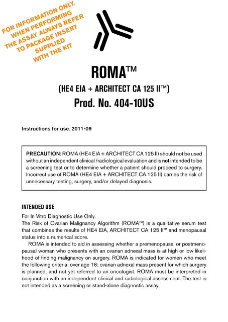
FOR INFORMATION ONLY.WHEN PERFORMINGTHE ASSAY ALWAYS REFERTO PACKAGE INSERTSUPPLIEDWITH THE KITROMA(HE4 EIA + ARCHITECT CA 125 II)Prod. No. 404-10USInstructions for use. 2011-09PRECAUTION: ROMA (HE4 EIA + ARCHITECT CA 125 II) should not be usedwithout an independent clinical /radiological evaluation and is not intended to bea screening test or to determine whether a patient should proceed to surgery.<strong>Inc</strong>orrect use of ROMA (HE4 EIA + ARCHITECT CA 125 II) carries the risk ofunnecessary testing, surgery, and/or delayed diagnosis.INTENDED USEFor In Vitro Diagnostic Use Only.The Risk of Ovarian Malignancy Algorithm (ROMA) is a qualitative serum testthat combines the results of HE4 EIA, ARCHITECT CA 125 II and menopausalstatus into a numerical score.ROMA is intended to aid in assessing whether a premenopausal or postmenopausalwoman who presents with an ovarian adnexal mass is at high or low likelihoodof finding malignancy on surgery. ROMA is indicated for women who meetthe following criteria: over age 18; ovarian adnexal mass present for which surgeryis planned, and not yet referred to an oncologist. ROMA must be interpreted inconjunction with an independent clinical and radiological assessment. The test isnot intended as a screening or stand-alone diagnostic assay.1
WARNINGS AND PRECAUTIONSFor In Vitro Diagnostic Use:• For professional use only• Follow the instructions in the package inserts for HE4 EIA and ARCHITECTCA 125 II, respectively. Reliability of assay results cannot be guaranteed ifthere are any deviations from the instructions in the package inserts.HE4 EIA KIT STORAGE AND HANDLINGFor detailed instructions on HE4 EIA Kit storage and handling including specificinstructions for all kit components, refer to the package insert.• When stored and handled as directed, the HE4 EIA Kit is stable until theexpiration date stated on the label outside of the kit box.• Do not use the HE4 EIA Kit beyond the expiration date.• The HE4 EIA Kit must be stored at 2-8°C. Do not freeze. Return to 2-8°Cimmediately after use.• The HE4 EIA Kit reagents should be allowed to reach room temperature(20–25°C) prior to use.• Do not mix identical reagents from kits having different lot numbers.ARCHITECT CA 125 II KIT STORAGE AND HANDLINGFor detailed instructions on ARCHITECT CA 125 II Kit storage and handlingincluding specific instructions for all kit components, refer to the package inserts.• When stored and handled as directed, ARCHITECT CA 125 II Kits are stableuntil the expiration date stated on the label outside of the kit box.• Do not use ARCHITECT CA 125 II Kits beyond the expiration date.• The ARCHITECT CA 125 II Kits must be stored at 2-8°C and may be usedimmediately after removal from 2-8°C storage.• The ARCHITECT CA 125 II Reagent Kit must be stored at 2-8°C in an uprightposition.• Do not mix identical reagents from kits having different lot numbers.SPECIMEN COLLECTION AND HANDLINGROMA is intended for use with serum (including serum collected in separatortubes (SST)). Plasma and other body fluids have not been validated for usewith ROMA. Collect blood by venipuncture and follow the tube manufacturer’sprocessing instructions for collection tubes.Serum can be stored at 2-8°C for 3 days before being tested. For longer periodsstore samples at -40°C or colder.5
a. ARCHITECT CA 125 II Value obtained from the ARCHITECT i2000SRNOTE: single assay measurements are used for ARCHITECT CA 125 IIValue entry.b. HE4 EIA Value obtained from the manual HE4 EIA method from <strong>Fujirebio</strong><strong>Diagnostics</strong>.NOTE: it is recommended that the assay is performed in duplicate. Use themean of the sample replicates for HE4 EIA Value entry.If non-numerical data or data out of range is entered, pop-up windows willappear as an alert to correct the associated data entry error.7. Once data points are correctly entered, click on the “Calculate Likelihood”button in the lower left corner of the Calculator Tool faceplate.8. Results from calculations will be displayed in the right side of the CalculatorTool faceplate:a. Inputs for ARCHITECT CA 125 II Value and HE4 EIA Value will beverified and repeatedb. Premenopausal LIKELIHOODc. Premenopausal ROMA Scored. Postmenopausal LIKELIHOODe. Postmenopausal ROMA Score9. Record the ROMA (HE4 EIA + ARCHITECT CA 125 II) calculated resultsas needed.Results are not saved by the Calculator Tool.10. To continue using the Calculator Tool, over-write the input values with newdata values as instructed in step 6 and continue through steps 7, 8, and 9.11. To close the ROMA (HE4 EIA + ARCHITECT CA 125 II) Calculator Toolapplication, click on the white X in the red box in the upper right corner ofthe faceplate.12. To re-open and use the ROMA (HE4 EIA + ARCHITECT CA 125 II) CalculatorTool, go to Step 2.LIMITATIONS OF THE PROCEDUREThe Risk of Ovarian Malignancy Algorithm (ROMA) uses the combination of HE4EIA and ARCHITECT CA 125 II assay values that depend on the premenopausalor postmenopausal status of a woman. The premenopausal or postmenopausalstatus must be based on ovarian function determined with information availablefrom clinical evaluation and medical history.• ROMA cannot be used as absolute evidence for the presence or absence ofmalignant disease.• ROMA should not be used as a cancer screening test.• ROMA has only been evaluated in women who will undergo a surgical interventionand is only intended for use in this population.7
• ROMA should not be used without an independent clinical evaluation andis not intended to determine whether a patient should proceed to surgery. Alow likelihood ROMA result, in the setting of a positive initial cancer risk assessment,should not preclude oncology referral.• ROMA has not been validated for the following groups: women previouslytreated for malignancy, women currently being treated with chemotherapy,pregnant women and women < 18 years of age.HE4 EIA results should not be used interchangeably with other manufacturers’methods for HE4 determinations in the ROMA calculation. Use only with the HE4 EIAassay value obtained from the manual HE4 EIA method from <strong>Fujirebio</strong> <strong>Diagnostics</strong>.ARCHITECT CA 125 II results should not be used interchangeably with other manufacturers’methods for CA 125 determinations in the ROMA calculation. Use onlywith the ARCHITECT CA 125 II assay value obtained from ARCHITECT i2000SR.Values obtained from non-designated methods or instrument platforms may produceincorrect ROMA results.An error in the calculation of results could lead to inaccurate likelihood of malignancyassessment and improper management of the patient.Anti-reagent antibodies (human anti-mouse antibody (HAMA) or heterophilicantibodies) in the patient sample may occasionally interfere with the assay, eventhough specific blocking agents are included in the buffers.Specimens containing levels of Rheumatoid Factor (RF) above 250 IU/mL mayinterfere with the ROMA result.Risk of ovarian malignancy algorithm (ROMA)Refer to the Calculation of Results section of this package insert.ROMA takes into account the results of HE4 EIA and the results of ARCHITECTCA 125 II as well as the menopausal status of the woman. The ROMA value isused to aid in assessing whether a woman is at high or low likelihood of findingmalignancy on surgery.The effectiveness of ROMA was determined in a prospective, multi-center,blinded clinical trial for premenopausal and postmenopausal women presentingwith an adnexal mass requiring surgical intervention.A total of 461 women were evaluable in the study. For each patient, an initialcancer risk assessment (ICRA) was completed by a non-gynecological oncologist,providing the assessment of the patient’s mass as benign (negative) or malignant(positive) based upon the information available to the non-gynecological oncologistduring their work-up of the patient. The corresponding histopathology reports werecollected after surgery.Using a preoperatively collected serum sample, ROMA was determined and thepatient was stratified into a low or a high likelihood group for finding malignancyon surgery.8
The histopathological classifications of the 461 evaluable patients are summarizedin the table below:HistopathologicalclassificationAlln=461Premenopausaln=240Premenopausaln=221N % N % N %Benign Pathology 375 81.3 220 91.7 155 70.1Low MalignantPotential (LMP)/BorderlineEpithelial OvarianCancerNon-EpithelialOvarian CancerOther GynecologicalCancer18 3.9 7 2.9 11 5.048 10.4 9 3.7 39 17.62 0.4 0 0.0 2 0.910 2.2 3 1.2 7 3.2Other Cancer 7 1.5 1 0.4 6 2.7Metastatic Cancer 1 0.2 0 0.0 1 0.5Use of ROMA for stratification into low likelihoodand high likelihood groups for finding malignancy on surgeryThe following cut-points were used in order to provide a specificity level of 75%:Premenopausal women:ROMA score ≥ 1.31 = High likelihood of finding malignancyROMA score < 1.31 = Low likelihood of finding malignancyPostmenopausal women:ROMA score ≥ 2.77 = High likelihood of finding malignancyROMA score < 2.77 = Low likelihood of finding malignancyThe reported result should include both the premenopausal and postmenopausallikelihood result and associated ROMA score on a scale of 0-10.9
The performance of ROMA for stratification into low likelihood and high likelihoodgroups for premenopausal and postmenopausal women with epithelial ovariancancer (EOC) only is shown in the table below:Premenopausal n=229 Postmenopausal n=194Estimate 95 % Cl Estimate 95 % ClSensitivity 100.0%92.3%70.1% – 99.2%(9/9)(36/39)79.7% – 97.2%Specificity 74.5%76.8%68.4% – 79.8%(164/220)(119/155)69.5% – 82.7%TP-FP 1 74.5% 68.7% – 80.4% 69.1% 58.2% – 80.0%PPV 2 13.8%50.0%7.5% – 24.2%(9/65)(36/72)38.7% – 61.2%NPV 3 100.0%97.5%97.7% – 99.9%(164/164)(119/122)93.0% – 99.1%Prevalence 3.9% (9/229) 20.1% (39/194)1TP-FP = True Positive rate – False Positive rate, 2 PPV = Positive PredictiveValue, 3 NPV = Negative Predictive ValueAdjunctive use of ROMA with Initial Cancer Risk Assessment (ICRA)for stratification into low likelihood and high likelihood groups of harboring malignancyThe performance for the adjunctive use of ROMA with ICRA (ROMA and/or ICRAbeing positive for high likelihood of finding malignancy on surgery) was evaluatedby calculating sensitivity, specificity, PPV (positive predictive value) and NPV (negativepredictive value). Adding ROMA to ICRA produced a statistically significantimprovement in the negative predictive value. The NPV for correctly classifying benignpatients into the low likelihood group increased from 93.2 to 96.2%, making theadjunctive use of ROMA with ICRA effective in ruling out malignancy.11
EXPECTED VALUESThe distribution of ROMA determined in 120 healthy premenopausal women and120 healthy postmenopausal women is shown in the table below:AllHealthy SubjectsPremenopausalHealthy SubjectsPostmenopausalHealthy SubjectsN 240 120 120ROMA ResultMean (SD) 1.19 (0.76) 0.94 (0.75) 1.44 (0.68)Median 0.98 0.72 1.30Range (min-max) 0.22-4.58 0.22-4.51 0.39-4.58Reference Interval(5 th –95 th percentile)0.39-2.75 0.33-2.36 0.61-2.75ROMA Likelihood (n, %)High Likelihood 25 (10.4%) 19 (15.8%) 6 (5.0%)Low Likelihood 215 (89.6%) 101 (84.2%) 114 (95.0%)In this study, 95% of the premenopausal healthy female subjects had ROMAresults equal to or below 2.36 and 95% of the postmenopausal healthy femalesubjects had ROMA results equal to or below 2.75.13
The distribution of ROMA determined in non-ovarian malignancy conditions isshown in the table below:BladderCancerBreastCancerEndometrialCancerGICancerLungCancerN 40 40 40 39 40ROMAMean (SD) 5.45 (3.09) 4.52 (3.07) 5.44 (2.99) 3.56 (2.81) 4.70 (2.45)Median 5.36 2.88 5.26 2.32 4.60Range (min-max) 0.38-10.0 0.60-9.93 0.67-9.99 0.55-9.24 0.74-9.63Reference Interval(5 th –95 th percentile)0.77-9.85 1.32-9.88 1.29-9.90 0.97-9.02 0.98-9.14ROMALikelihood (n, %)High Likelihood 31 (77.5%) 26 (65.0%) 33 (82.5%) 21 (53.8%) 31 (77.5%)Low Likelihood 9 (22.5%) 14 (35.0%) 7 (17.5%) 18 (46.2%) 9 (22.5%)14
The distribution of ROMA determined in benign conditions is shown in the table:OtherBenignDiseaseBenignGynecologicalDiseaseCongestiveHeartFailureHypertensionPregnantN 381 40 40 40 38ROMAMean (SD) 1.55 (1.20) 2.05 (1.47) 3.09 (1.82) 2.34 (1.72) 1.01 (0.59)Median 1.16 1.72 2.53 1.84 0.88Range (min-max) 0.19-8.56 0.15-6.97 0.83-7.93 0.33-8.38 0.28-3.47Reference Interval(5 th –95 th percentile)0.43-3.72 0.54-4.95 1.08-5.95 0.83-5.17 0.34-1.94ROMA Likelihood(n, %)High Likelihood 94 (24.7%) 15 (37.5%) 17 (42.5%) 12 (30.0%) 7 (18.4%)Low Likelihood 287 (75.3%) 25 (62.5%) 23 (57.5%) 28 (70.0%) 31 (81.6%)It is recommended that each laboratory establish its own reference value for thepopulation of interest.15
PERFORMANCE CHARACTERISTICSLot-to-Lot PrecisionA study was performed as described per the National Committee for ClinicalLaboratory Standards NCCLS (CLSI) guideline EP5-A2 (20). A panel of five serumsamples was tested and evaluated using both premenopausal and postmenopausalforms of the ROMA equation, using three lots of HE4 EIA Kits and three lots ofARCHITECT CA 125 II Reagent and Calibrator Kits, evaluating two measurementsof each panel, at two separate times per day for 5 days. Data from this study issummarized in the table below. 1Sample Menopausal n Mean ROMA Between Between Total TotalState Result Lots (SD) Lots (CV %) (SD) (CV %)1 Pre 60 0.66 0.023 3.5 0.051 7.7Post 60 1.05 0.014 1.3 0.043 4.12 Pre 60 1.32 0.023 1.8 0.060 4.5Post 60 2.55 0.012 0.5 0.043 1.73 Pre 60 2.81 0.000 0.0 0.150 5.2Post 60 4.83 0.007 0.1 0.085 1.84 Pre 60 1.28 0.025 1.9 0.051 4.0Post 60 2.39 0.022 0.9 0.046 1.95 Pre 60 8.66 0.000 0.0 0.071 0.8Post 60 8.73 0.018 0.2 0.043 0.51Representative data; results in individual laboratories may vary from these data.16
ReproducibilityA study was performed as described per the National Committee for Clinical LaboratoryStandards NCCLS (CLSI) guideline EP5-A2 (20). A panel of five serum sampleswas tested and evaluated using both premenopausal and postmenopausal forms ofthe ROMA equation, using one lot of HE4 EIA Kits and one lot of ARCHITECT CA125 II Reagent and Calibrator Kits, at three sites, evaluating two measurements ofeach panel, at two separate times per day for 6 days. Data from this study is summar -ized in the table below. 1Sample Menopausal n Mean ROMA Between Between Total TotalState Result Sites (SD) Sites (CV %) (SD) (CV %)1 Pre 72 0.56 0.107 19.0 0.143 25.9Post 72 0.96 0.083 8.6 0.107 11.22 Pre 72 1.16 0.168 14.6 0.195 16.9Post 72 2.39 0.124 5.2 0.153 6.43 Pre 72 2.66 0.143 5.4 0.297 11.2Post 72 4.75 0.116 2.5 0.186 3.94 Pre 72 1.13 0.180 16.0 0.232 20.7Post 72 2.25 0.149 6.6 0.183 8.15 Pre 72 8.59 0.046 0.5 0.178 2.1Post 72 8.72 0.048 0.6 0.086 1.01Representative data; results in individual laboratories may vary from these data.17
Analytical specificityStudies were performed to compare sera containing the listed substances at theindicated concentrations with control sera to determine if potential interferenceis observed impacting ROMA. Data from this study is summarized in the tablebelow. 1 Percent Difference From Control (%)Interferent Concentration ROMA (Low) ROMA (Med) ROMA (High)PRE 2 POST 2 PRE 2 POST 2 PRE 2 POST 2Hemoglobin 5 mg/mL -6.7 -3.4 0.5 0.6 4.0 1.0Bilirubin 20 mg/dL 2.3 1.2 -3.9 0.0 -5.5 -1.6(Conjugated)Bilirubin 20 mg/dL 2.8 2.3 3.9 1.9 -4.7 -1.2(Unconjugated)Protein 12 g/dL -3.7 -2.0 1.4 -0.8 -0.3 -1.1Lipid 3 g/dL 8.8 3.2 -4.0 -4.3 -1.0 -1.5Human 1000 ng/mL 7.7 9.2 -3.2 -0.9 0.9 -0.3Anti-MouseAntibodies (HAMA)Rheumatoid 1000 IU/mL 1.9 28.2 3 -1.0 6.0 2.7 -0.6Factor (RF) 500 IU/mL -6.9 12.6 3 2.6 3.6 -2.5 -1.5250 IU/mL -0.6 -0.6 0.6 0.8 2.6 0.01Representative data; results in individual laboratories may vary from these data.2Analyzed using both premenopausal and postmenopausal forms of the ROMAequation3Specimens with Rheumatoid factor greater than 250 IU/mL interference withthe test by more than 10%WARRANTYThe performance data presented here were obtained using the assay procedureindicated. Any change or modification of the procedure not recommended by<strong>Fujirebio</strong> <strong>Diagnostics</strong> may affect the results, in which event <strong>Fujirebio</strong> <strong>Diagnostics</strong>disclaims all warranties expressed, implied or statutory including the implied warrantyof merchantability and fitness for use.18
REFERENCES1. Hellstrom I, Raycraft J, et al. The HE4 (WFDC2) protein is a biomarker forovarian cancer. Cancer Res 2003;63:3695-3700.2. Moore RG, Brown AK, Miller MC, Skates S, Allard WJ, Verch T, et al. The useof multiple novel tumor biomarkers for the detection of ovarian carcinoma inpatients with a pelvic mass. Gynecologic oncology 2008 Feb;108(2):402-8.3. Montagnana M, Lippi G, Ruzzenente O, Bresciani V, Danese E, ScevarolliS, et al. The utility of serum human epididymis protein 4 (HE4) in patientswith a pelvic mass. J Clin Lab Anal 2009;23(5):331-5.4. Huhtinen K, Suvitie P, H<strong>ii</strong>ssa J, Junnila J, Huvila J, Kujari H, et al. Serum HE4concentration differentiates malignant ovarian tumours from ovarian endometrioticcysts. British journal of cancer 2009 Apr 21;100(8):1315-9.5. Montagnana M, Lippi G, Danese E, Franchi M, Guidi GC. Usefulness of serumHE4 in endometriotic cysts. British journal of cancer 2009 Aug 4;101(3):548.6. Andersen MR, Goff BA, Lowe KA, Scholler N, Bergan L, Drescher CW, et al.Use of a Symptom Index, CA125, and HE4 to predict ovarian cancer. Gynecologiconcology 2010 Mar;116(3):378-83.7. Moore RG, McMeekin SD, Brown AK, DiSilvestro P, Miller CM, Allard JW,Gajewski W, Kurman R, Bast RC Jr., Skates SJ. A novel multiple marker bioassayutilizing HE4 and CA125 for the prediction of ovarian cancer in patientswith a pelvic mass. Gynecologic oncology 2009 112:40-6.8. Havrilesky LJ, Whitehead CM, Rubatt JM, Cheek RL, Groelke J, He Q, et al.Evaluation of biomarker panels for early stage ovarian cancer detection andmonitoring for disease recurrence.Gynecologic oncology 2008 Sep;110(3):374-82.9. Nolen B, Velikokhatnaya L, Marrangoni A, De Geest K, Lomakin A, Bast RC, Jr.,et al. Serum biomarker panels for the discrimination of benign from malignantcases in patients with an adnexal mass. Gynecologic oncology 2010Jun;117(3):440-5.10. Yurkovetsky Z, Skates S, Lomakin A, Nolen B, Pulsipher T, Modugno F, et al.Development of a multimarker assay for early detection of ovarian cancer.J Clin Oncol 2010 May 1;28(13):2159-66.11. Anderson GL, McIntosh M, Wu L, Barnett M, Goodman G, Thorpe JD, et al.Assessing lead time of selected ovarian cancer biomarkers: a nested casecontrolstudy. J Natl Cancer Inst 2010 Jan 6;102(1):26-38.12. Moore RG, Jabre-Raughley M, Brown AK, Robison KM, Miller MC, Allard WJ,et al. Comparison of a novel multiple marker assay vs the Risk of MalignancyIndex for the prediction of epithelial ovarian cancer in patients with a pelvicmass. Am J Obstet Gynecol 2010 Sep;203(3):228 e1-6.19
13. National Institutes of Health Consensus Development Conference Statement.Ovarian Cancer: Screening, treatment and follow-up. Gynecol Oncol1994;55:S4-14.14. ACOG Practice Bulletin. Clinical Management Guideline for Obstetrician-Gynecologists. Management of Adnexal Masses. Obstet Gynecol 2007;110:201-213.15. Finkler NJ, Benacerraf B, Lavin PT, Wojciechowski C, Knapp RC. Comparisonof serum CA 125, clinical impression and ultrasound in the preoperative evaluationof ovarian masses. Obstet Gynecol 1988;72:659-64.16. Maggino T, Gadducci A, D’Addario V, et al. Prospective Multicenter Study onCA 125 in postmenopausal pelvic masses. Gynecol Oncol 1994:54;117-123.17. Roman LD, Muderspach LI, Stein SM, et al. Pelvic Examination, Tumor markerlevel, and Gray-Scale and Doppler Sonography in the prediction of pelviccancer. Obstet Gynecol 1997:89;493-500.18. DePriest PD, Shenson D, Fried A, et al. A morphology index based on sonographicfindings in ovarian cancer. Gynecol Oncol 1993;51:7-11.19. Li J, Dowdy S, Tipton T, Podratz K, Lu WG, Xie X, et al. HE4 as a biomarkerfor ovarian and endometrial cancer management. Expert Rev Mol Diagn 2009Sep;9(6):555-66.20. National Committee for Clinical Laboratory Standards (NCCLS/CLSI), Evaluationof Precision Performance of Clinical Chemistry Devices; ApprovedGuideline – Second Edition. EP5-A2 (2004).CanAg ® is a registered trademark of <strong>Fujirebio</strong> <strong>Diagnostics</strong> AB<strong>Fujirebio</strong> <strong>Diagnostics</strong> ABElof Lindälvs gata 13SE-414 58 GöteborgSwedenPhone + 46 31 85 70 30Fax + 46 31 85 70 40info@fdab.comwww.fdab.com20ROMA HE4 EIA Prod. No. 404-10US, 2011-09. F6054