Spotlight 3: Optical Techniques in Medical Imaging - Institute of ...
Spotlight 3: Optical Techniques in Medical Imaging - Institute of ...
Spotlight 3: Optical Techniques in Medical Imaging - Institute of ...

You also want an ePaper? Increase the reach of your titles
YUMPU automatically turns print PDFs into web optimized ePapers that Google loves.
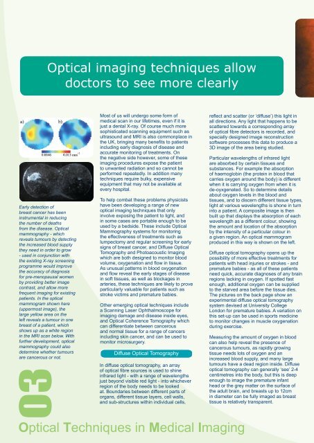
<strong>Optical</strong> imag<strong>in</strong>g techniques allowdoctors to see more clearlyEarly detection <strong>of</strong>breast cancer has been<strong>in</strong>strumental <strong>in</strong> reduc<strong>in</strong>gthe number <strong>of</strong> deathsfrom the disease. <strong>Optical</strong>mammography - whichreveals tumours by detect<strong>in</strong>gthe <strong>in</strong>creased blood supplythey need <strong>in</strong> order to grow- used <strong>in</strong> conjunction withthe exist<strong>in</strong>g X-ray screen<strong>in</strong>gprogramme would improvethe accuracy <strong>of</strong> diagnosisfor pre-menopausal womenby provid<strong>in</strong>g better imagecontrast, and allow morefrequent imag<strong>in</strong>g for exist<strong>in</strong>gpatients. In the opticalmammogram shown here(uppermost image), thelarge yellow area on theleft reveals a tumour <strong>in</strong> onebreast <strong>of</strong> a patient, whichshows up as a white region<strong>in</strong> the MRI scan below. Withfurther development, opticalmammography could alsodeterm<strong>in</strong>e whether tumoursare cancerous or not.03Most <strong>of</strong> us will undergo some form <strong>of</strong>medical scan <strong>in</strong> our lifetimes, even if it isjust a dental X-ray. Of course much moresophisticated scann<strong>in</strong>g equipment such asultrasound and MRI is also commonplace <strong>in</strong>the UK, br<strong>in</strong>g<strong>in</strong>g many benefits to patients<strong>in</strong>clud<strong>in</strong>g early diagnosis <strong>of</strong> disease andaccurate monitor<strong>in</strong>g <strong>of</strong> treatments. Onthe negative side however, some <strong>of</strong> theseimag<strong>in</strong>g procedures expose the patientto unwanted radiation and so cannot beperformed repeatedly. In addition manytechniques require bulky, expensiveequipment that may not be available atevery hospital.To help combat these problems physicistshave been develop<strong>in</strong>g a range <strong>of</strong> newoptical imag<strong>in</strong>g techniques that only<strong>in</strong>volve expos<strong>in</strong>g the patient to light, and<strong>in</strong> some cases are portable enough to beused by a bedside. These <strong>in</strong>clude <strong>Optical</strong>Mammography systems for monitor<strong>in</strong>gthe effectiveness <strong>of</strong> treatments such aslumpectomy and regular screen<strong>in</strong>g for earlysigns <strong>of</strong> breast cancer, and Diffuse <strong>Optical</strong>Tomography and Photoacoustic Imag<strong>in</strong>gwhich are both designed to monitor bloodvolume, oxygenation and flow <strong>in</strong> tissue.As unusual patterns <strong>in</strong> blood oxygenationand flow reveal the early stages <strong>of</strong> disease<strong>in</strong> s<strong>of</strong>t tissues, as well as blockages <strong>in</strong>arteries, these techniques are likely to proveparticularly valuable for patients such asstroke victims and premature babies.Other emerg<strong>in</strong>g optical techniques <strong>in</strong>cludea Scann<strong>in</strong>g Laser Ophthalmoscope forimag<strong>in</strong>g damage and disease <strong>in</strong>side eyes,and <strong>Optical</strong> Coherence Tomography whichcan differentiate between cancerousand normal tissue for a range <strong>of</strong> cancers<strong>in</strong>clud<strong>in</strong>g sk<strong>in</strong> cancer, and can be used tomonitor microsurgery.Diffuse <strong>Optical</strong> TomographyIn diffuse optical tomography, an array<strong>of</strong> optical fibre sources is used to sh<strong>in</strong>e<strong>in</strong>frared light - with a range <strong>of</strong> wavelengthsjust beyond visible red light - <strong>in</strong>to whicheverregion <strong>of</strong> the body needs to be lookedat. Boundaries between different parts <strong>of</strong>organs, different tissue layers, cell walls,and sub-structures with<strong>in</strong> <strong>in</strong>dividual cells,<strong>Optical</strong> <strong>Techniques</strong> <strong>in</strong> <strong>Medical</strong> Imag<strong>in</strong>greflect and scatter (or ‘diffuse’) this light <strong>in</strong>all directions. Any light that happens to bescattered towards a correspond<strong>in</strong>g array<strong>of</strong> optical fibre detectors is recorded, andspecially designed image reconstructions<strong>of</strong>tware processes this data to produce a3D image <strong>of</strong> the area be<strong>in</strong>g studied.Particular wavelengths <strong>of</strong> <strong>in</strong>frared lightare absorbed by certa<strong>in</strong> tissues andsubstances. For example the absorption<strong>of</strong> haemoglob<strong>in</strong> (the prote<strong>in</strong> <strong>in</strong> blood thatcarries oxygen around the body) is differentwhen it is carry<strong>in</strong>g oxygen from when it isde-oyxgenated. So to determ<strong>in</strong>e detailsabout oxygen levels <strong>in</strong> the blood andtissues, and to discern different tissue types,light at various wavelengths is shone <strong>in</strong> turn<strong>in</strong>to a patient. A composite image is thenbuilt up that displays the absorption <strong>of</strong> eachwavelength as a different colour, show<strong>in</strong>gthe amount and location <strong>of</strong> the absorptionby the <strong>in</strong>tensity <strong>of</strong> a particular colour <strong>in</strong>a given region. An optical mammogramproduced <strong>in</strong> this way is shown on the left.Diffuse optical tomography opens up thepossibility <strong>of</strong> more effective treatments forpatients with head <strong>in</strong>juries or strokes - andpremature babies - as all <strong>of</strong> these patientsneed quick, accurate diagnoses <strong>of</strong> any bra<strong>in</strong>regions lack<strong>in</strong>g <strong>in</strong> oxygen. If spotted fastenough, additional oxygen can be suppliedto the starved area before the tissue dies.The pictures on the back page show anexperimental diffuse optical tomographysystem devised at University CollegeLondon for premature babies. A variation onthis set-up can be used <strong>in</strong> sports medic<strong>in</strong>eto monitor changes <strong>in</strong> muscle oxygenationdur<strong>in</strong>g exercise.Measur<strong>in</strong>g the amount <strong>of</strong> oxygen <strong>in</strong> bloodcan also help reveal the presence <strong>of</strong>cancerous tumours, as rapidly grow<strong>in</strong>gtissue needs lots <strong>of</strong> oxygen and an<strong>in</strong>creased blood supply, and many largetumours have a dead region <strong>in</strong>side. Diffuseoptical tomography can generally ‘see’ 2-4centimetres <strong>in</strong>to the body, but this is deepenough to image the premature <strong>in</strong>fanthead or the grey matter on the surface <strong>of</strong>the adult bra<strong>in</strong>, and breasts up to 12cm<strong>in</strong> diameter can be fully imaged as breasttissue is relatively transparent.