Spotlight 3: Optical Techniques in Medical Imaging - Institute of ...
Spotlight 3: Optical Techniques in Medical Imaging - Institute of ...
Spotlight 3: Optical Techniques in Medical Imaging - Institute of ...

Create successful ePaper yourself
Turn your PDF publications into a flip-book with our unique Google optimized e-Paper software.
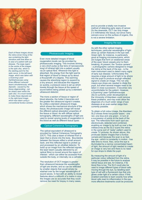
Each <strong>of</strong> these images showsthe ret<strong>in</strong>a (<strong>in</strong>ner l<strong>in</strong><strong>in</strong>g <strong>of</strong> theeye conta<strong>in</strong><strong>in</strong>g the lightsensitivecells that allow usto see) <strong>of</strong> a patient with eyedisease. The large region<strong>in</strong> the centre <strong>of</strong> the imagesis the optic disc, which isthe start<strong>in</strong>g po<strong>in</strong>t for theoptic nerve. In the left-handimage, which was taken witha Scann<strong>in</strong>g LaserOphthalmoscope us<strong>in</strong>gthree different colouredlasers simultaneously, whitedeposits - caused by thetissue degenerat<strong>in</strong>g - canbe clearly seen with<strong>in</strong> theoptic disc. It is much harderto make these deposits out<strong>in</strong> the right-hand image,which was taken us<strong>in</strong>g aconventional fundus camera.Photoacoustic Imag<strong>in</strong>gEven more detailed images <strong>of</strong> bloodoxygenation levels can be provided byphotoacoustic imag<strong>in</strong>g. This <strong>in</strong>volves sh<strong>in</strong><strong>in</strong>ga very short pulse (a few tens <strong>of</strong> nanosecondslong) <strong>of</strong> <strong>in</strong>frared light <strong>in</strong>to a patient usuallyvia an optical fibre. Wherever this light isabsorbed, the energy from the light warmsthat region <strong>of</strong> blood or tissue up by abouta thousandth <strong>of</strong> a degree. This warm<strong>in</strong>gcauses the absorb<strong>in</strong>g region to expand bya t<strong>in</strong>y amount, and because this happensso quickly, a pressure wave is set up whichtravels through the tissue at the speed <strong>of</strong>sound before be<strong>in</strong>g picked up by a standardultrasound detector.The more a section <strong>of</strong> tissue or region <strong>of</strong>blood absorbs, the hotter it becomes andthe greater the ultrasound signal it creates.So unlike a standard ultrasound image,which shows the mechanical structure <strong>of</strong> thetissue, the photoacoustic image will revealthe different optical absorption properties <strong>of</strong>the tissue or blood. As with diffuse opticaltomography, different wavelengths <strong>of</strong> light areused to reveal vary<strong>in</strong>g levels <strong>of</strong> oxygenation <strong>in</strong>the blood as well as different tissue types.<strong>Optical</strong> Coherence TomographyThe optical equivalent <strong>of</strong> ultrasound isprovided by <strong>Optical</strong> Coherence Tomography(OCT). This uses a beam <strong>of</strong> <strong>in</strong>frared from alaser that is shone <strong>in</strong>to the body. Boundariesbetween the tissues reflect a small amountback, and this reflected signal is picked upand processed by an ultrafast detector. Tobuild up an image from the reflected signals,the laser beam (usually delivered by anoptical fibre) is moved around the region <strong>of</strong><strong>in</strong>terest - which can either be accessed fromoutside the body, or <strong>in</strong>ternally via a catheter.The resolution <strong>of</strong> OCT images is greaterthan ultrasound because the wavelengths<strong>of</strong> light are shorter, and so can be reflectedby smaller objects that would simply bewashed over by the longer wavelengths <strong>of</strong>sound waves. In fact with an ability to revealfeatures about a millionth <strong>of</strong> a metre <strong>in</strong> size,the images are so accurate that they couldremove the need for biopsy <strong>in</strong> many cases,and so provide a totally non-<strong>in</strong>vasivemethod <strong>of</strong> detect<strong>in</strong>g early signs <strong>of</strong> cancer.On the downside, OCT can only image2-3 millimetres <strong>in</strong>to tissue, but s<strong>in</strong>ce manycancers occur on the surface <strong>of</strong> organs, thisis not a severe limitation.Imag<strong>in</strong>g the eyeAs with the other optical imag<strong>in</strong>gtechniques, particular wavelengths <strong>of</strong> lightshow up certa<strong>in</strong> features <strong>in</strong> the eye well.Green light, for <strong>in</strong>stance, is particularlygood at imag<strong>in</strong>g microaneurysms, whichare bulges that form on weakened areas<strong>of</strong> the eyes’ blood vessels prior to thembreak<strong>in</strong>g. At present, the “fundus camera”- a photographic camera designed to imagethe ret<strong>in</strong>a at the back <strong>of</strong> the eye - is thema<strong>in</strong> <strong>in</strong>strument used to detect many signs<strong>of</strong> early eye disease. Unfortunately thisrequires a large amount <strong>of</strong> light to be shone<strong>in</strong>to the pupil to produce enough reflectedsignal to create an image. This not onlymeans eye drops are needed to dilate thepupil, but when several images need to betaken <strong>in</strong> close succession, it becomes veryuncomfortable for the patient. However,a Scann<strong>in</strong>g Laser Ophthalmoscope(SLO) currently under development atthe University <strong>of</strong> Aberdeen could removethese problems, and may even allow thediagnosis <strong>of</strong> a much wider range <strong>of</strong> eyediseases at an even earlier stage thancurrently possible.To obta<strong>in</strong> a full colour image, the AberdeenSLO sh<strong>in</strong>es three low-power lasers - onered, one blue and one green - <strong>in</strong> turn ata succession <strong>of</strong> po<strong>in</strong>ts at the back <strong>of</strong> theeye. The reflections from each spot areelectronically detected and comb<strong>in</strong>ed,enabl<strong>in</strong>g an image to be built up over a fewmilliseconds as the SLO scans the ret<strong>in</strong>a<strong>in</strong> the same sort <strong>of</strong> ‘raster’ pattern used tocreate TV pictures. As shown above, thisproduces clearer images than a funduscamera and because the scan is so fast,the eye - which is constantly <strong>in</strong> motion - haslittle time to move. Also, s<strong>in</strong>ce the eye isillum<strong>in</strong>ated by a narrow concentrated beam<strong>of</strong> light, the amount <strong>of</strong> light needed to createan image is very low and there is no needfor eye drops to dilate the pupil.By measur<strong>in</strong>g the amount <strong>of</strong> light <strong>of</strong> aparticular colour reflected from the ret<strong>in</strong>a,it may be possible <strong>in</strong> the future to analyseret<strong>in</strong>al tissue and see, for <strong>in</strong>stance, if itsblood flow is low. The SLO could also beused to image different types <strong>of</strong> blood cellsmov<strong>in</strong>g through the eye, by labell<strong>in</strong>g eachtype <strong>of</strong> cell with a fluorescent dye that onlyglows under light <strong>of</strong> a certa<strong>in</strong> colour. If thisenabled scientists to learn exactly how whiteblood cells react to <strong>in</strong>flammation <strong>in</strong> the eye,more effective treatments could be produced.