

Dental Press
Dental Press
Dental Press

You also want an ePaper? Increase the reach of your titles
YUMPU automatically turns print PDFs into web optimized ePapers that Google loves.
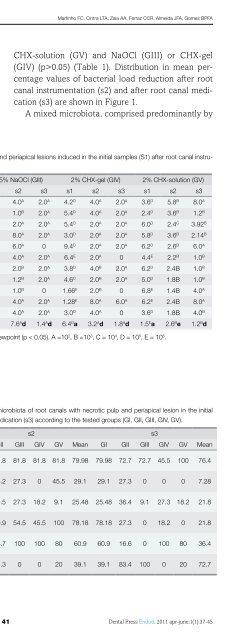
Souza MA, Menin MLF, Montagner F, Cecchin D, Farina APIntroductionThe success of endodontic therapy is groundednot only in the correct diagnosis, but also the properplanning and technical implementation, and especiallyin caring for the maintenance of the aseptic chainduring the patients care.The endodontic instruments are used to removethe remnants of pulp tissue during the procedures ofcleaning and shaping of the root canal system. Theseinstruments can be recycled for reuse after its firstuse. In a large study was reported that 88% of generalpractitioners dentists in the UK re-process the endodonticfiles after use 1 .The mandatory conduct of biosecurity recognizesthat the endodontic instruments, to be reused, theymust go through a cleaning process before sterilization2 , since the presence of organic matter and/ordebris on the instruments may interfere with the sterilizationprocess. These organic compounds createsbarriers to protect the microorganisms, which mayprevent the penetration of the sterilizing agent 3 .The procedures of pre-cleaning and autoclave canbe used to sterilize endodontic instruments 4,5 . However,the complex architecture of endodontic filescould difficult these procedures 5 . <strong>Dental</strong> structuresand organic debris have been observed on the surfaceof rotary instruments, especially in the cracks 6 .According to a previous study, 66% of endodonticfiles retrieved dental general practitioners remainedvisibly contaminated 7 . Thus, there is the possibility ofcross contamination associated with the inability toproperly clean and sterilize them, and suggested thatthese instruments should be single-use devices.Therefore, the aim of this study was to determinethe presence of debris left on the surface of endodonticfiles after performing a cleaning process and analyzeits influence on the sterilization process.Materials and methodsFifty endodontic files K #25 were selected for thisstudy, regardless of their trademark. The sampleswere divided into two groups (n = 25) according tothe method of analysis, prior use and performance ofdisinfection protocol, according to Table 1.The endodontic instruments were obtained directlyfrom students of the School of Dentistry ofPontifical Catholic University of Rio Grande do SulTable 1. Distribution of samples in groups.Group Method N Prior use Clean SterilizationG1 SEM 25 Yes Yes YesG2 Culture 25 Yes Yes Yes(PUC-RS, Porto Alegre, Brazil). The cleaning protocolused consisted of brushing with chlorhexidine gluconate2% (Globomedia, Sacomã, SP, Brazil), washingin water and drying. Prior to analysis, sampleswere placed in plastic Eppendorf tubes (EppendorfAG, São Paulo, Brazil) and sterilized by autoclaving(Dabi Atlante, Ribeirão Preto, Brazil), for 30 minutesat a temperature of 120ºC.Analysis in Scanning Electron MicroscopyThe first group of endodontic files was removedfrom the Eppendorf plastic tubes with clinical tweezersand manipulated only by the cable, avoidingany contact of the active part of the instrument. Thecables of instruments were removed through a wirecutter and its metal rods, made by the blade and theintermediate portion, were fixed in stubs for furtherobservation.After this process, samples were taken to a scanningelectron microscope. The initial portion of theactive blade of each instrument was evaluated undermagnification of 150 X and 15 kV, recording the imagesfor each instrument.The images were evaluated by four examinersprevious calibrated by Kappa test for inter-examineragreement. A numeric score was assigned for eachimage, representing its degree of dirt for each instrument:1 = no residues in the file, 2 = file almost cleansurface, i.e., with low residue, 3 = surface file with anaverage amount of waste, and 4 = the surface of thefile with a large amount of waste.The data were submitted to Kruskal-Wallis test,using the mode to qualitative assessment on a significancelevel of 1%.Microbiological Analysis ofContamination of FilesAll procedures were performed under strict asepticconditions inside a laminar flow camera. Each endodonticfile was removed from the Eppendorf plastictube with a sterile tweezers and then introduced into© 2011 <strong>Dental</strong> <strong>Press</strong> Endodontics 83<strong>Dental</strong> <strong>Press</strong> Endod. 2011 apr-june;1(1):82-6


