Lynette Masters Neuroradiologist
Lynette Masters Neuroradiologist
Lynette Masters Neuroradiologist
- No tags were found...

You also want an ePaper? Increase the reach of your titles
YUMPU automatically turns print PDFs into web optimized ePapers that Google loves.
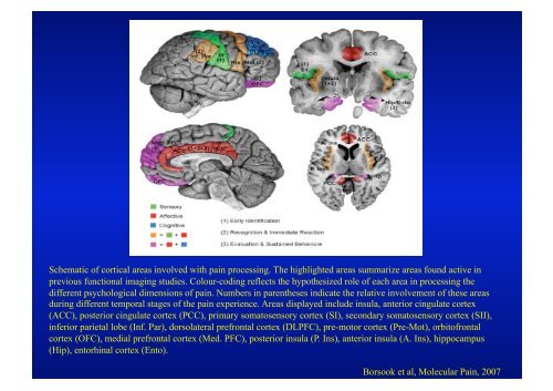
Schematic of cortical areas involved with pain processing. The highlighted areas summarize areas found active inprevious functional imaging studies. Colour-coding reflects the hypothesized role of each area in processing thedifferent psychological dimensions of pain. Numbers in parentheses indicate the relative involvement of these areasduring different temporal stages of the pain experience. Areas displayed include insula, anterior cingulate cortex(ACC), posterior cingulate cortex (PCC), primary somatosensory cortex (SI), secondary somatosensory cortex (SII),inferior parietal lobe (Inf. Par), dorsolateral prefrontal cortex (DLPFC), pre-motor cortex (Pre-Mot), orbitofrontalcortex (OFC), medial prefrontal cortex (Med. PFC), posterior insula (P. Ins), anterior insula (A. Ins), hippocampus(Hip), entorhinal cortex (Ento).Borsook et al, Molecular Pain, 2007