Bioluminescence in the Deep-Sea Cirrate Octopod ... - Duke Biology
Bioluminescence in the Deep-Sea Cirrate Octopod ... - Duke Biology
Bioluminescence in the Deep-Sea Cirrate Octopod ... - Duke Biology
Create successful ePaper yourself
Turn your PDF publications into a flip-book with our unique Google optimized e-Paper software.
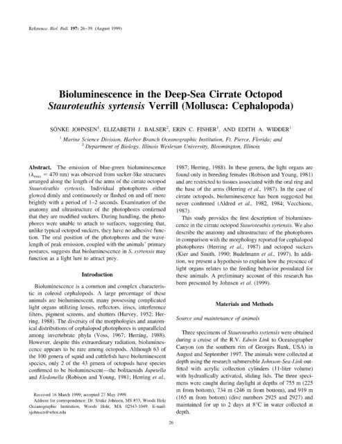
Reference: Viol. Bull. 197: 26-39. (August 1999)<br />
<strong>Biolum<strong>in</strong>escence</strong> <strong>in</strong> <strong>the</strong> <strong>Deep</strong>-<strong>Sea</strong> <strong>Cirrate</strong> <strong>Octopod</strong><br />
Stauroteuthis syrtensis Verrill (Mollusca: Cephalopoda)<br />
SGNKE JOHNSEN’, ELIZABETH J. BALSER’, ERIN C. FISHER’, AND EDITH A. WTDDER’<br />
’ A4ar<strong>in</strong>e Science Division, Harlx~r Branch Oceanographic Institution, Ft. Pierce, Florida; and<br />
’ Department of <strong>Biology</strong>, Ill<strong>in</strong>ois Wesleyan University, Bloom<strong>in</strong>gton, Ill<strong>in</strong>ois<br />
Abstract. The emission of blue-green biolum<strong>in</strong>escence<br />
(A,,,,, = 470 nm) was observed from sucker-like structures<br />
arranged along <strong>the</strong> length of <strong>the</strong> arms of <strong>the</strong> cirrate octopod<br />
Stauroteuthis syrtensis. Individual photophores ei<strong>the</strong>r<br />
glowed dimly and cont<strong>in</strong>uously or flashed on and off more<br />
brightly with a period of 1-2 seconds. Exam<strong>in</strong>ation of <strong>the</strong><br />
anatomy and ultrastructure of <strong>the</strong> photophores conhrmed<br />
that <strong>the</strong>y are modified suckers. Dur<strong>in</strong>g handl<strong>in</strong>g. <strong>the</strong> photo-<br />
phorcs were unable to attach to surfaces, suggest<strong>in</strong>g that,<br />
unlike typical octopod suckers, <strong>the</strong>y have no adhesive func-<br />
tion. The oral position of <strong>the</strong> photophores and <strong>the</strong> wave-<br />
length of peak emission, coupled with <strong>the</strong> animals’ primary<br />
postures, suggests that biolum<strong>in</strong>escence <strong>in</strong> S. syrtensis may<br />
function as a light lure to attract prey.<br />
Introduction<br />
<strong>Biolum<strong>in</strong>escence</strong> is a common and complex characteris-<br />
tic <strong>in</strong> coleoid cephalopods. A large percentage of <strong>the</strong>se<br />
animals arc biolum<strong>in</strong>escent, many possess<strong>in</strong>g complicated<br />
light organs utiliz<strong>in</strong>g lenses, reflectors, irises. <strong>in</strong>terference<br />
filters, pigment screens, and shutters (Harvey, 1952: Her-<br />
r<strong>in</strong>g, 1988). The diversity of <strong>the</strong> morphologies and anatom-<br />
ical distributions of cephalopod photophorcs is unparalleled<br />
among <strong>in</strong>vertebrate phyla (Voss, 1967; Herr<strong>in</strong>g, 1988).<br />
However. despite this extraord<strong>in</strong>ary radiation, biolum<strong>in</strong>es-<br />
ccncc appears to be rare among octopods. Although 63 of<br />
<strong>the</strong> 100 genera of squid and cuttlefish have biolum<strong>in</strong>escent<br />
species, only 2 of <strong>the</strong> 43 genera of octopods have species<br />
confirmed to be biolum<strong>in</strong>escent-<strong>the</strong> bolitaenids Japetella<br />
and Eledonella (Robison and Young, 1981; Herr<strong>in</strong>g et al.,<br />
Received 16 March 1999; accepted 27 May 1999.<br />
Address for correspondence: Dr. S(<strong>in</strong>ke Johnsen, MS #33, Woods 1101~<br />
Oceanographic Institution, Woods IIole, MA 025431049. E-mail:<br />
sjohnsen@whoi.edu<br />
26<br />
1987; Herr<strong>in</strong>g, 1988). In <strong>the</strong>se genera, <strong>the</strong> light organs arc<br />
found only <strong>in</strong> breed<strong>in</strong>g females (Robison and Young, 1981)<br />
and arc restricted to tissues associated with <strong>the</strong> oral r<strong>in</strong>g and<br />
<strong>the</strong> base of <strong>the</strong> arms (Herr<strong>in</strong>g et al., 1987). In <strong>the</strong> case of<br />
cirrate octopods, biolum<strong>in</strong>escence has been suggested but<br />
never confirmed (hldred et al., 1982, 1984; Vecchionc,<br />
1987).<br />
This study provides <strong>the</strong> first description of biolum<strong>in</strong>es-<br />
cence <strong>in</strong> <strong>the</strong> cirratc octopod Stauroteuthis syrtensis. We also<br />
describe <strong>the</strong> anatomy and ultrastructure of <strong>the</strong> photophores<br />
<strong>in</strong> comparison with <strong>the</strong> morphology reported for cephalopod<br />
photophores (Herr<strong>in</strong>g et al., 1987) and octopod suckers<br />
(Kier and Smith, 1990; Budelmann et czl., 1997). In addi-<br />
tion, we present a hypo<strong>the</strong>sis to expla<strong>in</strong> how <strong>the</strong> presence of<br />
light organs relates to <strong>the</strong> feed<strong>in</strong>g behavior postulated for<br />
<strong>the</strong>se animals. A prelim<strong>in</strong>ary account of this research has<br />
been presented by Johnsen et al. (1999).<br />
Materials and Methods<br />
Source and ma<strong>in</strong>tenance of animals<br />
Three specimens of Stauroteuthis syrtensis were obta<strong>in</strong>ed<br />
dur<strong>in</strong>g a cruise of <strong>the</strong> R.V. Edw<strong>in</strong> L<strong>in</strong>k to Oceanographer<br />
Canyon (on <strong>the</strong> sou<strong>the</strong>rn rim of Georges Bank, USA) <strong>in</strong><br />
August and September 1997. The animals were collected at<br />
depth us<strong>in</strong>g <strong>the</strong> research submersible Johnson-<strong>Sea</strong>-L<strong>in</strong>k out-<br />
fitted with acrylic collection cyl<strong>in</strong>ders (11 -liter volume)<br />
with hydraulically activated, slid<strong>in</strong>g lids. The three speci-<br />
mens were caught dur<strong>in</strong>g daylight at depths of 755 m (225<br />
m from bottom), 734 m (246 m from bottom), and 919 m<br />
(165 m from bottom) (dive numbers 2925 and 2927) and<br />
ma<strong>in</strong>ta<strong>in</strong>ed for up to 2 days at 8°C <strong>in</strong> water collected at<br />
depth.
Video and photography<br />
Specimens were videotaped <strong>in</strong> two situations. First, <strong>the</strong><br />
behavior of two animals was recorded from <strong>the</strong> submersible.<br />
Second, <strong>the</strong> captured animals were filmed aboard ship <strong>in</strong> <strong>the</strong><br />
dark by us<strong>in</strong>g an <strong>in</strong>tensified video camera (Intevac’s Nite-<br />
Mate 1305/1306 CCTV <strong>in</strong>tensifier coupled to a Panasonic<br />
Charge Coupled Device). Dur<strong>in</strong>g shipboard film<strong>in</strong>g, <strong>the</strong><br />
animals were gently prodded to <strong>in</strong>duce biolum<strong>in</strong>escence.<br />
Representative video frames were digitized (ITSCE capture<br />
board, Eyeview Software, Coreco Inc.). The animals were<br />
also placed <strong>in</strong> a plankton kreisel (Hamner, 1990) and pho-<br />
tographed with a Nikon SLR camera with Kodak Elite 100<br />
color film. Data from a previously recorded <strong>in</strong> situ video of<br />
a specimen of S. syrtensis from <strong>the</strong> slope waters near Cape<br />
Hatteras at 840 m (35 m from bottom; August 1996; R.V.<br />
Edw<strong>in</strong> L<strong>in</strong>k; dive 2777) are also reported <strong>in</strong> this paper.<br />
Spectrophotometry<br />
Biolum<strong>in</strong>escent spectra were measured us<strong>in</strong>g an <strong>in</strong>tensi-<br />
fied optical multichannel analyzer (OMA-detector model<br />
1420, detector <strong>in</strong>terface model 1461, EG&G Pr<strong>in</strong>ceton Ap-<br />
plied Research) coupled to a 2-mm-diameter fiber optic<br />
cable. The detector was wavelength calibrated us<strong>in</strong>g a low-<br />
pressure mercury spectrum lamp (Model 6047, Oriel Inc.)<br />
and <strong>in</strong>tensity calibrated us<strong>in</strong>g a NIST referenced low-<strong>in</strong>ten-<br />
sity source (Model 310, Optronics Laboratories) <strong>in</strong>tended<br />
for <strong>the</strong> calibration of detectors from 350 nm to 800 nm. For<br />
fur<strong>the</strong>r details on <strong>the</strong> <strong>the</strong>ory of operation and calibration of<br />
<strong>the</strong> OMA detector, see Widder et al. (1983). Three emission<br />
spectra were recorded from one animal, and an average<br />
spectrum was calculated.<br />
Microscopy of photophores<br />
The fixation, dehydration, and <strong>in</strong>filtration procedures<br />
were performed at room temperature aboard <strong>the</strong> R. V.<br />
Edw<strong>in</strong> L<strong>in</strong>k. The animals were sacrificed by over-anes<strong>the</strong>sia<br />
with MS222 (Sigma Chemicals Inc.). One specimen was<br />
fixed <strong>in</strong> 10% formal<strong>in</strong> <strong>in</strong> seawater and dissected to confirm<br />
species identification. One arm of a different specimen was<br />
fixed <strong>in</strong> 2.5% glutaraldehyde <strong>in</strong> 0.2 it4 Millonig’s buffer at<br />
pH 7.4, adjusted to an osmolarity of 1000 mOs with NaCl.<br />
After an <strong>in</strong>itial l-h fixation, several photophores were dis-<br />
sected from <strong>the</strong> arm and placed <strong>in</strong> fresh fixative for an<br />
additional 5.5 h. Postfixation of <strong>the</strong> dissected photophores <strong>in</strong><br />
1% osmium tetroxide <strong>in</strong> Millonig’s buffer for 70 m<strong>in</strong> was<br />
followed by dehydration through a graded series of ethyl<br />
alcohols. Over a period of 6 days, <strong>the</strong> specimens were<br />
slowly <strong>in</strong>filtrated with propylene oxide and Polybed 812<br />
(Polysciences) and <strong>the</strong>n embedded <strong>in</strong> Polybed 812.<br />
Semith<strong>in</strong> (1 pm) and th<strong>in</strong> sections of embedded material<br />
were cut with a diamond knife (Diatome) and a Sorvall<br />
MT2 rotary ultramicrotome. Semith<strong>in</strong> sections were sta<strong>in</strong>ed<br />
BIOLUMINESCENCE IN A DEEP-SEA OCTOPOD 27<br />
with 2% toluid<strong>in</strong>e blue <strong>in</strong> 1% sodium borate and photo-<br />
graphed with a Zeiss Photomicroscope II us<strong>in</strong>g Kodak<br />
Tmax 100 black-and-white film. Intact arms fixed <strong>in</strong> 10%<br />
formal<strong>in</strong> <strong>in</strong> seawater were photographed with a Tessovar<br />
photographic system. Th<strong>in</strong> sections for ultrastructural eval-<br />
uation were sta<strong>in</strong>ed with aqueous 3% uranyl acetate and<br />
0.3% lead citrate. Sta<strong>in</strong>ed sections were viewed and photo-<br />
graphed with a Zeiss EM 9 transmission electron micro-<br />
scope.<br />
For scann<strong>in</strong>g electron microscopy, an arm with suckers<br />
was fixed <strong>in</strong> 2.5% gluteraldehyde (as described above),<br />
dehydrated with ethyl alcohol, <strong>in</strong>filtrated with hexa-<br />
methyldisalizane (Pellco), and air-dried. Micrographs were<br />
obta<strong>in</strong>ed with a JEOL JSM 5800LV scann<strong>in</strong>g electron mi-<br />
croscope us<strong>in</strong>g Kodak Polapan 400 film.<br />
Results<br />
General description of animal and distribution of<br />
photophores<br />
Figures 1A and 1B show <strong>the</strong> largest of <strong>the</strong> three captured<br />
specimens of S. syrtensis. The appearance of <strong>the</strong> specimen<br />
is typical for <strong>the</strong> species (Vecchione and Young, 1997). The<br />
mantle length is about 9 cm (mantle lengths of o<strong>the</strong>r two<br />
specimens - 6 cm), suggest<strong>in</strong>g that all three animals were<br />
immature (Coll<strong>in</strong>s, unpubl. data). The measurements are<br />
highly approximate because <strong>the</strong> mantle <strong>in</strong> <strong>the</strong> liv<strong>in</strong>g animal<br />
is easily deformed. The primary webb<strong>in</strong>g extends for about<br />
three-quarters of <strong>the</strong> length of <strong>the</strong> arms. The arms are oral to<br />
<strong>the</strong> primary web and attached to it by a secondary web. The<br />
photophores are arranged <strong>in</strong> a s<strong>in</strong>gle row along <strong>the</strong> oral<br />
surface of each arm, situated between successive pairs of<br />
cirri (Fig. 1B). Each arm supports about 40 photophores.<br />
The distance between photophores decreases from <strong>the</strong> base<br />
to <strong>the</strong> tip of <strong>the</strong> arm, with <strong>the</strong> greatest distance be<strong>in</strong>g 4 mm<br />
and <strong>the</strong> smallest less than 0.1 mm. The diameter and <strong>the</strong><br />
degree of development of <strong>the</strong> photophores located at <strong>the</strong> tip<br />
are less than those located at <strong>the</strong> base of <strong>the</strong> arm. The fresh<br />
tissue of <strong>the</strong> entire animal had a gelat<strong>in</strong>ous consistency<br />
typical of many deep-sea cephalopods (Voss, 1967). Al-<br />
though orange-red under <strong>the</strong> photo-floodlights, <strong>the</strong> color of<br />
<strong>the</strong> animal was closer to reddish-brown <strong>in</strong> daylight.<br />
<strong>Biolum<strong>in</strong>escence</strong><br />
When mechanically stimulated, S. syrtensis emitted mod-<br />
erately bright, blue-green light (A,,,,, = 470 nm) from <strong>the</strong><br />
sucker-like photophores along <strong>the</strong> length of each arm (Fig.<br />
2). With cont<strong>in</strong>uous stimulation, <strong>the</strong>se photophores pro-<br />
duced light for up to 5 m<strong>in</strong>, though <strong>the</strong> <strong>in</strong>tensity of biolu-<br />
m<strong>in</strong>escence decreased over time. Individual photophores<br />
ei<strong>the</strong>r glowed dimly and cont<strong>in</strong>uously or bl<strong>in</strong>ked on and off<br />
brightly at 0.5 to 1 Hz. The bl<strong>in</strong>k<strong>in</strong>g photophores cycled<br />
asynchronously, produc<strong>in</strong>g a tw<strong>in</strong>kl<strong>in</strong>g effect. All suckers
28 S. JOHNSEN ET AL<br />
Figure 1. Photographs under artificial light of <strong>the</strong> deep-sea f<strong>in</strong>ned<br />
octopod Srauroteufhis syrfensis with <strong>the</strong> webbed arms <strong>in</strong> swimm<strong>in</strong>g pos-<br />
ture (A) and spread (B) display<strong>in</strong>g <strong>the</strong> photophores/suckers (arrowheads)<br />
that appear as white spheres along <strong>the</strong> length of <strong>the</strong> <strong>in</strong>ner surface of <strong>the</strong><br />
arms. The posture shown <strong>in</strong> (B) may be one of extreme withdrawal<br />
<strong>in</strong>tended to startle <strong>in</strong>truders with <strong>the</strong> sudden appearance of biolum<strong>in</strong>escent<br />
suckers. ar, arm; ey, eye; ti, f<strong>in</strong>; wb, webb<strong>in</strong>g between arms. Scale bars =<br />
4 cm.<br />
(except possibly <strong>the</strong> very small ones at <strong>the</strong> tips of <strong>the</strong> arms)<br />
appeared capable of lum<strong>in</strong>escence. No o<strong>the</strong>r portion of <strong>the</strong><br />
body was observed to emit light.<br />
Morphology of photophores<br />
Each photophore is a raised papilla-like structure partially<br />
embedded <strong>in</strong> <strong>the</strong> connective tissue of <strong>the</strong> arm. The photo-<br />
phores are composed of three layers of cells: an outer<br />
epi<strong>the</strong>lium modified to form a collar, <strong>in</strong>fundibulum, and<br />
acetabulum; a capsule-like mass of muscle and neural tissue<br />
beneath <strong>the</strong> epi<strong>the</strong>lium; and a th<strong>in</strong> layer separat<strong>in</strong>g <strong>the</strong><br />
capsule from <strong>the</strong> dermis of <strong>the</strong> arm (Figs. 3,4,5). The collar<br />
epi<strong>the</strong>lium is cont<strong>in</strong>uous with <strong>the</strong> epidermis and is folded<br />
<strong>in</strong>ward, form<strong>in</strong>g a rim around <strong>the</strong> central portion of <strong>the</strong><br />
photophore (Figs. 3B, C; 4A). In both formal<strong>in</strong>- and glut-<br />
araldehyde-preserved specimens, <strong>the</strong> photophores appear to<br />
be ei<strong>the</strong>r everted above (Fig. 3B) or retracted below (Fig.<br />
3C) <strong>the</strong> outer edge of <strong>the</strong> collar.<br />
Outer epi<strong>the</strong>lium<br />
The outer and <strong>in</strong>ner folds of <strong>the</strong> collar epi<strong>the</strong>lium are<br />
morphologically dist<strong>in</strong>ct and are different from <strong>the</strong> epider-<br />
mis cover<strong>in</strong>g <strong>the</strong> arm (Figs. 5,6). The epidermis of <strong>the</strong> arm<br />
is squamous to cuboidal <strong>in</strong> character and consists of epi<strong>the</strong>-<br />
lial cells possess<strong>in</strong>g scattered apical microvilli (Fig. 6A).<br />
The outer edge of <strong>the</strong> collar is composed of columnar cells<br />
with apical microvilli, numerous electron-lucent and elec-<br />
tron-dense vesicles, and large, apically placed, elongated<br />
nuclei (Fig. 6B). Like <strong>the</strong> epidermis, this region of <strong>the</strong> collar<br />
is not covered by a cuticle.<br />
The <strong>in</strong>ner edge of <strong>the</strong> collar is similar <strong>in</strong> cellular mor-<br />
phology to <strong>the</strong> outer collar epi<strong>the</strong>lium except that <strong>the</strong> mi-<br />
crovilli are more densely arranged and are covered by a<br />
cuticle (Figs. 6C, D). In this region, <strong>the</strong> cuticle is composed<br />
of at least three layers: an outer lam<strong>in</strong>a 0.3 pm thick with<br />
irregular projections; a second electron-dense lam<strong>in</strong>a, also<br />
0.3 pm thick; and an <strong>in</strong>ner layer approximately 1 km thick<br />
consist<strong>in</strong>g of amorphous material.<br />
The epi<strong>the</strong>lium and <strong>the</strong> overly<strong>in</strong>g cuticle of <strong>the</strong> <strong>in</strong>ner edge<br />
of <strong>the</strong> collar cont<strong>in</strong>ue as <strong>the</strong> epi<strong>the</strong>lium of a flat recessed<br />
region of <strong>the</strong> photophore correspond<strong>in</strong>g to <strong>the</strong> <strong>in</strong>fundibulum<br />
of typical octopod suckers (Figs. 3B; 4A; 5A, B). The outer<br />
edge of <strong>the</strong> <strong>in</strong>fundibulum is r<strong>in</strong>ged by hook-shaped den-<br />
titles (Fig. 4B-D), which are elaborations of <strong>the</strong> cuticle<br />
(Fig. 6C, D). In addition to <strong>the</strong> presence of denticles, <strong>the</strong><br />
cuticle cover<strong>in</strong>g <strong>the</strong> <strong>in</strong>fundibular epitbelium differs from<br />
that described for <strong>the</strong> <strong>in</strong>ner part of <strong>the</strong> collar <strong>in</strong> that <strong>the</strong> outer<br />
layer conta<strong>in</strong>s more irregular projections and <strong>the</strong> <strong>in</strong>nermost<br />
lam<strong>in</strong>a is greatly expanded. The cuticle <strong>in</strong> this region is<br />
apparently secreted by <strong>the</strong> <strong>in</strong>fundibulum and, as supported<br />
by Figure 6C and D, is periodically molted and replaced by<br />
a new, pre-formed cuticle. Subcuticular spaces were ob-<br />
served <strong>in</strong> association with what appear to be newly form<strong>in</strong>g<br />
denticles.<br />
Three cell types-gland cells, columnar epi<strong>the</strong>lial cells,<br />
and multiciliated cells-were observed <strong>in</strong> <strong>the</strong> <strong>in</strong>fundibular<br />
epi<strong>the</strong>lium. Gland cells with narrow apical necks and a<br />
reduced number of apical microvilli are situated between<br />
columnar epitbelial cells, which are characterized by a brush<br />
border of branched microvilli, rounded apical nuclei, apical<br />
endocytic vesicles, and mitochondria (Fig. 7A). Both co-<br />
lumnar cells and gland cells have a f<strong>in</strong>e granular cytoplasm<br />
replete with Golgi bodies and electron-dense and electron-<br />
lucent vesicles of vary<strong>in</strong>g sizes (Fig. 7B, C). Electron-dense<br />
granules, not bounded by a membrane, were observed be-<br />
tween microvilli. These presumably orig<strong>in</strong>ate from <strong>the</strong> <strong>in</strong>-<br />
fundibular cells and are <strong>in</strong>corporated <strong>in</strong>to <strong>the</strong> cuticle (Fig.<br />
7C). Multiciliated columnar cells were <strong>in</strong>frequently ob-<br />
served as part of <strong>the</strong> <strong>in</strong>fundibulum. Cilia were not found <strong>in</strong><br />
epidermal or collar cells. The cilia of <strong>the</strong> <strong>in</strong>fundibular cells<br />
have two nearly parallel striated rootlets and appear to have<br />
reduced axonemes that do not project above <strong>the</strong> level of <strong>the</strong>
Figure 2. Digitized frames from a video sequence of light emission (white spots) from photophoreslsuckers<br />
taken from video of an animal filmed <strong>in</strong> <strong>the</strong> dark us<strong>in</strong>g an <strong>in</strong>tensified video camera (Intevac’s NiteMate<br />
1305/1306 CCTV Intensifier coupled to a Panasonic CCD). Two arms are shown. For scale, <strong>the</strong>ir closest<br />
approach is approximately 1 cm,<br />
microvilli (Fig. 7C). All three cell types are <strong>in</strong>terconnected<br />
by apical adherens and subapical septate junctions (Fig.<br />
7D).<br />
At <strong>the</strong> center of <strong>the</strong> light organ, <strong>the</strong> <strong>in</strong>fundibular epi<strong>the</strong>-<br />
lium <strong>in</strong>vag<strong>in</strong>ates to form <strong>the</strong> acetabulum, which is seen<br />
externally as an open<strong>in</strong>g, or pore (Figs. 3B, 4A). This<br />
central open<strong>in</strong>g cont<strong>in</strong>ues <strong>in</strong>ternally as a bl<strong>in</strong>d canal (Fig.<br />
5C). The acetabular cells differ from those of <strong>the</strong> <strong>in</strong>fundib-<br />
ulum primarily <strong>in</strong> <strong>the</strong> basal position of <strong>the</strong> nuclei, <strong>the</strong> highly<br />
<strong>in</strong>terdigitated lateral membranes, and <strong>the</strong> dim<strong>in</strong>ution of <strong>the</strong><br />
outer two layers of <strong>the</strong> cuticle (Figs. 5C; 8A, B).<br />
The <strong>in</strong>fundibulum and <strong>the</strong> acetabulum rest on a basal<br />
lam<strong>in</strong>a beneath which is located an expanded layer of con-<br />
nective tissue with a maximum thickness of 1.5 pm (Figs.<br />
5C; 8C, D). Fibers, presumably collagen, although confir-<br />
mation of this is not provided by <strong>the</strong> data, are arranged <strong>in</strong><br />
alternat<strong>in</strong>g directions <strong>in</strong> multiple layers, giv<strong>in</strong>g <strong>the</strong> tissue a<br />
herr<strong>in</strong>gbone appearance. Occasional breaks, traversed by<br />
nerve axons, were observed <strong>in</strong> this o<strong>the</strong>rwise cont<strong>in</strong>uous<br />
connective tissue sheath.<br />
Muscle and neural tissue<br />
Beneath <strong>the</strong> connective tissue underly<strong>in</strong>g <strong>the</strong> epi<strong>the</strong>lium<br />
of <strong>the</strong> <strong>in</strong>fundibulum and acetabulum is a mass of tissue<br />
consist<strong>in</strong>g of muscle and neural cells; this surrounds and<br />
encapsulates <strong>the</strong> outer epi<strong>the</strong>lium (Figs. 5; 8A, E; 9). The<br />
myofilaments, which <strong>in</strong>clude thick filaments (25 and 50 nm<br />
<strong>in</strong> diameter) and th<strong>in</strong> filaments consistent with <strong>the</strong> size of<br />
myos<strong>in</strong> and act<strong>in</strong>, are oriented <strong>in</strong> three planes-circular,<br />
29<br />
radial, and longitud<strong>in</strong>al with respect to <strong>the</strong> axis of <strong>the</strong><br />
photophore. Although all sections were taken <strong>in</strong> <strong>the</strong> longi-<br />
tud<strong>in</strong>al plane of <strong>the</strong> photophore, <strong>the</strong> precise plane of each<br />
section for <strong>the</strong>se transmission electron micrographs was not<br />
known. Thus, <strong>the</strong> differentiation of <strong>the</strong> fibers seen <strong>in</strong> Figure<br />
8C (shown <strong>in</strong> cross-section) and Figure 9A (horizontal<br />
fibers shown <strong>in</strong> longitud<strong>in</strong>al section) as circular or radial<br />
cannot be determ<strong>in</strong>ed.<br />
Interm<strong>in</strong>gled with <strong>the</strong> muscle cells are nerve cells char-<br />
acterized by electron-dense granules 0.1 pm <strong>in</strong> diameter.<br />
Nerve axons are located throughout <strong>the</strong> capsule and espe-<br />
cially <strong>in</strong> <strong>the</strong> basal region closest to <strong>the</strong> dermis (Fig 9B).<br />
Although a direct connection was not documented, fluores-<br />
cent images of <strong>the</strong> photophores <strong>in</strong>dicate that axons orig<strong>in</strong>at-<br />
<strong>in</strong>g from <strong>the</strong> large branchial nerve traverse <strong>the</strong> dermis and<br />
connect to <strong>the</strong> photophore.<br />
The <strong>in</strong>nermost layer of <strong>the</strong> photophores is an epi<strong>the</strong>lium<br />
that separates <strong>the</strong> muscular capsule from <strong>the</strong> dermis of <strong>the</strong><br />
arm. The cells of this layer have <strong>in</strong>terdigitated lateral mem-<br />
branes and a cytoplasm that appears more granular than that<br />
of <strong>the</strong> outer epi<strong>the</strong>lium. This layer is associated with extr<strong>in</strong>-<br />
sic (to <strong>the</strong> photophore) muscle cells (Fig. 9A) and a blood<br />
vessel located <strong>in</strong> <strong>the</strong> dermis (Fig 9B).<br />
In situ behavior<br />
Animal I (from Cape Hatteras) was first seen <strong>in</strong> a bell<br />
posture with its f<strong>in</strong>s scull<strong>in</strong>g (Fig. 10A). It <strong>the</strong>n moved away<br />
from <strong>the</strong> submersible, us<strong>in</strong>g a slow medusoid locomotion.<br />
After one contraction/expansion cycle, <strong>the</strong> animal closed its
30 S. JOHNSEN ET AL<br />
Figure 3. (A) Photograph of part of an arm of Stauroteuthis syrtensis with <strong>the</strong> webbmg removed.<br />
Photophores (arrowheads) are arranged <strong>in</strong> a s<strong>in</strong>gle row along <strong>the</strong> length of <strong>the</strong> arm and are unequally spaced with<br />
decreas<strong>in</strong>g distance between light organs at <strong>the</strong> proximal tip of <strong>the</strong> arm. The positions of <strong>the</strong> photophores<br />
alternate with <strong>the</strong> positions of <strong>the</strong> cirri (ci). Scale bar = 0.5 cm. (B) Light micrograph of a fluorescent image of<br />
a s<strong>in</strong>gle formal<strong>in</strong>-fixed photophore <strong>in</strong> <strong>the</strong> extended position. Like octopus suckers, <strong>the</strong> photophore is elevated<br />
above <strong>the</strong> epidermis (ep), is surrounded by a collar of epidermal cells (co), and consists of an <strong>in</strong>fundibulum (<strong>in</strong>)<br />
and central acetabular canal (ac). (C) Light micrograph of a retracted photophore that has been bisected<br />
longitudmally. Internally, a capsule-like mass of tissue (ca) underlies <strong>the</strong> epi<strong>the</strong>lium of <strong>the</strong> <strong>in</strong>fundibulum and<br />
acetabulum. ct, dermal connective tissue of <strong>the</strong> arm. Scale bar for B and C = 0.1 mm.<br />
web and assumed a highly distended balloon posture with<br />
motionless f<strong>in</strong>s (Fig. 10B). After several m<strong>in</strong>utes <strong>in</strong> this<br />
posture, <strong>the</strong> arms opened to a bell posture, and <strong>the</strong>n closed<br />
to a considerably smaller balloon posture (Fig. 1OC) re-<br />
ferred to as <strong>the</strong> “pumpk<strong>in</strong> posture” by Vecchione (pers.<br />
comm.). After 2 m<strong>in</strong>, <strong>the</strong> f<strong>in</strong>s began scull<strong>in</strong>g and <strong>the</strong> animal<br />
made one more medusoid contraction and <strong>the</strong>n aga<strong>in</strong> closed<br />
its web to <strong>the</strong> pumpk<strong>in</strong> posture with f<strong>in</strong>s scull<strong>in</strong>g. After a<br />
m<strong>in</strong>ute, <strong>the</strong> animal made about seven more medusoid con-<br />
tractions and <strong>the</strong>n closed to <strong>the</strong> pumpk<strong>in</strong> posture with f<strong>in</strong>s<br />
scull<strong>in</strong>g and head down.<br />
Animal 2 (from Oceanographer Canyon) was first seen<br />
with its arms spread <strong>in</strong> <strong>the</strong> horizontal plane with <strong>the</strong> mouth<br />
oriented upwards (Fig. IOD). It underwent one medusoid<br />
contraction and <strong>the</strong>n <strong>in</strong>flated to a highly distended balloon<br />
posture with f<strong>in</strong>s motionless and cirri extended and pressed<br />
aga<strong>in</strong>st <strong>the</strong> primary web. After several m<strong>in</strong>utes, <strong>the</strong> f<strong>in</strong>s<br />
began scull<strong>in</strong>g and <strong>the</strong> animal simultaneously twisted its<br />
body and opened its arms (Fig. 10E).<br />
AnimaZ 3 (from Oceanographer Canyon) was first seen <strong>in</strong><br />
a bell posture. Then, us<strong>in</strong>g slow medusoid locomotion,<br />
moved away from <strong>the</strong> submersible. Dur<strong>in</strong>g <strong>the</strong> escape, its<br />
f<strong>in</strong> sculled cont<strong>in</strong>uously and sometimes vigorously. Dur<strong>in</strong>g<br />
expansion of <strong>the</strong> primary web, <strong>the</strong> cirri could be seen and<br />
were extended perpendicular to <strong>the</strong> arms and pressed<br />
aga<strong>in</strong>st <strong>the</strong> primary web.<br />
Discussion<br />
Morphology of photophores and homology with octopus<br />
suckers<br />
Although <strong>the</strong> anatomical position and morphology of <strong>the</strong><br />
light organs of S. syrtensis <strong>in</strong>dicate <strong>the</strong>ir homology with<br />
octopod suckers, o<strong>the</strong>r aspects of <strong>the</strong>ir structure are consis-
BIOLUMINESCENCE IN A DEEP-SEA OCTOPOD 31<br />
Figure 4. Scann<strong>in</strong>g electron micrographs of photophores. (A) Externally, each photophore has three ma<strong>in</strong><br />
recognizable parts: outer wall or collar (co), <strong>in</strong>fundibulum (<strong>in</strong>), and acetabulum (arrow <strong>in</strong>dicates <strong>the</strong> open<strong>in</strong>g to<br />
<strong>the</strong> acetabular canal). Scale bar = 100 pm. (B) The junction between <strong>the</strong> <strong>in</strong>fundibulum and <strong>the</strong> <strong>in</strong>folded collar<br />
is r<strong>in</strong>ged by a row of denticles (arrowheads). Scale bar = 10 pm. (C-D) These hook-like denticles (de), which<br />
are atypical of octopod suckers, appear to be elaborations of <strong>the</strong> cuticle cover<strong>in</strong>g <strong>the</strong> <strong>in</strong>fundibulum and<br />
acetabulum. Scale bars for C and D = 1 pm. A and B adapted from Johnsen er al. (1999), with permission from<br />
Nature, copyright 1999 Macmillan Magaz<strong>in</strong>es Ltd.<br />
tent with those reported for simple photophores <strong>in</strong> o<strong>the</strong>r<br />
cephalopods (Young and Arnold, 1982; Herr<strong>in</strong>g et al., 1987,<br />
1994). Def<strong>in</strong>itive structural characteristics of octopod suck-<br />
ers are given by Kier and Smith (1990) and Budelmann et<br />
al. (1997). Like <strong>the</strong> suckers of o<strong>the</strong>r cirrate octopods, <strong>the</strong><br />
photophores of S. sytiensis are arranged <strong>in</strong> a s<strong>in</strong>gle row<br />
along <strong>the</strong> oral surface of <strong>the</strong> arm with <strong>the</strong> largest, most<br />
developed organs located at <strong>the</strong> base of <strong>the</strong> arm, nearest <strong>the</strong><br />
mouth. Suckers and <strong>the</strong>se photophores both consist of three<br />
layers of tissue: an outer epi<strong>the</strong>lium, an <strong>in</strong>tr<strong>in</strong>sic muscular<br />
layer, and an extr<strong>in</strong>sic layer associated with muscle cells.<br />
The outer epi<strong>the</strong>lium is covered by a cuticle that, as <strong>in</strong><br />
suckers, appears to be periodically molted. Moreover, <strong>the</strong><br />
epidermis associated with <strong>the</strong> photophore is modified to<br />
form <strong>the</strong> columnar epi<strong>the</strong>lial cells of <strong>the</strong> recessed <strong>in</strong>fundib-<br />
ulum and <strong>the</strong> <strong>in</strong>vag<strong>in</strong>ated acetabulum. The arrangement of<br />
myofilaments <strong>in</strong> <strong>the</strong> muscular capsule are consistent with<br />
<strong>the</strong> three-dimensional array of contractile fibers typical of<br />
suckers. Although this may be an artifact of fixation, <strong>the</strong><br />
morphology and <strong>the</strong> arrangement of myofilaments would<br />
allow for <strong>the</strong> retraction and extension, as well as a change <strong>in</strong><br />
<strong>the</strong> diameter, of <strong>the</strong> photophore and may be important <strong>in</strong><br />
regulat<strong>in</strong>g <strong>the</strong> <strong>in</strong>tensity of <strong>the</strong> emitted light.<br />
Although denticles are not common <strong>in</strong> octopod suckers<br />
(Nixon and Dilly, 1977; Budelmann et al., 1997), hooks and<br />
denticles of various sizes are found <strong>in</strong> decapod cephalopods.<br />
The functional significance, especially with <strong>the</strong> apparent<br />
loss of an adhesive function for <strong>the</strong> suckers, of <strong>the</strong> denticles<br />
on <strong>the</strong> photophores of S. syrtensis is unknown. They may,<br />
however, be vestigial structures <strong>in</strong>dicat<strong>in</strong>g an evolutionary<br />
connection to <strong>the</strong> decapods.<br />
Although def<strong>in</strong>itive morphological characteristics are
32 S. JOHNSEN ET AL.<br />
Figure 5. Light micrographs of a series of semith<strong>in</strong> sections from <strong>the</strong><br />
outer edge of <strong>the</strong> <strong>in</strong>fundibulum (A), through <strong>the</strong> middle region of <strong>the</strong><br />
<strong>in</strong>fundibulum (B), to <strong>the</strong> center of <strong>the</strong> acetabular canal (C). Each photo-<br />
phore consists of an outer epi<strong>the</strong>lium that is recessed below <strong>the</strong> level of a<br />
support<strong>in</strong>g epidermal collar (co). This epi<strong>the</strong>lium forms <strong>the</strong> <strong>in</strong>fundibulum<br />
(<strong>in</strong>) and <strong>the</strong> acetabulum (ac) and is covered by a cuticle (cu). A capsule-like<br />
mass of tissue (ca) is located below <strong>the</strong> outer epi<strong>the</strong>lium and is separated<br />
from <strong>the</strong> connective tissue (ct) of <strong>the</strong> arm by a third layer of cells (tl).<br />
Arrowheads, denticles; arrow, putative reflector. Scale bars = 0.2 mm.<br />
lack<strong>in</strong>g for photocytes <strong>in</strong> general (Herr<strong>in</strong>g, 1988), <strong>the</strong> epi-<br />
<strong>the</strong>lium of <strong>the</strong> acetabulum (and possibly of <strong>the</strong> <strong>in</strong>fundibu-<br />
lum) is presumed to be <strong>the</strong> biolum<strong>in</strong>escent region of <strong>the</strong><br />
photophores <strong>in</strong> S. syrtensis. Characteristics that identify<br />
photocytes <strong>in</strong> <strong>the</strong> octopod Jupetella diaphana (Herr<strong>in</strong>g et al.<br />
1987) and <strong>the</strong> squid Abralia trigonura (Young and Arnold,<br />
1982) and are also found <strong>in</strong> <strong>the</strong> photophores of S. syrtensis<br />
<strong>in</strong>clude <strong>the</strong> presence of an amorphous, f<strong>in</strong>ely granular cy-<br />
toplasmic ground substance conta<strong>in</strong><strong>in</strong>g numerous electron-<br />
dense vesicles, large basal nuclei, highly <strong>in</strong>terdigitated lat-<br />
eral plasma membranes, ciliary rootlets, and abundant Golgi<br />
bodies. To some degree, this cellular morphology is found<br />
<strong>in</strong> <strong>the</strong> cells of both <strong>the</strong> <strong>in</strong>fundibulum and <strong>the</strong> acetabulum.<br />
S<strong>in</strong>ce thcsc ultrastructural traits are also typical of secretory<br />
epi<strong>the</strong>lia, one hypo<strong>the</strong>sis is that <strong>the</strong> <strong>in</strong>fundibular epi<strong>the</strong>lium<br />
secretes <strong>the</strong> cuticle, and <strong>the</strong> acetabular epi<strong>the</strong>lium is <strong>in</strong>-<br />
volved <strong>in</strong> light production.<br />
Reflectors <strong>in</strong> cephalopod photophores are typically com-<br />
posed of collagen fibers arranged <strong>in</strong> layers beneath <strong>the</strong><br />
photocytes (Young and Arnold, 1982; Herr<strong>in</strong>g et al., 1994).<br />
The layer of connective tissue separat<strong>in</strong>g <strong>the</strong> outer epi<strong>the</strong>-<br />
hum and <strong>the</strong> underly<strong>in</strong>g capsule of muscle and nerve tissue<br />
<strong>in</strong> S. syrtensis conforms to this description. As <strong>in</strong> J. di-<br />
aphana (Herr<strong>in</strong>g et al., 1987), <strong>the</strong> fibrous layers appear<br />
concentric with respect to <strong>the</strong> axes of <strong>the</strong> photophore and<br />
alternate <strong>in</strong> orientation. In a longitud<strong>in</strong>al tangential section,<br />
this arrangement would produce <strong>the</strong> herr<strong>in</strong>gbone effect seen<br />
<strong>in</strong> <strong>the</strong> putative reflector of S. syrtensis.<br />
The functional significance of biolum<strong>in</strong>escence<br />
The replacement of adhesive suckers with photophores<br />
might have occurred dur<strong>in</strong>g colonization of <strong>the</strong> pelagic deep<br />
sea from <strong>the</strong> shallow-water benthos. However, <strong>the</strong> signifi-<br />
cance of this transition and <strong>the</strong> function of <strong>the</strong> light emis-<br />
sions are, at present, unknown. One function of light emis-<br />
sion common to almost all biolum<strong>in</strong>escent animals is<br />
defense (reviewed <strong>in</strong> Widder, 1999). The vast majority of<br />
biolum<strong>in</strong>escent animals emit light when disturbed, perhaps<br />
to startle predators or to attract larger animals that may prey<br />
upon <strong>the</strong> predator. Feed<strong>in</strong>g experiments have shown that<br />
feed<strong>in</strong>g rates of predators are dim<strong>in</strong>ished <strong>in</strong> <strong>the</strong> presence of<br />
biolum<strong>in</strong>escence (see Widder, 1999, for fur<strong>the</strong>r details).<br />
S<strong>in</strong>ce S. syrtensis emitted light <strong>in</strong> response to physical<br />
disturbance, it is likely that defense is at least one function<br />
of its light emission.<br />
The functions of biolum<strong>in</strong>escence <strong>in</strong> deep-sea animals are<br />
difficult to determ<strong>in</strong>e because <strong>the</strong>se animals are seldom<br />
observed <strong>in</strong> <strong>the</strong>ir own environment, and <strong>the</strong>n only under<br />
bright lights that both mask natural light emissions and<br />
affect behavior. Therefore, <strong>the</strong> follow<strong>in</strong>g discussion is based<br />
on circumstantial ra<strong>the</strong>r than direct evidence and is neces-<br />
sarily speculative. On <strong>the</strong> basis of <strong>the</strong> anatomical distribu-<br />
tion of <strong>the</strong> photophores, <strong>the</strong> optical characteristics of <strong>the</strong><br />
emitted light, and <strong>the</strong> behavior of <strong>the</strong> animal, we propose<br />
two more functions for biolum<strong>in</strong>escnce <strong>in</strong> S. syrtensis:<br />
sexual signal<strong>in</strong>g and light lur<strong>in</strong>g. Visual communication is
BIOLUMINESCENCE IN A DEEP-SEA OCTOPOD 33<br />
Figure 6. Transmission electron micrographs of tangential longitud<strong>in</strong>al sections through <strong>the</strong> epidermis (A);<br />
outer rim of <strong>the</strong> collar (B), and <strong>the</strong> <strong>in</strong>ner rim of <strong>the</strong> collar and outer edge of <strong>the</strong> <strong>in</strong>ftmdibulum (D and C). These,<br />
as do all TEM images, represent sections taken <strong>in</strong> <strong>the</strong> longitud<strong>in</strong>al axis of <strong>the</strong> photophore. The epidermis is<br />
composed of flat cells with scattered apical microvilli (mv). The <strong>in</strong>ner rim of <strong>the</strong> collar (co) and <strong>the</strong> <strong>in</strong>fundibulum<br />
(<strong>in</strong>) are covered externally by a cuticle (cu), which at <strong>the</strong> edge of <strong>the</strong> <strong>in</strong>fundibulum is modified to form denticles<br />
(de). Like <strong>the</strong> cuticle of suckers, <strong>the</strong> photophore cuticle is apparently‘shed and replaced by a new prc-formed<br />
cuticle (nd). ct, dermal connective tissue of <strong>the</strong> arm; ne, nucleolus; nu, nucleus. Scale bars for A, B, and C =<br />
1 km. Scale bar for C = 3 pm.<br />
extremely common among cephalopods (Hanlon and Mes-<br />
senger, 1996) and <strong>the</strong> suckers of certa<strong>in</strong> shallow-water<br />
octopods have been implicated <strong>in</strong> (non-biolum<strong>in</strong>escent) sex-<br />
ual signal<strong>in</strong>g (Packard, 1961). Indeed, sexual signal<strong>in</strong>g is<br />
<strong>the</strong> proposed function of biolum<strong>in</strong>escence <strong>in</strong> <strong>the</strong> o<strong>the</strong>r<br />
biolum<strong>in</strong>escent genera of octopods (Robison and Young,<br />
1981). In <strong>the</strong>se animals, light organs are found only <strong>in</strong><br />
mature breed<strong>in</strong>g females. Males, immature females, and<br />
brood<strong>in</strong>g females all lack this organ. In addition, Robison<br />
and Young (1981) noted that <strong>the</strong> emitted light is dist<strong>in</strong>ctly<br />
green (ra<strong>the</strong>r than <strong>the</strong> far more common blue-green) and<br />
suggested that <strong>the</strong> biolum<strong>in</strong>escence may be a private l<strong>in</strong>e of<br />
communication.<br />
However, although <strong>the</strong> octoradial pattern of tw<strong>in</strong>kl<strong>in</strong>g<br />
biolum<strong>in</strong>escence <strong>in</strong> S. syrtensis makes a highly species-<br />
specific signal, several factors contradict its use as a sexual<br />
signal. First, <strong>the</strong> photophores appear to be found <strong>in</strong> mem-<br />
bers of <strong>the</strong> species that, based on mantle length, are imma-<br />
ture (Coll<strong>in</strong>s, unpubl. data). None of <strong>the</strong> three collected<br />
specimens were sexed by dissection. However, S. syrtensis<br />
can be externally sexed by a sexual dimorphism <strong>in</strong> suckers<br />
9-22. In mature males, suckers 9-22 are considerably en-<br />
larged; <strong>in</strong> immature males, <strong>the</strong> enlargement is less notice-<br />
able (Coll<strong>in</strong>s, unpubl. data). By this method, two of <strong>the</strong><br />
three animals were sexed as female; <strong>the</strong> sex of <strong>the</strong> third<br />
animal is unknown. All, <strong>in</strong>clud<strong>in</strong>g <strong>the</strong> animal observed from<br />
<strong>the</strong> submersible at Cape Hatteras, had <strong>the</strong> same character-<br />
istically reflective suckers. Second, <strong>the</strong> light organs’ wave-
34 S. JOHNSEN ET AL<br />
Figure 7. Transmission electron micrographs show<strong>in</strong>g <strong>the</strong> details of <strong>the</strong> epi<strong>the</strong>lial cells of <strong>the</strong> <strong>in</strong>fundibuhtm.<br />
This epi<strong>the</strong>lium conta<strong>in</strong>s gland cells (gc) and columnar epi<strong>the</strong>lial cells (A) and multiciliated columnar cells (C).<br />
(A, B) Numerous electron-dense (ev) vesicles, vesicles that are less opaque (ve), and Golgi bodies (arrowheads)<br />
are found <strong>in</strong> f<strong>in</strong>ely granular cytoplasm (cy) of all cell types. Scale bars = 1 pm. (C) Both types of columnar<br />
epi<strong>the</strong>lial cells are characterized by hav<strong>in</strong>g mitochondria (mi) and a brush-border of branched microvilli (mv).<br />
A few multiciliated cells were identified by <strong>the</strong> presence of ciliary rootlets (cr) and short apical axonemes. Scale<br />
bar = 2km. (D) Infundibular cells are <strong>in</strong>terconnected by apicolateral adherens (ad) and subapical septate (sp)<br />
junctions. Scale bar = 0.5 pm. nu, nucleus.<br />
length of peak emission closely approximates <strong>the</strong> wave-<br />
length of maximum light transmission <strong>in</strong> <strong>the</strong> ocean (475<br />
nm) and <strong>the</strong> usual peak wavelengths of biolum<strong>in</strong>escence<br />
and visual sensitivity <strong>in</strong> deep-sea animals (Jerlov, 1976;<br />
Herr<strong>in</strong>g, 1983; Widder et al., 1983; Frank and Case, 1988;<br />
Partridge et al., 1992; Kirk, 1983). Therefore, though <strong>the</strong><br />
biolum<strong>in</strong>escent signal would be visible at long distances to<br />
o<strong>the</strong>r members of S. syrtensis, it would also be visible at<br />
long distances to potential predators. In addition, s<strong>in</strong>ce <strong>the</strong><br />
octoradial pattern would be distorted by light scatter<strong>in</strong>g<br />
after a short distance underwater (Mertens, 1970), it would<br />
be difficult to dist<strong>in</strong>guish from <strong>the</strong> many o<strong>the</strong>r biolum<strong>in</strong>es-<br />
cent signals of similar wavelength.<br />
A more attractive hypo<strong>the</strong>sis is that biolum<strong>in</strong>escence <strong>in</strong> S.<br />
syrtensis functions to lure potential prey. Voss (1967) has<br />
previously suggested that <strong>the</strong> photophores <strong>in</strong> some cepha-<br />
lopods function as light lures, and this function <strong>in</strong> S. syr-<br />
tensis is supported by <strong>the</strong> follow<strong>in</strong>g l<strong>in</strong>e of evidence. The<br />
citrate genera Stauroteuthis, Cirroteuthis, and Cirrothauma<br />
feed on small planktonic crustaceans, and o<strong>the</strong>r researchers
BIOLUMINESCENCE IN A DEEP-SEA OCTOPOD<br />
Figure 8. Transmission electron micrographs of <strong>the</strong> acetabular epi<strong>the</strong>lium (A-C), <strong>the</strong> putative reflector (D),<br />
and distally positioned cells <strong>in</strong> <strong>the</strong> capsule of tissue beneath <strong>the</strong> outer epi<strong>the</strong>lium (E). (A) A montage show<strong>in</strong>g<br />
<strong>the</strong> presumptive photocytic epi<strong>the</strong>lium (ph), reflector (re), and <strong>the</strong> underly<strong>in</strong>g capsule (ca) of muscle and nerve<br />
cells. Scale bar = 3 pm. (B) Like photocytes of o<strong>the</strong>r cephalopods, <strong>the</strong> cells of <strong>the</strong> acetabulum have highly<br />
digitized lateral membranes (arrowheads) and a f<strong>in</strong>ely granular cytoplasm (cy). Scale bar = 0.5 pm. (C, D) A<br />
layer of connective tissue separates <strong>the</strong> acetabular epi<strong>the</strong>lium from <strong>the</strong> underly<strong>in</strong>g muscle (mu) and nerve cells.<br />
Breaks <strong>in</strong> <strong>the</strong> connective tissue layer are bridged by neurons (nv). Fibers (fi) <strong>in</strong> <strong>the</strong> connective tissue are arranged<br />
<strong>in</strong> layers with alternat<strong>in</strong>g orientation, giv<strong>in</strong>g <strong>the</strong> tissue a herr<strong>in</strong>gbone appearance. Scale bars = 0.5 km. (E)<br />
Presumptive neurons with electron-dense granules (gr) are found throughout <strong>the</strong> capsule. Scale bar = 0.5 pm.<br />
have suggested that <strong>the</strong>se are trapped with<strong>in</strong> a mucous web<br />
produced by buccal secretory glands and handled by <strong>the</strong><br />
elongated cirri (Vecchione, 1987; Vecchione and Young,<br />
1997). S<strong>in</strong>ce all three genera appear to have nonfunctional<br />
suckers (Aldred et al., 1983; Voss and Pearcy, 1990), this<br />
method of feed<strong>in</strong>g seems likely. In all three genera, <strong>the</strong>
36 S. JOHNSEN ET AL<br />
Figure 9. (A, B) Transmtssion electron micrographs of <strong>the</strong> media1 region of <strong>the</strong> capsule and of <strong>the</strong> third<br />
<strong>in</strong>nermost cell layer of <strong>the</strong> photophore. The capsule, like <strong>the</strong> <strong>in</strong>tr<strong>in</strong>sic musculature of suckers, consists, <strong>in</strong> part,<br />
of cells with myofilaments arranged m three planes (also see <strong>in</strong>set). Longitud<strong>in</strong>al (Im) and horizontal fibers (mu)<br />
alternate throughout <strong>the</strong> capsule. Inset shows thick and th<strong>in</strong> filaments consrstent with <strong>the</strong> size and appearance of<br />
myos<strong>in</strong> and act<strong>in</strong>. Neural axons (nv) are Interm<strong>in</strong>gled wrth muscle cells. The capsule is separated from <strong>the</strong><br />
connective ttssue of <strong>the</strong> arm (ct) by a third layer of cells (*), which rs associated with extr<strong>in</strong>sic muscle cells (em).<br />
bv, lumen of a blood vessel; nu, nucleus. Scale bars = 1 Frn.<br />
shape of <strong>the</strong> primary web precludes any flow-through feed- et al., 1975), <strong>the</strong> photophores of S. syrtensis may provide<br />
<strong>in</strong>g current. Therefore, <strong>the</strong> prey cannot be filtered from <strong>the</strong> <strong>the</strong> lure.<br />
water column, but must somehow be attracted to <strong>the</strong> mucous With <strong>the</strong> exception of <strong>the</strong> twist<strong>in</strong>g behavior follow<strong>in</strong>g<br />
web. S<strong>in</strong>ce many deep-sea crustaceans have well-devel- balloon<strong>in</strong>g (observed only once), <strong>the</strong> <strong>in</strong> situ behaviors of <strong>the</strong><br />
oped, sensitive eyes and are attracted to light sources (Mor<strong>in</strong> three specimens of S. syrtensis reported here are similar to
Figure 10. Digitized frames of <strong>in</strong> situ video of Stuuroteuthis syrrensis:<br />
(A) bell posture; (B) distended balloon posture; (C) pumpk<strong>in</strong> posture<br />
(dist<strong>in</strong>guished from <strong>the</strong> balloon posture by <strong>the</strong> fact <strong>the</strong> web is not fully<br />
<strong>in</strong>flated); (D) <strong>in</strong>verted umbrella posture; (E) animal twist<strong>in</strong>g and open<strong>in</strong>g<br />
its arms after balloon<strong>in</strong>g. The highlights on <strong>the</strong> mantle <strong>in</strong> A, C, and E are<br />
reflections from <strong>the</strong> submersible lights.<br />
those described from videos of two o<strong>the</strong>r specimens ob-<br />
served by Vecchione and Young (1997). When first ap-<br />
proached, S. syrfensis is generally found with its arms<br />
spread <strong>in</strong> an umbrella or bell posture, with <strong>the</strong> mouth<br />
oriented ei<strong>the</strong>r upwards or downwards (Roper and Brund-<br />
age, 1972; Vecchione and Young, 1997; Villeneuva et al.,<br />
1997). S<strong>in</strong>ce <strong>the</strong> animal almost certa<strong>in</strong>ly detects <strong>the</strong> rela-<br />
tively large and well-lit submersible well before itself be<strong>in</strong>g<br />
captured on video, it is difficult to know whe<strong>the</strong>r this is <strong>the</strong><br />
natural posture or a defensive reaction. Given <strong>the</strong> assump-<br />
tion that <strong>the</strong> animals are <strong>in</strong> an undisturbed, non-withdrawal<br />
state when first filmed, <strong>the</strong>ir posture and <strong>the</strong> location of <strong>the</strong>ir<br />
photophores are consistent with <strong>the</strong> use of biolum<strong>in</strong>escence<br />
as a lure. As mentioned above, <strong>the</strong> wavelength of peak<br />
emission approximates <strong>the</strong> wavelength of maximum light<br />
transmission. This suggests that <strong>the</strong> emission spectrum of<br />
<strong>the</strong> photophores has been selected for maximum visibility.<br />
BIOLUMINESCENCE IN A DEEP-SEA OCTOPOD 37<br />
F<strong>in</strong>ally, because <strong>the</strong> <strong>in</strong>tensity of upwell<strong>in</strong>g light is only a<br />
small percentage of that of downwell<strong>in</strong>g light (Denton,<br />
1990), animals biolum<strong>in</strong>esc<strong>in</strong>g <strong>in</strong> <strong>the</strong> mouth-up posture<br />
would be highly visible to potential prey <strong>in</strong> shallower<br />
depths. Collectively, <strong>the</strong>se observations give credence to <strong>the</strong><br />
idea that S. syrtensis uses photophores to attract prey.<br />
Existence and evolution of photophores <strong>in</strong> octopods<br />
Aside from <strong>the</strong> present study, <strong>the</strong> only o<strong>the</strong>r conclusive<br />
evidence of biolum<strong>in</strong>escence <strong>in</strong> octopods is restricted to <strong>the</strong><br />
breed<strong>in</strong>g females of <strong>the</strong> family Bolitaenidae (Herr<strong>in</strong>g,<br />
1988). However, <strong>the</strong> existence of light organs with<strong>in</strong> suck-<br />
ers (at <strong>the</strong> base of <strong>the</strong> peduncle) has been suggested <strong>in</strong> <strong>the</strong><br />
cm-ate octopod C. murrayi (Chun, 1910, 1913). As <strong>in</strong> <strong>the</strong><br />
photophores of S. syrtensis, <strong>the</strong>se organs have a bright white<br />
appearance due to reflection from a connective tissue layer<br />
and are found <strong>in</strong> suckers that have many reduced traits<br />
compared to typical octopod suckers (Aldred et al., 1983).<br />
Unlike <strong>the</strong> photophores of S. syrtensis, <strong>the</strong> postulated light<br />
organs of C. murrayi are not found with<strong>in</strong> <strong>the</strong> sucker itself.<br />
In addition, <strong>the</strong> connective tissue layer is situated such that<br />
<strong>the</strong> produced light would be reflected <strong>in</strong>to <strong>the</strong> tissue of <strong>the</strong><br />
arm. After a complex subsequent study (Aldred et al., 1978,<br />
1982, 1983), Aldred et al. (1984) tentatively <strong>in</strong>terpreted <strong>the</strong><br />
organs as unusual nerve ganglia (see also Vecchione, 1987).<br />
Because photophores and photocytes have a bewilder<strong>in</strong>g<br />
variety of morphologies (Buck, 1978; Herr<strong>in</strong>g, 1988), con-<br />
clusive determ<strong>in</strong>ation of <strong>the</strong> presence or absence of light<br />
organs (which often emit only dim light) requires observa-<br />
tion of a healthy specimen <strong>in</strong> near-total darkness by a<br />
thoroughly dark-adapted observer (i.e., <strong>in</strong> near-total dark-<br />
ness for a m<strong>in</strong>imum of 10 m<strong>in</strong>) (Widder et al., 1983). Ow<strong>in</strong>g<br />
to <strong>the</strong> bright lights of submersibles and remotely operated<br />
vehicles, biolum<strong>in</strong>escence is seldom observed <strong>in</strong> situ. In<br />
addition, s<strong>in</strong>ce most biolum<strong>in</strong>escent animals produce light<br />
when disturbed, deep-sea cephalopods collected <strong>in</strong> nets<br />
generally have exhausted <strong>the</strong>ir light production by <strong>the</strong> time<br />
<strong>the</strong>y reach <strong>the</strong> surface. F<strong>in</strong>ally, observations of spontaneous<br />
lum<strong>in</strong>escence are rare; most biolum<strong>in</strong>escent animals must<br />
be physically stimulated (often for a considerable period of<br />
time) before light is observed (Widder et al., 1983).<br />
The evolutionary history of photophores <strong>in</strong> any animal<br />
group is extremely difficult to determ<strong>in</strong>e because biolumi-<br />
nescence has no fossil record (Buck, 1978). The evolution<br />
of biolum<strong>in</strong>escence <strong>in</strong> <strong>the</strong> coleoid cephalopods is particu-<br />
larly <strong>in</strong>trigu<strong>in</strong>g because of <strong>the</strong> extraord<strong>in</strong>ary diversity and<br />
complexity of photophores <strong>in</strong> deep-sea decapods and<br />
vampyromorphs and <strong>the</strong>ir apparent rarity and simplicity <strong>in</strong><br />
deep-sea octopods (Herr<strong>in</strong>g, 1988). However, biolum<strong>in</strong>es-<br />
cence <strong>in</strong> <strong>the</strong> deep-sea octopods may not be as rare as<br />
previously assumed. For <strong>the</strong> reasons given <strong>in</strong> <strong>the</strong> previous<br />
paragraph, biolum<strong>in</strong>escence may be under-reported <strong>in</strong> <strong>the</strong><br />
deep-sea octopods. Cirrothauma murrayi and Cirroteuthis
38 S. JOHNSEN ET AL.<br />
are found at abyssal depths (except <strong>in</strong> polar regions, where<br />
<strong>the</strong>y can be found at <strong>the</strong> surface) (Voss, 1988), mak<strong>in</strong>g<br />
capture of healthy specimens extremely difficult. Opistoteu-<br />
this is found at shallower depths and has been ma<strong>in</strong>ta<strong>in</strong>ed <strong>in</strong><br />
aquaria (Pereyra, 1965), but it is not known whe<strong>the</strong>r it was<br />
observed under <strong>the</strong> specialized conditions necessary to de-<br />
tect biolum<strong>in</strong>escence. However, given that Opistoteuthis<br />
feeds on a variety of benthic prey that it apparently captures<br />
us<strong>in</strong>g functional suckers (Villaneuva and Guerra, 1991), if it<br />
is biolum<strong>in</strong>escent, its photophores are probably <strong>in</strong> a differ-<br />
ent site.<br />
<strong>Octopod</strong> biolum<strong>in</strong>escence may exist only <strong>in</strong> S. syrtensis<br />
and <strong>the</strong> bolitaenids. A cladistic analysis of <strong>the</strong> <strong>Octopod</strong>a<br />
<strong>in</strong>volv<strong>in</strong>g 66 morphological characters places <strong>the</strong> curates<br />
basal to <strong>the</strong> <strong>in</strong>cirrates and <strong>the</strong> bolitaenids basal among <strong>the</strong><br />
<strong>in</strong>&rates (Voight, 1997). This analysis also supports <strong>the</strong><br />
monophyly of <strong>the</strong> bolitaenids and <strong>the</strong> two clades (Cirroteu-<br />
thidae and Stauroteuthidae) compos<strong>in</strong>g <strong>the</strong> genera Stauro-<br />
teuthis, Cirrothauma, and Cirroteuthis. The homology of<br />
<strong>the</strong> photophores <strong>in</strong> S. syrtensis and <strong>the</strong> bolitaenids is un-<br />
likely. The photophores of S. syrtensis appear to exhibit <strong>the</strong><br />
rare trait of muscle derivation. The only o<strong>the</strong>r example of<br />
muscle-derived light organs has been found <strong>in</strong> <strong>the</strong> scopel-<br />
archid fish Benthalbella <strong>in</strong>fans (Johnston and Herr<strong>in</strong>g,<br />
1985). Although <strong>the</strong> lum<strong>in</strong>ous circumoral r<strong>in</strong>g <strong>in</strong> <strong>the</strong> boli-<br />
taenid Japetella diaphana is <strong>in</strong>itially a muscular band, <strong>the</strong><br />
great <strong>in</strong>crease <strong>in</strong> <strong>the</strong> size of <strong>the</strong> r<strong>in</strong>g <strong>in</strong> adult females<br />
apparently requires de novo syn<strong>the</strong>sis of lum<strong>in</strong>ous tissue<br />
(Herr<strong>in</strong>g et al., 1987). In addition, <strong>the</strong> light organs differ <strong>in</strong><br />
almost all o<strong>the</strong>r anatomical and morphological characteris-<br />
tics. F<strong>in</strong>ally, only mature female bolitaenids have light<br />
organs, which appear to have a sexual function, whereas <strong>the</strong><br />
light organs of S. syrtensis are found <strong>in</strong> immature animals<br />
and may be <strong>in</strong>volved <strong>in</strong> feed<strong>in</strong>g. Multiple <strong>in</strong>dependent evo-<br />
lutions of photophores are common <strong>in</strong> decapods, at least at<br />
taxonomic ranks of subfamily or higher (Young and Ben-<br />
nett, 1988; Herr<strong>in</strong>g et al., 1992, 1994). Therefore, despite<br />
<strong>the</strong> close evolutionary relationship between <strong>the</strong> curates and<br />
<strong>the</strong> bolitaenids, photophores <strong>in</strong> <strong>the</strong>se two groups seem to<br />
have evolved <strong>in</strong>dependently. However, <strong>the</strong> monophyly of<br />
Stauroteuthis, Cirroteuthis, and Cirrothauma and <strong>the</strong> fact<br />
that <strong>the</strong>y all have suckers with reduced traits suggest <strong>the</strong><br />
possibility of light production by modified suckers <strong>in</strong> <strong>the</strong><br />
latter two genera.<br />
Acknowledgments<br />
We thank <strong>the</strong> capta<strong>in</strong> and crew of <strong>the</strong> R.V. Edw<strong>in</strong> L<strong>in</strong>k<br />
and <strong>the</strong> Johnson-<strong>Sea</strong>-L<strong>in</strong>k pilots, Phil Santos and Scott<br />
Olsen, for assistance with all aspects of animal collection.<br />
We also thank Dr. Tamara Frank for a critical read<strong>in</strong>g of <strong>the</strong><br />
manuscript, Dr. Michael Vecchione for aid with identify<strong>in</strong>g<br />
<strong>the</strong> specimens, Dr. Janet Voight for helpful discussions on<br />
octopod evolution, and Dr. Mart<strong>in</strong> Coll<strong>in</strong>s for use of un-<br />
published data. The authors are grateful to <strong>the</strong> Smithsonian<br />
Mar<strong>in</strong>e Station at Fort Pierce, Florida, for allow<strong>in</strong>g <strong>the</strong> use<br />
of <strong>the</strong> photomicroscopes. Our thanks are also extended to<br />
Dr. William Jaeckle for his assistance with <strong>the</strong> scann<strong>in</strong>g<br />
electron microscopy and to Julie Piriano for help with <strong>the</strong><br />
transmission electron microscope. This work was funded by<br />
a grant from <strong>the</strong> National Oceanic and Atmospheric Ad-<br />
m<strong>in</strong>istration (subgrant UCAP-95-02b, University of Con-<br />
necticut, Award No. NA76RU0060) to Drs. Tamara Frank<br />
and EAW, a grant from <strong>the</strong> National Science Foundation<br />
(OCE-9313872) to Drs. Tamara Frank and EAW, and by a<br />
Harbor Branch Institution Postdoctoral Fellowship to SJ.<br />
This is Harbor Branch Contribution No. 1287 and Contri-<br />
bution No. 479 of <strong>the</strong> Smithsonian Station at Fort Pierce,<br />
Florida.<br />
Literature Cited<br />
Aldred, R. G., M. Nixon, and J. Z. Young. 1978. The bl<strong>in</strong>d octopus<br />
Cirrothauma. Nature 275: 541-549.<br />
Aldred, R. G., M. Nixon, and J. Z. Young. 1982. Possible light organs<br />
<strong>in</strong> f<strong>in</strong>ned octopods. J. Molluscan Stud. 48: 100-101.<br />
Aldred, R. G., M. Nixon, and J. Z. Young. 1983. Cirrothauma murrayi<br />
Chun, a f<strong>in</strong>ned octopod. Philos. Trans. R. Sot. Land. B 301: l-54.<br />
Aldred, R. G., M. Nixon, and J. Z. Young. 1984. Ganglia not light<br />
organs <strong>in</strong> <strong>the</strong> suckers of octopods. J. Molluscan Sfud. 50: 67-69.<br />
Buck, J. 1978. Functions and evolutions of biolum<strong>in</strong>escence. Pp. 419-<br />
460 <strong>in</strong> <strong>Biolum<strong>in</strong>escence</strong> <strong>in</strong> Action, P. J. Herr<strong>in</strong>g, ed. Academic Press,<br />
London.<br />
Budelmann, B. U., R. Schipp, and S. Boletzky. 1997. Cephalopoda. Pp.<br />
119-414 <strong>in</strong> Microscopic Anatomy of Invertebrates Vol. 6A, F. W.<br />
Harrison and A. J. Kobn, eds. Wiley-Liss Publications, New York.<br />
Chun, C. 1910. Die Cephalopoden. Wiss. Ergebn. dt. Thiefsee-Exped.<br />
“Valdivia” 18: l-552.<br />
Chun, C. 1913. Cephalopoda. Pp. 1-21 <strong>in</strong> Report on <strong>the</strong> Scientijc<br />
Results of <strong>the</strong> “Michael Sars” North Atlantic <strong>Deep</strong>-<strong>Sea</strong> Expedition<br />
1910 Vol. III, Part 1, J. Murray and J. Hjort, eds. The Trustees of <strong>the</strong><br />
Bergen Museum, Bergen.<br />
Denton, E. J. 1990. Light and vision at depths greater than 200 meters.<br />
Pp. 127-148 <strong>in</strong> Light and Life <strong>in</strong> <strong>the</strong> <strong>Sea</strong>, P. J. Herr<strong>in</strong>g, A. K. Campbell,<br />
M. Whitfield, and L. Maddock, eds. Cambridge University Press, New<br />
York.<br />
Frank, T. M., and J. F. Case. 1988. Visual spectral sensitivities of<br />
biolum<strong>in</strong>escent deep-sea crustaceans. Biol. Bull. 175: 261-273.<br />
Hamner, W. M. 1990. Design developments <strong>in</strong> <strong>the</strong> planktonkreisel, a<br />
plankton aquarium for ships at sea. J. Plankton Res. 12: 397-402.<br />
Hanlon, R. T., and J. B. Messenger. 1996. Cephalopod Behaviour.<br />
Cambridge University Press, Cambridge.<br />
Harvey, E. N. 1952. <strong>Biolum<strong>in</strong>escence</strong>. Academic Press, New York.<br />
Herr<strong>in</strong>g, P. J. 1983. The spectral characteristics of lum<strong>in</strong>ous mar<strong>in</strong>e<br />
organisms. Proc. R. Sot. Land. B 220: 183-217.<br />
Herr<strong>in</strong>g, P. J. 1988. Lum<strong>in</strong>escent organs. Pp. 449-489 <strong>in</strong> The Mollusca<br />
Vol. II, E. R. Trueman and M. R. Clarke, eds. Academic Press, New<br />
York.<br />
Herr<strong>in</strong>g, P. J., P. N. Dilly, and C. Cope. 1987. The morphology of <strong>the</strong><br />
biolum<strong>in</strong>escent tissue of <strong>the</strong> cephalopod Japetella diaphana (Cepha-<br />
lopoda: Bolitaenidae). J. Zool. Land. 212: 245-254.<br />
Herr<strong>in</strong>g, P. J., P. N. Dilly, and C. Cope. 1992. Different types of<br />
photophore <strong>in</strong> <strong>the</strong> oceanic squids, Octopoteuthis and Tan<strong>in</strong>gia (Cepha-<br />
lopoda: Octopoteuthidae). J. Zool. Land. 227: 479-491.<br />
Herr<strong>in</strong>g, P. J., P. N. Diy, and C. Cope. 1994. The biolum<strong>in</strong>escent
organs of <strong>the</strong> deep-sea cephalopod Vampyroteuthis <strong>in</strong>fernalis (Cepha-<br />
lopoda: Vampyromorpha). J. ZooI. Land. 233: 45-55.<br />
Jerlov, N. G. 1976. Mar<strong>in</strong>e oprics. Elscvicr Scientitic Publish<strong>in</strong>g, New York.<br />
Johnsen, S., E. J. BaIser, and E. A. Widder. 1999. Light-emitt<strong>in</strong>g<br />
suckers <strong>in</strong> an octopus. Nature 398: 113-l 14.<br />
Johnston, I. A., and P. J. Herr<strong>in</strong>g. 1985. The transformation of muscle<br />
<strong>in</strong>to biolum<strong>in</strong>escent tissue <strong>in</strong> <strong>the</strong> fish Benthalbella <strong>in</strong>fans Zugmayer.<br />
Proc. R. Sot. Land. B 225: 213-218.<br />
Kier, W. M., and A. M. Smith. 19%. The morphology and mechanics<br />
of octopus suckers. BioZ. BUZZ. 178: 126-136.<br />
Kirk, J. T. 0. 1983. Light and Photosyn<strong>the</strong>sis <strong>in</strong> Aquatic Ecosystems.<br />
Cambridge University Press, Cambridge.<br />
Mertens, L. E. 1970. In-water Photography: Theory and Practice.<br />
Wiley Interscience, New York.<br />
Mor<strong>in</strong>, J. G., A. Harr<strong>in</strong>gton, K. NeaIson, N. KrIeger, T. 0. Baldw<strong>in</strong>,<br />
and J. W. Hast<strong>in</strong>gs. 1975. Light for all reasons: versatility <strong>in</strong> <strong>the</strong><br />
behavioral repertoire of <strong>the</strong> flashlight fish. Science 190: 74-76.<br />
Nixon, M., and P. N. Dilly. 1977. Sucker surfaces and prey capture.<br />
Symp. ZooZ. Sot. Land. 38: 447-5 11.<br />
Packard, A. 1961. Sucker display of Octopus vulgaris. Nature 190:<br />
736-737.<br />
Partridge, J. C., S. N. Archer, and J. Van Oostrmn. 1992. S<strong>in</strong>gle and<br />
multiple visual pigments <strong>in</strong> deep-sea fishes. J. Mar. BioZ. Assoc. U.K.<br />
72: 113-130.<br />
Pereyra, W. T. 1965. New records and observations on <strong>the</strong> flapjack<br />
devilfish Opistoteuthis californiana Berry. Pac. Sci. 19: 427-441.<br />
Robison, B. H., and R. E. Young. 1981. <strong>Biolum<strong>in</strong>escence</strong> <strong>in</strong> pelagic<br />
octopods. Pac. Sci. 35: 39-44.<br />
Roper, C. F. E., and W. L. Brundage. 1972. Cite octopods with<br />
associated deep-sea organisms: new biological data based on deep benthic<br />
photographs (Cephalopoda). Smithson. Contrib. ZooZ. 121: l-45.<br />
Vecchione, M. 1987. A multispecies aggregation of cirrate octopods<br />
trawled from north of <strong>the</strong> Bahamas. Bull. Mar. Sci. 40: 78-84.<br />
BIOLUMINESCENCE IN A DEEP-SEA OCTOPOD 39<br />
Vecchione, M., and R. E. Young. 1997. Aspects of <strong>the</strong> functional<br />
morphology of citrate octopods: locomotion and feed<strong>in</strong>g. Vie Milieu<br />
47: 101-l 10.<br />
Viianeuva, R., and A. Guerra. 1991. Food and prey detection <strong>in</strong> two<br />
deep-sea cephalopods: Opistoteuthis agassizi and 0. vossi. Bull. Mar.<br />
Sci. 49: 288-299.<br />
ViIlaneuva, R., M. Segonzac, and A. Guerra. 1997. Locomotion modes<br />
of deep-sea citrate octopods (Cephalopoda) based on observations from<br />
video record<strong>in</strong>gs on <strong>the</strong> Mid-Atlantic Ridge. Mar. BioZ. 129: 113-122.<br />
Voigbt, J. R. 1997. Cladistic analysis of <strong>the</strong> octopods based on anatom-<br />
ical characters. J. MoZZuscan Stud. 63: 31 l-325.<br />
Voss, G. L. 1967. The biology and bathymetric distribution of deep-sea<br />
cephalopods. Stud. Trop. Oceanogr. 5: 511-535.<br />
Voss, G. L. 1988. The biogeography of <strong>the</strong> deep-sea octopoda. MaZaco-<br />
Zogia 29: 295-307.<br />
Voss, G. L., and W. G. Pearcy. 1990. <strong>Deep</strong>-water octopods (Mollusca:<br />
Cephalopoda) of <strong>the</strong> nor<strong>the</strong>astern Pacific. Proc. CaZ$ Acad. Sci. 47:<br />
47-94.<br />
Widder, E. A. 1999. <strong>Biolum<strong>in</strong>escence</strong>. Pp. 555-581 <strong>in</strong> Adaptive Mech-<br />
anisms <strong>in</strong> <strong>the</strong> Ecology of Vision, S. N. Archer, M. B. A. Djamgoz, E.<br />
Loew, J. C. Partridge, and S. Vallerga, eds. Kluwer Academic Pub-<br />
lishers, Dordrecht, The Ne<strong>the</strong>rlands.<br />
Widder, E. A., M. I. Latz, and J. F. Case. 1983. Mar<strong>in</strong>e biolum<strong>in</strong>es-<br />
cence spectra measured with an optical multichannel detection system.<br />
BioZ. Bull. 165: 791-810.<br />
Young, R. E., and J. M. Arnold. 1982. The functional morphology of a<br />
ventral photophore from <strong>the</strong> mesopelagic squid, Abralia trigonura.<br />
Malacologia 23(l): 135-163.<br />
Young, R. E., and T. M. Bennett. 1988. Photophore structure and<br />
evolution with<strong>in</strong> <strong>the</strong> Enoploteuthidae (Cephalopoda). Pp. 241-251 <strong>in</strong><br />
The MoZZusca Vol. XII, E. R. Trueman and M. R. Clarke, eds. Aca-<br />
demic Press, New York.


