Application Note DiUb K63 - tebu-bio
Application Note DiUb K63 - tebu-bio
Application Note DiUb K63 - tebu-bio

You also want an ePaper? Increase the reach of your titles
YUMPU automatically turns print PDFs into web optimized ePapers that Google loves.
The use of <strong>K63</strong> linked diUbiquitin<br />
containing an internally quenched<br />
fluorescent (IQF) dye pair as a<br />
substrate for deubiquitylases.<br />
Steven J. Orcutt and<br />
Christian M. Loch<br />
Senior Scientists<br />
LifeSensors, Inc.<br />
June 2010<br />
BACKGROUND<br />
Ubiquitin and polyubiquitylation<br />
Ubiquitin is a small polypeptide that can be conjugated via its<br />
C-terminus to the ε-amino groups of lysine residues in target<br />
proteins. Formation of this isopeptide bond between a single<br />
ubiquitin and the target substrate is termed<br />
monoubiquitylation. Conjugation of additional ubiquitin<br />
moieties to this initial ubiquitin via isopeptide bond through<br />
one of the seven lysine residues present in ubiquitin leads to<br />
the formation of polyubiquitin chains. Polyubiquitin chains<br />
resulting from the conjugation of ubiquitin moeities through<br />
Lys63 (<strong>K63</strong>-linked chains) have been widely characterized.<br />
Known roles of <strong>K63</strong> polyubiquitylation include regulation of<br />
DNA repair and cellular vesicle trafficking.<br />
Deubiquitylating enzymes<br />
Protein ubiquitylation is reversible through the action of<br />
deubiquitylating enzymes (DUBs). These enzymes are<br />
capable of recognizing and cleaving the isopeptide bond<br />
between ubiquitin moeities or between ubiquitin and the<br />
target protein. These isopeptidases have been divided into<br />
five families based on sequence homology. These families<br />
include the ubiquitin-C terminal hydrolases (UCH), the<br />
ubiquitin specific processing proteases (USPs), the<br />
Machado-Joseph Disease domain proteases, the Otubain<br />
proteases (Otu), and JAMM domain proteases. Although a<br />
number of DUBs can cleave ubiquitin molecules with small<br />
adducts at the C-terminus, the true substrate for most of<br />
these enzymes is the isopeptide bond. The mechanistic<br />
basis for recognition and cleavage of an isopeptide bond is<br />
widely considered fundamentally different from normal amide<br />
bonds. Removal of ubiquitin or polyubiquitin can affect<br />
cellular physiology in a number of ways, and several<br />
isopeptidases have been linked to pathologies such as<br />
cancer, cardiovascular disease, and neurodegeneration.<br />
About the <strong>DiUb</strong>iquitin (<strong>DiUb</strong>) IQF substrates<br />
The assay is based on the gain in fluorescent signal that is<br />
produced following DUB cleavage of the isopeptide bond<br />
present between two ubiquitin moieties. In this instance, the<br />
two ubiquitins are linked via an isopeptide bond between the<br />
C-terminal glycine of one ubiquitin and specific lysine of the<br />
second ubiquitin. Each of the two ubiquitins is singly labeled<br />
with a different dye molecule, one of which is a reporter<br />
fluorophore (TAMRA) and the other is an efficient<br />
fluorescence quencher. As the diubiquitin is cleaved at the<br />
isopeptide bond, the proximity of the quencher to the<br />
reporting fluorophore is reduced, resulting in an increased<br />
fluorescence signal. Currently, LifeSensors provides IQF<br />
diUbiquitin substrates linked via either K48 or <strong>K63</strong>. In<br />
addition, LifeSensors is expanding this product line to<br />
include K11 and K29 linkages, with the goal of having a<br />
comprehensive panel of IQF substrates for all linkage types<br />
in Q4 of 2010.<br />
METHODOLOGY FOR ASSAYING USP2<br />
core WITH <strong>DiUb</strong> <strong>K63</strong><br />
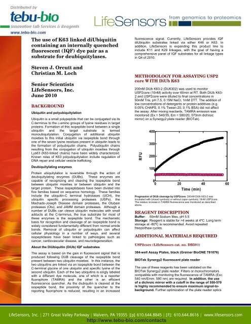
200nM <strong>DiUb</strong> <strong>K63</strong>-2 (DU6302) was used to monitor<br />
USP2core (10nM) activity over 60min at RT. Both <strong>DiUb</strong> <strong>K63</strong>-<br />
2 and USP2core were diluted to final concentrations in<br />
50mM Tris, pH 7.5, 0.15M NaCl, 1mM DTT. The addition of<br />
low concentrations of detergents or protein additives (e.g.<br />
0.05% CHAPS, 0.1% Tween-20, 0.1% BSA) did not affect<br />
the assay. After mixing reactants, TAMRA emission was<br />
monitored (Ex = 540/35, Em = 580/20, 570nm dichroic<br />
mirror) on a Synergy2 plate reader (BIOTek).<br />
RFU<br />
400<br />
300<br />
200<br />
100<br />
0<br />
0 20 40 60<br />
Time (min)<br />
Progression of <strong>DiUb</strong> cleavage by USP2core: 200nM <strong>DiUb</strong> <strong>K63</strong>-2 was<br />
incubated with (closed symbols) or without (open symbols) 10nM USP2core.<br />
The relative increase in TAMRA fluorescence was monitored as described<br />
above.<br />
REAGENT DESCRIPTION<br />
Buffer: 50mM Sodium Mes, pH 5.5<br />
Storage: Reagent is stable for >4 weeks at 4ºC. Long term<br />
storage at -80ºC is recommended. Avoid repeated<br />
freeze/thaw cycles.<br />
ADDITIONAL MATERIALS REQUIRED<br />
USP2core (LifeSensors cat. no. DB501)<br />
384-well Assay Plates, black (Greiner BioONE 781076)<br />
BIOTek Synergy2 fluorescent plate reader<br />
The use of these reagents has been validated on the<br />
BIOTek Synergy2 plate reader. Filters or monochromators<br />
compatible with monitoring the fluorescence of TAMRA (Exc.<br />
540 nm/Emm. 580 nm) are required. In addition, the use<br />
of a dichroic mirror with a cutoff in the range of 550-570<br />
is highly recommended to ensure maximum signal-tobackground.<br />
Further optimization of the plate reader optics<br />
LifeSensors, Inc. | 271 Great Valley Parkway | Malvern, PA 19355 |[p] 610.644.8845 | [F]: 610.644.8616 | www.lifesensors.com<br />
http://www.<strong>tebu</strong>-<strong>bio</strong>.com/contacts
(e.g. signal gain, plate height reads, etc.) is also<br />
recommended. Any fluorescence or multimode plate reader<br />
capable of the configuration described above should be<br />
suitable for this assay.<br />
SUGGESTED PROTOCOL (96 well plate)<br />
1. Dilute <strong>DiUb</strong> <strong>K63</strong> substrate to 400nM, or 2x the desired<br />
final concentration, in assay buffer of choice (e.g.<br />
50mM Tris, pH 8.0, 0.05% CHAPS, 10mM DTT).<br />
2. Dilute the USP2core to 2X desired final concentration in<br />
buffer of choice. A range of enzyme concentrations,<br />
spanning at least three orders of magnitude, is<br />
recommended.<br />
3. Dispense 50µL of <strong>DiUb</strong> <strong>K63</strong> substrate (or assay buffer<br />
as no enzyme control) into black assay plate wells.<br />
4. Add 50µL of USP2core and read immediately in a preconfigured<br />
fluorescence plate reader (see above) for<br />
30min to 1hour.<br />
Representative Data for <strong>DiUb</strong> panel<br />
Beyond differential linkages (e.g. K48 versus <strong>K63</strong>),<br />
LifeSensors has created subpanels of IQF diubiquitin<br />
substrates within each linkage. Because each DUB is likely<br />
to recognize and cleave substrate with unique steric<br />
considerations, these subpanels vary in location of reporter<br />
fluorophore and quencher. It is recommended that each<br />
DUB be empirically evaluated against the entire panel to<br />
select the fluorophore/quencher combination that provides<br />
the optimal signal:background ratio. Shown below is<br />
representative data from such testing.<br />
Signal:Background<br />
20<br />
15<br />
10<br />
5<br />
0<br />
<strong>DiUb</strong>K48-1<br />
<strong>DiUb</strong>K48-2<br />
<strong>DiUb</strong>K48-3<br />
USP2core<br />
<strong>DiUb</strong><strong>K63</strong>-1<br />
<strong>DiUb</strong><strong>K63</strong>-2<br />
<strong>DiUb</strong><strong>K63</strong>-3<br />
<strong>DiUb</strong><strong>K63</strong>-4<br />
<strong>DiUb</strong>K48-2<br />
AMSHcore<br />
<strong>DiUb</strong>K48-3<br />
<strong>DiUb</strong><strong>K63</strong>-1<br />
<strong>DiUb</strong><strong>K63</strong>-2<br />
<strong>DiUb</strong><strong>K63</strong>-3<br />
<strong>DiUb</strong><strong>K63</strong>-4<br />
LifeSensors, Inc. | 271 Great Valley Parkway | Malvern, PA 19355 |[p] 610.644.8845 | [F]: 610.644.8616 | www.lifesensors.com<br />
http://www.<strong>tebu</strong>-<strong>bio</strong>.com/contacts<br />
Signal:Background<br />
12<br />
10<br />
8<br />
6<br />
4<br />
2<br />
0<br />
100nM enzyme (USP2core and AMSHcore) was incubated for 1 hour with<br />
100nM substrate. Fluorescence was measured and compared with no enzyme<br />
control to determine signal:background. While USP2c exibits less linkage<br />
specificity, it nonetheless prefers certain fluor/quencher location combinations.<br />
AMSHc displays both linkage and location specificity.<br />
REFERENCES<br />
1. Pickart, C.M. and Eddins, M.J. (2004). "Ubiquitin:<br />
structures, functions, mechanisms." Biochim <strong>bio</strong>phys Acta.<br />
1695 (1-3): 55-72.<br />
2. Wilkinson, K. D. (2000). "Ubiquitination and<br />
deubiquitination: targeting of proteins for degradation by the<br />
proteasome." Semin Cell Dev Biol 11(3): 141-8.<br />
3. Ciechanover, A. (2003) "The ubiquitin proteolytic system<br />
and pathogenesis of human diseases: a novel platform for<br />
mechanism-based drug targeting." Biochem Soc Trans<br />
31(2): 474-81.<br />
4. Shahri, R. (2005) “Diverse polyubiquitin interaction<br />
properties of ubiquitin-associated domains.” Nature<br />
Structural & Molecular Biology 12(8): 708-14.<br />
.
LifeSensors, Inc.<br />
LifeSensors is a <strong>bio</strong>technology company located 35 miles<br />
west of Philadelphia, Pennsylvania, USA. Founded in 1996,<br />
LifeSensors has developed a number of innovative protein<br />
expression technologies that enable efficient translation of<br />
the genome into proteome.<br />
LifeSensors is well-known for its innovations in an important<br />
family of proteins consisting of ubiquitin and ubiquitin-like<br />
proteins (UBL) such as SUMO (Small Ubiquitin-like<br />
MOdifier).<br />
LifeSensors has been granted several patents to cover the<br />
use of SUMO and other UBLs as gene fusion tags to<br />
improve the expression and purification of recombinant<br />
proteins. Additional patent applications are in various stages<br />
of review. Currently, LifeSensors is expanding its protein<br />
production capabilities and is developing protein micro array<br />
for drug discovery and diagnostics.<br />
This reagent is intended for research purposes only. For<br />
information on obtaining a license for commercial<br />
purposes, contact: Marc Scholtyssek, Sales Manager,<br />
LifeSensors, Inc., Malvern PA 19355, Phone:<br />
610.644.8845 ext 305.<br />
all products are for research use only<br />
not intended for human or animal diagnostic or therapeutic uses<br />
LifeSensors, Inc., 271 Great Valley Parkway, Malvern PA 19355<br />
(p) 610.644.8845 (f) 610.644.8616<br />
techsupport@lifesensors.com • www.lifesensors.com • sales@lifesensors.com<br />
Copyright © 2010 LifeSensors, Inc. All Rights Reserved<br />
Your one-stop European source<br />
– find all you need and locate<br />
your nearest <strong>tebu</strong>-<strong>bio</strong> office at<br />
www.<strong>tebu</strong>-<strong>bio</strong>.com<br />
LifeSensors, Inc. | 271 Great Valley Parkway | Malvern, PA 19355 |[p] 610.644.8845 | [F]: 610.644.8616 | www.lifesensors.com<br />
http://www.<strong>tebu</strong>-<strong>bio</strong>.com/contacts