Molecular Biology - The Scripps Research Institute
Molecular Biology - The Scripps Research Institute
Molecular Biology - The Scripps Research Institute

Create successful ePaper yourself
Turn your PDF publications into a flip-book with our unique Google optimized e-Paper software.
168 MOLECULAR BIOLOGY 2005<br />
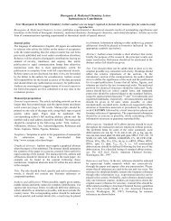
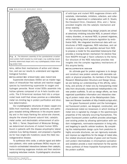
Fig. 2. <strong>The</strong> tetrameric assembly of the cyanobacterial circadian<br />
clock protein KaiB revealed by small-angle x-ray scattering (experimentally<br />
determined shape) and x-ray crystallography (ribbon showing<br />
protein fold).<br />
teins, define their mechanisms of action and interaction,<br />
and use our results to understand and regulate<br />
biological function.<br />
METALLOENZYME STRUCTURE AND FUNCTION<br />
Superoxide dismutases (SODs) act as master regulators<br />
of intracellular free radicals and reactive oxygen<br />
species by transforming superoxide to oxygen and<br />
hydrogen peroxide. Novel nickel SODs assemble into<br />
hollow spheres composed of six 4-helix bundle subunits.<br />
<strong>The</strong> 9 N-terminal residues fold into a unique<br />
nickel hook motif that shows promise as a detectable<br />
metal ion–binding tag in protein purification and structure<br />
determination.<br />
Our crystallographic structures of classic copper-zinc<br />
SODs from mammals, bacterial symbionts, and pathogens<br />
revealed striking differences in the enzyme assembly<br />
and in the loops flanking the active-site channel,<br />
despite the shared β-barrel subunit fold, catalytic<br />
metal center, and electrostatic enhancement of activity.<br />
With J. Tainer, Department of <strong>Molecular</strong> <strong>Biology</strong>,<br />
we determined structures of mutant human SODs<br />
found in patients with the disease amyotrophic lateral<br />
sclerosis (Lou Gehrig disease), and proposed a hypothesis<br />
for how single-site mutations cause this fatal neurodegenerative<br />
disease.<br />
To synthesize nitric oxide, a cellular signal and defensive<br />
cytotoxin, nitric oxide synthases (NOSs) require calmodulin-orchestrated<br />
interactions between their catalytic,<br />
heme-containing oxygenase module and their electronsupplying<br />
reductase module. Crystallographic structures<br />
Published by TSRI Press ®. ©Copyright 2005,<br />
<strong>The</strong> <strong>Scripps</strong> <strong>Research</strong> <strong>Institute</strong>. All rights reserved.<br />
of wild-type and mutant NOS oxygenase dimers with<br />
substrate, intermediate, inhibitors, cofactors, and cofactor<br />
analogs, determined in collaboration with D. Stuehr,<br />
the Cleveland Clinic, Cleveland, Ohio, and J. Tainer,<br />
provided insights into the catalytic mechanism and<br />
dimer stability.<br />
Our structure-based drug design projects are aimed<br />
at selectively inhibiting inducible NOS, to prevent inflammatory<br />
disorders, or neuronal NOS, to prevent migraines,<br />
while maintaining blood pressure regulation by endothelial<br />
NOS. We integrated biochemical data with our<br />
structures of NOS oxygenase, NOS reductase, and calmodulin<br />
in complex with peptides derived from NOS<br />
to propose a model for the assembled holoenzyme that<br />
provides a moving-domain mechanism for electron flow<br />
from NADPH through 2 flavin cofactors to the heme.<br />
Our structure of the NOS reductase provides new<br />
insights into the complex regulatory mechanisms of<br />
this enzyme family.<br />
METALLOPROTEIN DESIGN<br />
An ultimate goal for protein engineers is to design<br />
and construct new protein variants with desirable catalytic<br />
or physical properties. As members of the <strong>Scripps</strong><br />
<strong>Research</strong> Metalloprotein Structure and Design Group,<br />
we are testing our understanding of the affinity, selectivity,<br />
and activity of metal ions by transplanting metal<br />
sites from structurally characterized metalloproteins into<br />
new protein scaffolds. To aid our design efforts, we have<br />
organized quantitative information and interactive viewing<br />
of protein metal sites at the Metalloprotein Database<br />
and Browser (available at http://metallo.scripps.edu).<br />
For green fluorescent protein and the homologous<br />
red fluorescent protein, we designed, constructed, and<br />
characterized metal-ion biosensors in which binding of<br />
metal ions is signaled by changes in the spectroscopic<br />
properties of the naturally occurring fluorophores. <strong>The</strong><br />
green fluorescent protein scaffold provides advantages<br />
over existing probes by allowing optimization with random<br />
mutagenesis, noninvasive expression in living cells,<br />
and targeting to specific cellular locations. By completing<br />
the metalloprotein design cycle from prediction to<br />
highly accurate structures, we can rigorously evaluate<br />
and improve our algorithms for the design of metal sites.<br />
Our related structural studies of green and red fluorescent<br />
protein intermediates in chromophore cyclization<br />
and oxidation provide a novel mechanism for the spontaneous<br />
synthesis of these tripeptide fluorophores within<br />
the protein scaffold.