Molecular Biology - The Scripps Research Institute
Molecular Biology - The Scripps Research Institute
Molecular Biology - The Scripps Research Institute

You also want an ePaper? Increase the reach of your titles
YUMPU automatically turns print PDFs into web optimized ePapers that Google loves.
172 MOLECULAR BIOLOGY 2005<br />
olution structures of our transporters. Through these<br />
united efforts, we will gain a broader understanding of<br />
the structure and function of drug transporters that<br />
cause MDR in cancer and bacterial infection.<br />
Recently, we determined a new structure of the MDR<br />
ATP-binding cassette transporter homolog MsbA in complex<br />
with magnesium, adenosine diphosphate, inorganic<br />
vanadate, and rough-chemotype lipopolysaccharide.<br />
This structure supports a model involving a rigid-body<br />
torque of the 2 transmembrane domains during ATP<br />
hydrolysis and suggests a mechanism by which the<br />
nucleotide-binding domain communicates with the transmembrane<br />
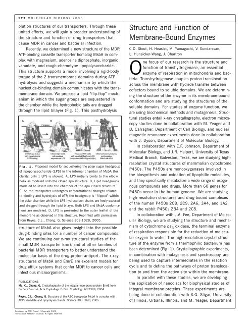
domain. We propose a lipid “flip-flop” mechanism<br />
in which the sugar groups are sequestered in<br />
the chamber while the hydrophobic tails are dragged<br />
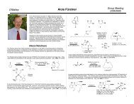
through the lipid bilayer (Fig. 1). This posthydrolysis<br />
Fig. 1. Proposed model for sequestering the polar sugar headgroup<br />
of lipopolysaccharide (LPS) in the internal chamber of MsbA (for<br />
clarity, only 1 LPS is shown). A, LPS initially binds to the elbow<br />
helix as modeled onto the closed apo structure. B, Lipid headgroups<br />
modeled to insert into the chamber of the apo closed structure.<br />
C, As the transporter undergoes conformational changes related<br />
to binding and hydrolysis of ATP, the headgroup is “flipped” within<br />
the polar chamber while the LPS hydrocarbon chains are freely exposed<br />
and dragged through the lipid bilayer. Both LPS and MsbA conformations<br />
are modeled. D, LPS is presented to the outer leaflet of the<br />
membrane as observed in this structure. Reprinted with permission<br />
from Reyes, C.L., Chang, G. Science 308:1028, 2005.<br />
structure of MsbA also gives insight into the possible<br />
drug-binding sites for a number of cancer compounds.<br />
We are continuing our x-ray structural studies of the<br />
small MDR transporter EmrE and of other families of<br />
bacterial MDR transporters to better understand the<br />
molecular basis of the drug-proton antiport. <strong>The</strong> x-ray<br />
structures of MsbA and EmrE are excellent models for<br />
drug efflux systems that confer MDR to cancer cells and<br />
infectious microorganisms.<br />
PUBLICATIONS<br />
Ma, C., Chang, G. Crystallography of the integral membrane protein EmrE from<br />
Escherichia coli. Acta Crystallogr. D Biol. Crystallogr. 60:2399, 2004.<br />
Reyes, C.L., Chang, G. Structure of the ABC transporter MsbA in complex with<br />
ADP•vanadate and lipopolysaccharide. Science 308:1028, 2005.<br />
Published by TSRI Press ®. ©Copyright 2005,<br />
<strong>The</strong> <strong>Scripps</strong> <strong>Research</strong> <strong>Institute</strong>. All rights reserved.<br />
Structure and Function of<br />
Membrane-Bound Enzymes<br />
C.D. Stout, H. Heaslet, M. Yamaguchi, V. Sundaresan,<br />
L. Hunsicker-Wang, J. Chartron<br />
One focus of our research is the structure and<br />
function of transhydrogenase, an essential<br />
enzyme of respiration in mitochondria and bacteria.<br />
Transhydrogenase couples proton translocation<br />
across the membrane with hydride transfer between<br />
cofactors bound to soluble domains. We are determining<br />
the structure of the enzyme in its membrane-bound<br />
conformation and are studying the structures of the<br />
soluble domains. For studies of enzyme function, we<br />
are using biochemical methods and mutagenesis. Structural<br />
studies entail x-ray crystallography, electron microscopy<br />
studies done in collaboration with M. Yeager and<br />
B. Carragher, Department of Cell <strong>Biology</strong>, and nuclear<br />
magnetic resonance experiments done in collaboration<br />
with J. Dyson, Department of <strong>Molecular</strong> <strong>Biology</strong>.<br />
In collaboration with E.F. Johnson, Department of<br />
<strong>Molecular</strong> <strong>Biology</strong>, and J.R. Halpert, University of Texas<br />
Medical Branch, Galveston, Texas, we are studying highresolution<br />
crystal structures of mammalian cytochrome<br />
P450s. <strong>The</strong> P450s are monooxygenases involved in<br />
the biosynthesis and oxidation of lipophilic molecules,<br />
and they specifically metabolize a wide range of exogenous<br />
compounds and drugs. More than 60 genes for<br />
P450s occur in the human genome. We are studying<br />
high-resolution structures and drug-bound complexes<br />
of the human P450s 2C8, 2C9, 2A6, 3A4, and 1A2<br />
and the rabbit P450s 2B4 and 2C5.<br />
In collaboration with J.A. Fee, Department of <strong>Molecular</strong><br />
<strong>Biology</strong>, we are studying the structure and mechanism<br />
of cytochrome ba 3 oxidase, the terminal enzyme<br />
of respiration responsible for the reduction of molecular<br />
oxygen to water. <strong>The</strong> high-resolution crystal structure<br />
of the enzyme from a thermophilic bacterium has<br />
been determined (Fig. 1). Crystallographic experiments,<br />
in combination with mutagenesis and spectroscopy, are<br />
being used to capture intermediates in the reaction<br />
cycle and to define the pathways of proton translocation<br />
to and from the active site within the membrane.<br />
In parallel with these studies, we are developing<br />
the application of nanodiscs for biophysical studies of<br />
integral membrane proteins. <strong>The</strong>se experiments are<br />
being done in collaboration with S.G. Sligar, University<br />
of Illinois, Urbana, Illinois, and M. Yeager, Department