Histology Drawings - School of Medical Sciences
Histology Drawings - School of Medical Sciences
Histology Drawings - School of Medical Sciences

You also want an ePaper? Increase the reach of your titles
YUMPU automatically turns print PDFs into web optimized ePapers that Google loves.
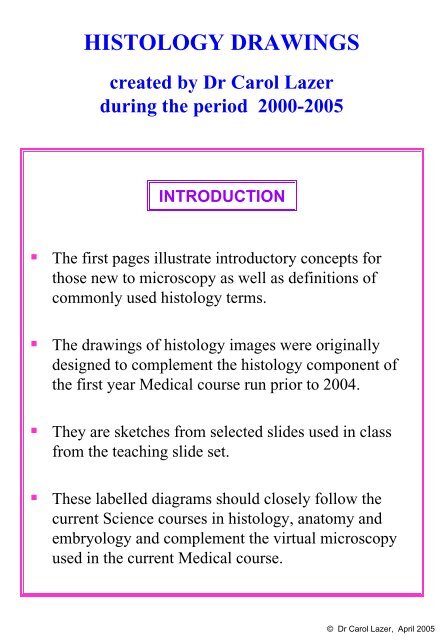
HISTOLOGY DRAWINGScreated by Dr Carol Lazerduring the period 2000-2005INTRODUCTION• The first pages illustrate introductory concepts forthose new to microscopy as well as definitions <strong>of</strong>commonly used histology terms.• The drawings <strong>of</strong> histology images were originallydesigned to complement the histology component <strong>of</strong>the first year <strong>Medical</strong> course run prior to 2004.• They are sketches from selected slides used in classfrom the teaching slide set.• These labelled diagrams should closely follow thecurrent Science courses in histology, anatomy andembryology and complement the virtual microscopyused in the current <strong>Medical</strong> course.© Dr Carol Lazer, April 2005
STEREOLOGY: SLICING A 3-D OBJECTSIMPLE TUBECROSS SECTION = TRANSVERSE SECTION(XS)(TS)OBLIQUE SECTION3-DLONGITUDINAL SECTION(LS)2-DBENDING AND BRANCHING TUBE3-D2-Dbranch <strong>of</strong>f a tube2 sections from 2 tubescut at different anglessection at the beginning<strong>of</strong> a branch3 sections from one tubeen face view = asseen from above1 section and thegrazed wall <strong>of</strong> a tubeCOMPLEX STRUCTURE (gland)COMPOUND ( = branched ducts)ACINAR ( = bunches <strong>of</strong> secretory cells)GLANDduct(XS =TS)acinus (cluster <strong>of</strong> cells) (TS)3-Dduct and acinus2-D(LS)Do microscope images <strong>of</strong> 2-D slices represent asingle plane <strong>of</strong> section <strong>of</strong> a 3-D structure?No, 2-D slices have a thickness which can varyfrom a sliver <strong>of</strong> one cell to several cells deep.With the limited depth <strong>of</strong> field <strong>of</strong> high power lensesit is possible to focus through the various levelswithin a slice.Do all microscope slides show 2-D slices <strong>of</strong> 3-Dstructures?No, slides can also be smears, where entire cellslie on the surface <strong>of</strong> the slide, or whole tissuemounts <strong>of</strong> very thin structures, such as mesentery.© Dr Carol Lazer, April 2005
LININGS, COVERINGS & TERMINOLOGYKEYlamina proprialumenepitheliumconnective tissuebeneath epitheliumconnective tissue,muscle, glands, etcmesothelium(simple squamous epithelium)ORIGIN: mesodermlining epitheliumORIGIN: endodermor mesodermMUCOSA orMUCOUS MEMBRANEhas a wet surface (mucus).Lines organs that open tothe outside.SEROSA orSEROUS MEMBRANEhas a wet surface (watery).Lines body cavities.Covers organs in body cavities.GENERALISED SECTIONOF THE BODYepidermis(keratinised stratifiedsquamous epithelium)ORIGIN: ectodermdermismesenterySKINCovers theexternal surface.ADVENTITIAConnective tissue <strong>of</strong>one structure meetsconnective tissue <strong>of</strong>another structure.blood vessel lined by endothelium(simple squamous epithelium)ORIGIN: mesoderm© Dr Carol Lazer, September 2000 – April 2005
EPITHELIUM:non-keratinisedstratified squamousTISSUE / ORGAN:oesophagusCOVERING AND LINING EPITHELIASTRATIFIED EPITHELIASTRATA:squamousspinosumgerminativummitotic figurelamina propriaEPITHELIUM:keratinisedstratified squamousTISSUE / ORGAN:skin (epidermis)squamesSTRATA:corneumgranulosumspinosumgerminativummitotic figuredermisvacuoles (folded plasma membrane)binucleate celladluminal or facet cellEPITHELIUM:transitionalTISSUE / ORGAN:bladder (relaxed)TISSUE / ORGAN:bladder (stretched)© Dr Carol Lazer, August 2000 – April 2005
SIMPLE EPITHELIAEPITHELIUM:simple cuboidalcolloidTISSUE / ORGAN:thyroidlamina propriaEPITHELIUM:simple columnarTISSUE / ORGAN:gall bladderlamina propriablood vessel2EPITHELIUM:simple squamousTISSUES / ORGANS:11 endothelium lining bloodvessel2 mesothelium <strong>of</strong> serosacovering lungedge onterminal baraboveciliated columnar cellciliacolumnar goblet cellEPITHELIUM:simple columnar orpseudostratified columnarTISSUE / ORGAN:seminal vesicle (ox)EPITHELIUM:basal cellpseudostratified columnar(respiratory epithelium)basement membraneMORE FULLY: pseudostratified ciliatedcolumnar epithelium with goblet cellsTISSUE / ORGAN: trachea© Dr Carol Lazer, August 2000 – April 2005
EXOCRINE GLANDS & DUCTSSIMPLE GLANDShairepidermisdying innerlarge cellsouter smallbasal cells(dividing)FEATURE: sebaceous glandTISSUE / ORGAN: around hair folliclein dermis <strong>of</strong> skincoiled ductportionhair folliclebranched alveolar (acinar) glandsebaceous gland opening into hairfolliclecoiled tubesecretory portion(TS)FEATURE: eccrine sweat glandTISSUE / ORGAN: dermis <strong>of</strong> skin(also in hypodermis)arrectorpilimuscleductsecretoryportionduct portion with darkstaining stratified cuboidalepithelium (bi-layer)secretory portion withthicker, pale stainingbi-layer <strong>of</strong> cellsbasement membranedermiscoiled tubular eccrinesweat gland with ducttwisting to open onthe skin surfacemyoepithelial cells (TS)within basement membranemyoepithelial cellswrapped in a spiralaround the coiled tubeFEATURE: duct and secretory portions,myoepithelial cellsTISSUE / ORGAN: eccrine sweat glandpalisade <strong>of</strong>myoepithelial cells (LS)Are myoepithelial cells only present around sweatglands and what is their function?They are found around the secretory acini and someducts <strong>of</strong> many glands. They contract under autonomicnervous control to expel the glandular secretions.© Dr Carol Lazer, November 2001 – April 2005
GLAND STRUCTURElobelobulelayers <strong>of</strong>connective tissueconnectivetissue septum(between lobules)secretory tubeand acinusCOMPOUND GLANDDUCT ORGANISATIONinterlobar duct = excretory duct[drains the lobes <strong>of</strong> a gland]interlobular duct = intralobar duct[drains many lobules in a lobe]intralobular duct(e.g. striated duct)[drains many acini in a lobule]acinus = alveolus = adenomereFEATURE: lobe, lobules and ductsTISSUE / ORGAN: compound tubulo-alveolar (tubulo-acinar) glandintercalated duct[drains each acinus]part <strong>of</strong> lobelobuleconnectivetissue septumFEATURE: lobe, lobules and ducts (TS)TISSUE / ORGAN: part <strong>of</strong> a lobe <strong>of</strong> salivary glandmucous secretory tubeintercalated ductssmall lumeninterlobular duct= intralobar ductintralobular duct(striated duct insalivary gland)blood vesselsartery & veininterlobar ductstriated ductlarge lumenand simplecolumnarepithelium withbasal striationsserousdemilunemucous acinuswedge-shaped cells filledwith pale staining remnant<strong>of</strong> mucous secretion andflattened basal nucleiserous acinuswedge-shapedcells packed withzymogen granulesand round nucleusFEATURE: serous and mucous acini and ductsTISSUE / ORGAN: submandibular salivary gland(LS)(TS)intercalatedductsDo all compound glands have striated ducts?No, they all have intralobular ducts but thesehave a characteristic appearance in the salivarygland and so have a different name.© Dr Carol Lazer, November 2001 – April 2005
DENSE CONNECTIVE TISSUECONNECTIVE TISSUEcollagenfibre (LS)fibrocytenucleus (LS)CLASSIFICATION: dense regular connective tissue (showing crimp pattern)TISSUE / ORGAN: tendon or ligament fascicleground substancecollagen fibre in theplane <strong>of</strong> section (LS)collagen fibre perpendicularto the plane <strong>of</strong> section (TS)fibroblast nucleus (TS)fibroblast nucleus (LS)CLASSIFICATION: dense irregular connective tissueTISSUE / ORGAN: dermis <strong>of</strong> skinground substancecollagen fibre in theplane <strong>of</strong> section (LS)collagen fibre perpendicularto the plane <strong>of</strong> section (TS)elastic fibre (LS)elastic fibre (TS)fibroblast nucleus (TS)fibroblast nucleus (LS)CLASSIFICATION: dense irregular connective tissueTISSUE / ORGAN: dermis <strong>of</strong> skinWhat is the difference between this specimenand the one before?It is stained to show elastic fibres. Thesewere always present but not seen without thespecial stain.© Dr Carol Lazer, August 2000 – April 2005
SPECIFIC TISSUES & FIBRESbrown fat cell (brown adipocyte)centrally located round nucleuslipid droplet (one <strong>of</strong> many -- multilocular)collagenous septum separating fat lobulesfibroblast nucleuswhite fat cell (white adipocyte)flattened nucleussingle large lipid droplet (unilocular)blood vesselCLASSIFICATION: brown and white adipose tissueTISSUE / ORGAN: fat stored in bodycollagen fibreanastomosing elastic fibremacrophagemast cellfibroblast nucleusCLASSIFICATION: loose connective tissueTISSUE / ORGAN: mesenteryendotheliummy<strong>of</strong>ibroblastcollagen fibreelastic fibreslymphocytecapsule with collagen andreticular fibresreticular fibretrabeculaCLASSIFICATION: reticular fibresTISSUE / ORGAN: lymph nodeCLASSIFICATION: elastic fibresTISSUE / ORGAN: elastic artery (brachial artery)fenestrationelastic fibre (TS)crenated elastic fibre (LS)© Dr Carol Lazer, August 2000 – April 2005
BLOOD CELLS (SMEAR)FEATURE: blood cell typesTISSUE / ORGAN: peripheral bloodMONONUCLEARLEUKOCYTES orAGRANULOCYTESPOLYMORPHONUCLEARLEUKOCYTES orGRANULOCYTESerythrocytebiconcave discwith pale centresmall lymphocytewith a large, roundnucleus and bluecytoplasmic rimneutrophilwith multilobednucleusand many smallgranulesrouleaux formationstack <strong>of</strong> erythrocytesplateletscytoplasmicfragments with adark chromomereand pale hyalomeremonocytewith a single nucleus that maybe irregular or bean shapedand blue cytoplasm which mayhave tiny granules or vacuolesIndentation <strong>of</strong> the nucleus(monocyte or lymphocyte)is caused by proximity tothe Golgi apparatus.eosinophilwith bi-lobednucleus andmany largered granules<strong>of</strong> similar sizebasophilwith manyblue granules<strong>of</strong> differentsize thatobscure thenucleusBLOOD-RELATED CELLS (SECTION)numerous redgranules incytoplasmoverlappinglobes <strong>of</strong> nucleusFEATURE: tissue eosinophil (and plasma cells)TISSUE / ORGAN: lamina propria <strong>of</strong> glands <strong>of</strong>stomach mucosaadipocytemarrowcellssingle, large,lobulatednucleuseosinophiliccytoplasmFEATURE: megakaryocyteTISSUE / ORGAN: bone marrownot to beconfused withbasophiliccytoplasm (rER)eosinophilic (or pale)perinuclear region(Golgi apparatus)eccentric nucleus(clock-faced)FEATURE: plasma cellTISSUE / ORGAN: lamina propria <strong>of</strong> salivarygland secretory acinibonetrabeculaosteocytemultiplenuclei inosteoclasteosinophiliccytoplasmshrinkageSchwann cell nucleusartefactFEATURE: osteoclastTISSUE / ORGAN: bone trabecula© Dr Carol Lazer, November 2001 – April 2005
CARTILAGECARTILAGE AND BONEthick perichondriumadventitia withadipose tissueblood vesselsperipheralnervefoldartefactfibrous layer (with fibroblasts)cellular layer (chondroblasts)territorial matrix(dark staining / recent)interterritorial matrix(pale staining / older)interstitial growth(from the middle)appositional growth(from the edge)thin perichondriumlamina propria with glandscell nests (recentlydivided chondrocytes)lacuna (space in the matrixonce occupied by a cell)chondrocytechondroblastadventitia = loose connective tissuefibroblastchondroblastchondrocytelacunathickperichondriumepithelium -- respiratory(pseudostratified ciliated columnar)FEATURE: hyaline cartilageTISSUE / ORGAN: tracheaattachment sidecell nests (recentlydivided chondrocytes)territorial matrix(dark staining / recent)interterritorial matrix(pale staining / older)interstitial growthfibrouslayercellularlayerelastic fibresappositional growththinperichondriumFEATURE: elastic cartilageTISSUE / ORGAN: pinna <strong>of</strong> earblood vesselsperipheral nervedermis and hypodermis <strong>of</strong> skinepithelium = epidermis <strong>of</strong> skin(keratinised stratified squamous)external ear side© Dr Carol Lazer, July 2002 – April 2005
COMPACT BONEskeletal musclefibrous periosteum (with fibroblasts)tendon orligament(TS)(LS)cellular periosteum (osteoblasts)outer circumferential lamellaeinterstitial lamellaeVolkmann’s canalconcentric lamellae <strong>of</strong> osteoncement line(outermost layer <strong>of</strong> osteon)Haversian canal(centre <strong>of</strong> osteon)osteocyte in lacuna(or empty lacuna)inner circumferential lamellae(absent in young bone)endosteum (osteoblasts)shrinkageartefactblood vessels inHaversian canalbone marrow cellsadipocyteFEATURE: Haversian systems ( = osteons) with lamellae (TS)TISSUE / ORGAN: diaphysis (shaft) <strong>of</strong> femurcanaliculus(channel in matrix)osteocyte in lacuna(or empty lacuna)concentric lamellaecement lineVolkmann’s canals (link Haversian canals)Haversian canal (centre <strong>of</strong> osteon)FEATURE: Haversian systems ( = osteons)with lamellae (LS)TISSUE / ORGAN: diaphysis <strong>of</strong> femur© Dr Carol Lazer, August 2002 – April 2005
BONE / MUSCLE FORMATION & JOINTS}}}}mesenchymeduraarachnoidpiabrainosteoblastHowship’s lacunaMeckel’s cartilageperichondrium1aFEATURE:intramembranous ossification examplesTISSUE / ORGAN: 1 dura mater and calvaria2 Meckel’s cartilage and mandibleYOUNG FETAL RAT HEAD(sagittal section)1a2duramarrow precursorosteoclast1b bone matrix in trabeculadurabraineyecartilageregion <strong>of</strong>bone growthteethtongueosteocyte osteoblastsosteoprogenitor cellINTRAMEMBRANOUS OSSIFICATION1bOLDER FETAL RAT HEAD(coronal section)2MYOBLASTS MYOTUBES MYOFIBRES(LS)(TS)(LS)(TS)(LS)elongated cells(central nuclei)aligned in onedirectionmesenchyme(undifferentiated mesoderm)multiplecentralnucleiperinuclearcytoplasmperipheralmy<strong>of</strong>ibrilsperipheralcytoplasmstellate-shaped cellscentral my<strong>of</strong>ibrilsspindle-shaped cellsyncytium <strong>of</strong> cells(fused multiple cells)STRIATED MUSCLE DEVELOPMENTFEATURE: spindle- and stellate-shaped cellsFEATURE: myoblasts and myotubesTISSUE / ORGAN:(developing striated muscle cells)TISSUE / ORGAN: fetal tonguemultipleperipheralnuclei© Dr Carol Lazer, September 2002 – April 2005
OSSIFICATION & JOINTSDISTALhyaline cartilage“model” <strong>of</strong> boneintervertebraldiscFEATURE: stages in long bone developmentTISSUE / ORGAN: fetal rat tailendochondralossificationmarrow cavityPROXIMALcollar <strong>of</strong> intramembranousbone in perichondriumnucleuspulposusJOINTSannulusfibrosusswelling, dyingchondrocytescollagen fibres and matrixchondrocytes and cartilage matrixingrowth <strong>of</strong> blood vessels,marrow cells and periostealosteoprogenitor cellsfibrous periosteumcellular periosteum withosteoprogenitor cellstrabeculamarrow cellformingHaversiancanalmarrowosteoblastosteocyteHowship’slacunaosteoclastligamentsynovialmembraneand villi1 - epiphysis2 - metaphysis3 - diaphysisperichondriumperiosteumFEATURE: developing symphysis jointTISSUE / ORGAN: intervertebral discperichondriallinedperforatingchannels withblood vessels(cartilage canals)pericondriumperiosteumaponeurosistendon orstriatedmusclelamina propria(loose c.t. oradipose tissue)312joint capsule(ligament )secondaryossificationcancellous(spongy) bonecompact boneFEATURE: generalised synovial jointTISSUE / ORGAN: knee (or elbow)FEATURE: bone growth and remodellingTISSUE / ORGAN: periosteal collar <strong>of</strong> vertebraENDOCHONDRAL OSSIFICATIONjoint internalligamentfibrocartilagemeniscussynovial cavityarticularcartilage1marrow2ZONES3fornixresting chondrocytesadvancingmarrow cellproliferating chondrocytescalcifiedcartilage matrixchondrocyte hypertrophycalcifying matrixresorptionbone trabeculaebonegrowthbone matrixFEATURE: epiphyseal disc (growth plate)TISSUE / ORGAN: growth zone <strong>of</strong> long bone© Dr Carol Lazer, October 2002 – April 2005
MUSCLECLASSIFICATION: fascicle <strong>of</strong> skeletal orstriated muscle (LS)fibroblast nucleusmuscle fibre(my<strong>of</strong>ibre)A bandI bandH bandZ lineendomysiumperimysiumCLASSIFICATION: fascicle<strong>of</strong> skeletal or striated muscle (TS)epimysiumperimysiumendomysiumblood vesselendotheliummuscle fibrecytoplasmmuscle fibre nucleiperimysiumendomysiumblood vesselendotheliummuscle fibre (TS)my<strong>of</strong>ibrils (TS)muscle fibre nucleifibroblast nucleussmooth muscle fascicle (LS)corkscrew nucleussmooth muscle cells (LS)blood vesselendotheliumfibroblast nucleussmooth muscle fascicle (TS)smooth muscle cell nucleiCLASSIFICATION: smooth muscle in fascicles(LS & TS) and arteriole wallWhy is there a large variation in smooth musclenuclei (TS) from absent to small to large?The cells are so long that sections cut the ovoidnucleus from centre to edge or even miss it.© Dr Carol Lazer, August 2000 – April 2005
CLASSIFICATION: single smoothmuscle cellsTISSUE / ORGAN:intestinal villussimple columnarepitheliumcolumnar cellgoblet cellsmooth muscle cell(my<strong>of</strong>ibroblast)blood vessel endotheliumlamina propriacardiac musclefascicle (TS)cardiac musclefascicle (LS)cardiac musclecell (LS)intercalated discA and I bandsmy<strong>of</strong>ibrilclearperinuclearregionnucleus(TS)(LS)Purkinje fibresconnectivetissue septa(perimysium)fibroblastnucleusendotheliumnucleuscardiac musclecell (TS)nucleusmy<strong>of</strong>ibrilsclear perinuclear regionCLASSIFICATION: cardiac muscle fasciclesTISSUE / ORGAN: heart (interventricular septum)interwovenmuscle fibresendocardium(endothelium +lamina propria)© Dr Carol Lazer, August 2000 – April 2005
CENTRAL NERVOUS SYSTEM(CNS)dura materarachnoidpia materseptum <strong>of</strong> pia matergrey matterNERVOUS TISSUEdorsal nerve rootletsmall neuron<strong>of</strong> dorsal hornependymal cellsaround central canalmotor neuron<strong>of</strong> ventral hornwhite mattermyelinated axons (TS)myelinated axons (LS)blood vesselsventral nerve rootletFEATURE: white matter and cells <strong>of</strong> grey matterTISSUE / ORGAN: spinal cord and meningesPERIPHERAL NERVOUS SYSTEM (PNS)neuropiloligodendrocytenucleusastrocyte nucleusaxonaxon hillocknucleusNissl granulesdendriteFEATURE: myelin sheathTISSUE / ORGAN: peripheral nerve (TS)blood vessel (vasa nervorum)myelinated nerve fibreendoneuriumperineuriumepineuriumepineuriumperineuriumendoneuriumparanodal swellingnode <strong>of</strong> RanvierFEATURE: Nodes <strong>of</strong> RanvierTISSUE / ORGAN: peripheral nerve (LS)Schmidt-Lanterman cleftmyelin sheathaxoplasmwhite adipocyteinternode© Dr Carol Lazer, August 2000 – April 2005
epineuriumperineurium around nerve fascicle (LS)perineurium around nerve fascicle (TS)FEATURE: Schwann cellsTISSUE / ORGAN: sciatic nerveblood vessel (vasa nervorum)white fat cell (white adipocyte)septum <strong>of</strong> perineuriumendoneuriumaxoplasmneurokeratin remnant <strong>of</strong> myelin sheathSchwann cell nucleusfibroblast nucleusaxonRanvier’s crossSchmidt-Lanterman cleftneurokeratin <strong>of</strong>myelin sheathNissl granulesaxon hillockganglion cellganglion cellnucleussatellite cell nucleusFEATURE:pseudounipolar neurons and myelinTISSUE / ORGAN: trigeminal sensory ganglionperineurium around nerve fascicle (LS)endoneuriummyelinated axonSchwann cell nucleusblood vessel endotheliumsmooth muscle (TS)FEATURE: peripheral nerve fasciclesTISSUE / ORGAN: connective tissue (tongue)perineurium around nerve fascicle (TS)endoneuriummyelinated axonSchwann cell nucleusfibroblast nucleus© Dr Carol Lazer, August 2000 – April 2005
FETAL MEMBRANESPLACENTAFEATURE: chorionic plate, villi, uterine wallTISSUE / ORGAN: placenta (3 months)branch <strong>of</strong> umbilicalblood vesselchorionic platechorionic villiwith trophoblastcovering and amesoblast(extraembryonicmesoderm) corematernal bloodspaces (lacunae)syncytiotrophoblastsprout (primary villus)FETAL SIDEMATERNAL SIDEstratum basale <strong>of</strong>endometrium withdarker peripheralregion, glands andsmall vesselscytotrophoblasticanchoring columnsmooth muscle<strong>of</strong> myometriumlarge vessels inthe myometriumSECONDARY VILLUStrophoblast + mesoblastPRIMARY VILLUStrophoblast onlysyncytiotrophoblastcytotrophoblastmesoblastrami <strong>of</strong>umbilicalvesselsTERTIARY VILLUStrophoblast + mesoblast + blood vesselscolumnar epithelium<strong>of</strong> tubular uterine(endometrial) glandbloodvesselUMBILICAL CORDFEATURE: vessels and Wharton’s jellyTISSUE / ORGAN: umbilical cord (TS)mucoid connectivetissue (Wharton’s jelly)umbilical veinWarton’s jelly is an artefact <strong>of</strong> “dead”umbilical cord. What should “living”cord look like and why is this different?Before birth the cord has three largevessels and very little connective tissue.After birth the blood flow stops andwithout blood pressure the vesselscollapse. The connective tissue swellswith fluid that leaks from the vessels.collapsedlumenumbilical arteries© Dr Carol Lazer, November 2001 – April 2005