lecture11 outline (print no images) - UNSW Cell Biology - The ...
lecture11 outline (print no images) - UNSW Cell Biology - The ...
lecture11 outline (print no images) - UNSW Cell Biology - The ...

Create successful ePaper yourself
Turn your PDF publications into a flip-book with our unique Google optimized e-Paper software.
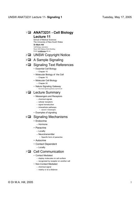
<strong>UNSW</strong> ANAT3231 Lecture 11- Signaling 1 Tuesday, May 17, 20051 ANAT3231 - <strong>Cell</strong> <strong>Biology</strong>Lecture 11School of Medical Sciences<strong>The</strong> University of New South WalesDr Mark Hill<strong>Cell</strong> <strong>Biology</strong> LaboratoryRoom G20 Wallace Wurth BuildingEmail: m.hill@unsw.edu.au2 <strong>UNSW</strong> Copyright Notice3 A Sample Signaling4 Signaling Text References• Essential <strong>Cell</strong> <strong>Biology</strong>– Chapter 15• Molecular <strong>Biology</strong> of the <strong>Cell</strong>– Chapter 15• Molecular <strong>Cell</strong> <strong>Biology</strong>– Chapter 20• Nature Signaling Gateway– http://www.signaling-gateway.org/molecule/5 Lecture Summary• Messengers and Receptors– chemical signals– cellular receptors– signal transduction– intracellular pathways• second messengers• Examples of signaling6 Signaling Mechanisms• Endocrine– Hormone• Paracrine– Locally– Neurotransmitter• Specific form of paracrine• Autocrine• Contact Dependent– Locally7 <strong>Cell</strong> Communication• Contact Mediated– display molecules on cell surface– recognized by receptor on a<strong>no</strong>ther cell• Non-Contact Mediated– chemical signal– nearby or at a distance© Dr M.A. Hill, 2005 1
<strong>UNSW</strong> ANAT3231 Lecture 11- Signaling 1 Tuesday, May 17, 200520 Second Messengers21 Activation of the GTPase Rac in living motile fibroblast22 Second Messengers• Cyclic nucleotides– cAMP, cGMP• Calcium Ions• Protein Kinase A• PKA, B, C• diacylglycerol (DAG)• modified lipid activates PKC• Kinase cascades• small GTP binding proteins• related to RAS which is G protein family23 Movie: Signaling IP3 / calcium24 2 Signal Transductions25 cAMP and Kinases26 Lipids in <strong>Cell</strong> Signaling• Arachidonic Acid (AA) pathway– generates many of the lipids involved as second messengers in cell signaling pathways27 Lipid Soluble-Steroids28 Steroid Responses29 Intracellular Receptors• Steroid Hormones– thyroxine– vitamin D3– reti<strong>no</strong>ic acid• Nuclear location• Cytosol location– translocates to nucleus on ligand binding• binds ligand and DNA– becomes transcription factor30 Steroid Receptors• steroid binding region– near C-terminus• DNA binding– central region• zinc finger motif• alpha helix and 2 beta sheets held in place by cysteine or histidine residues by a zinc atom• multiple fingers typical• DNA response element– Enhancer31 Steroid Receptor Pathway32 Steroid Receptor Pathway© Dr M.A. Hill, 2005 3
<strong>UNSW</strong> ANAT3231 Lecture 11- Signaling 1 Tuesday, May 17, 200533 Steroid Receptor Pathway34 Steroid Receptor Pathway35 Other Transcription Factors36 Membrane Receptors• embedded in plasma membrane• ligand binding– leads to conformational change in receptor– activation of intracellular pathway• G Protein Linked receptors37 <strong>Cell</strong> Surface Receptor Types38 G Protein Receptors39 G Protein-Coupled Signal Pathways• Transmembrane proteins transduce extracellular signals– common structural motif of 7 membrane spanning regions• Receptor binding promotes interaction– between receptor– G protein on interior surface of membrane40 G Protein-Coupled Signal Pathways• induces an exchange of GDP for GTP on G protein α subunit and dissociation of the α subunit fromthe βγ heterodimer• Depending on isoform, GTP-α subunit complex mediates intracellular signaling either– indirectly by acting on effector molecules• adenylyl cyclase (AC)• phospholipase C (PLC)– directly by regulating ion channel or kinase function41 G Protein Linked42 Receptor associated with Kinase• many growth factors use this pathway– Vascular Endothelial Growth Factor– Epidermal Growth Factor– Nerve Growth Factor– Bone Morphogenic Protein– Transforming Growth Factor-beta• Ligand binding• Receptor association• Phosphorylation• Kinase cascade43 VEGF Receptor and Ligands44 EGF Receptor Transduction Pathway45 Signaling Pathway of TGF-β46 TrkA Receptor© Dr M.A. Hill, 2005 4
<strong>UNSW</strong> ANAT3231 Lecture 11- Signaling 1 Tuesday, May 17, 2005• Trk proto-oncogenes– TrkA, TrkB, TrkC, TrkE• variably expressed in CNS and PNS• TrkA binds to nerve growth factor (NGF) and autophosphorylates– leading to activation of multiple downstream effector proteins47 Proto-oncogenes• proto-oncogenes– Normal cell proteins that have potential to cause uncontrolled growth when mutated• loss of receptor regulation• cells grow out of control• mutation in TK Receptor– receptor always activated• mutation of activating protein– always active• Oncogenes– Ras– mutants detected in 30% cervical cancers48 Movie: Methods Receptor/Ligand49 Movie: Receptor Internalization• HEK-293 cells express GFP tagged Beta-2 adrenergic receptors– treated with <strong>no</strong>radrenaline and imaged by time-lapse confocal microscopy at 5 s intervals over 30 minutes• movement underneath plasma membrane due to <strong>no</strong>radrenaline-evoked internalisation of receptors50 Movie: GLUT4 Dynamics• dynamics of glucose transporter isoform 4 (GLUT4)-containing vesicles in 3T3-L1 adipocytes microinjected with GFP-GLUT4• After 24h adipocytes were serum-starved for 3h prior to imaging• Insulin was added at t=0• cell was imaged at 1 frame/s– Two types of movement GLUT4 vesicles are evident:• rapid vibrational-type displacements• rapid movements over short distances51 Movie:• Expressed transiently in porcine aortic endothelial (PAE) cells• GFP tagged 32 kDa PtIns(3,4,5)P3-binding protein (DAPP1)• translocated from cytosol to plasma membrane in response to platelet-derived growth factor (PDGF)52 Movie: Agonist-induced translocation of EGFP-PHPLC• Agonist-induced translocation of EGFP-PHPLC _ in SH-SY5Y and CHO-lacmGlu1_cells• Single-cell imaging of graded Ins(1,4,5)P3 production following G-protein-coupledreceptoractivation53 Growth Factors54 Online References• ANAT3231 Lectures• http://cellbiology.med.unsw.edu.au/units/science/lectures.htm• Molecular <strong>Biology</strong> of the <strong>Cell</strong> (Ch15)• http://www.ncbi.nlm.nih.gov:80/books/bv.fcgi?call=bv.View..ShowSection&rid=cell.section.3834• Developmental <strong>Biology</strong> (Ch6)• http://www.ncbi.nlm.nih.gov:80/books/bv.fcgi?call=bv.View..ShowSection&rid=.TWQLjiW2xLXyWUOVemh0sDWJf2YbG1QHJC-• Molecular <strong>Cell</strong> <strong>Biology</strong> (Ch20)• http://www.ncbi.nlm.nih.gov:80/books/bv.fcgi?call=bv.View..ShowSection&rid=mcb.chapter.5687© Dr M.A. Hill, 2005 5
<strong>UNSW</strong> ANAT3231 Lecture 11- Signaling 1 Tuesday, May 17, 2005• <strong>The</strong> <strong>Cell</strong>- A molecular approach (Ch13)• http://www.ncbi.nlm.nih.gov:80/books/bv.fcgi?tool=bookshelf&call=bv.View..ShowSection&searchterm=cell&rid=cooper.chapter.2198• Sigma Apoptosis Brochure• http://www.sigmaaldrich.com/Area_of_Interest/Life_Science/<strong>Cell</strong>_Signaling.html55 Signal Transduction Research Labs1• Henry Bourne (Uni of California, San Francisco) MV, Y• Joan Brugge (Harvard Medical School) M, MV• Lewis Cantley (Beth Israel Hospital, Harvard Medical School) M, MV• David Capco (Arizona State Uni) * # M• Gwen V. Childs (Uni of Arkansas for Medical Sciences) * MV• Nam-Hai Chua (Rockefeller Uni) * P• David Clapham (Children's Hospital, Harvard Medical School) * M, MV• Peter Devreotes (Johns Hopkins Uni School of Medicine) * # Di• Catherine Dulac (Harvard Uni) M• Raymond Erikson (Harvard Uni) M, MV• Gerald Fink (MIT) Y• Richard Firtel (Uni of California, San Diego) * Di• John Flanagan (Harvard Medical School) M, MV• Elisabeth Ge<strong>no</strong>t (Uni of Bordeaux, France) MV• François Guesdon (Uni of Sheffield, UK) * H• Alan Hall (Uni College, London, UK) * M, MV• Ira Herskowitz (Uni of California, San Francisco) * # Y• Saul M. Honigberg (Uni of Missouri, Kansas City) * Y2• James Hurley (NIH) * Z• Rolf König (Uni of Texas Medical Branch at Galveston) M, H• Harvey Lodish (Massachusetts Institute of Tech<strong>no</strong>logy) * H, M, MV• Robert Messing (Uni of California, San Francisco) M• Danton H. O'Day (Uni of Toronto, Mississauga) * Di, MV• John H. Richburg (Uni of Texas at Austin) MV• Andrew M. Scharenberg (Uni of Washington) MV• John Scott (Vollum Institute, Oregon Health Sciences Uni) MV• Chris Stubbs (Thomas Jefferson Uni) * MV• David Thomas (National Research Council, Montreal, Québec, Canada) * Y• Jeremy Thorner (Uni of Calif., Berkeley) Y• Peter van Haastert (Uni of Groningen, <strong>The</strong> Netherlands) * Di• Dan Wang (Lineberger Cancer Center, Uni of North Carolina) * MV• Keith Yamamoto (Uni of California, San Francisco) M, MV• Bruce Zetter (Children's Hospital, Harvard Medical School) * # H, M, MV56 Reference: Molecular <strong>Biology</strong> of <strong>Cell</strong>• III. Internal Organization of the <strong>Cell</strong>– 15. <strong>Cell</strong> Signaling• Introduction• General Principles of <strong>Cell</strong> Signaling• Signaling via G-Protein-linked <strong>Cell</strong>-Surface Receptors• Signaling via Enzyme-linked <strong>Cell</strong>-Surface Receptors• Target-<strong>Cell</strong> Adaptation• <strong>The</strong> Logic of Intracellular Signaling: Lessons from Computer-based "Neural Networks"• References57 Reference: Molecular <strong>Cell</strong> <strong>Biology</strong>• 20. <strong>Cell</strong>-to-<strong>Cell</strong> Signaling: Hormones and Receptors– 20.1 Overview of Extracellular Signaling– 20.2 Identification and Purification of <strong>Cell</strong>-Surface Receptors– 20.3 G Protein –Coupled Receptors and <strong>The</strong>ir Effectors– 20.4 Receptor Tyrosine Kinases and Ras– 20.5 MAP Kinase Pathways– 20.6 Second Messengers– 20.7 Interaction and Regulation of Signaling Pathways– 20.8 From Plasma Membrane to Nucleus– PERSPECTIVES• Future• Literature58 Reference: <strong>The</strong> <strong>Cell</strong>• IV. <strong>Cell</strong> Regulation– 13. <strong>Cell</strong> Signaling• Signaling Molecules and <strong>The</strong>ir Receptors• Functions of <strong>Cell</strong> Surface Receptors• Pathways of Intracellular Signal Transduction• Signal Transduction and the Cytoskeleton© Dr M.A. Hill, 2005 6
<strong>UNSW</strong> ANAT3231 Lecture 11- Signaling 1 Tuesday, May 17, 2005• Signaling in Development and Differentiation• Regulation of Programmed <strong>Cell</strong> Death• Summary• Questions• References and Further Reading59 Reference: Developmental <strong>Biology</strong>• Part 1. Principles of development in biology– 6. <strong>Cell</strong>-cell communication in development• Induction and Competence• Paracrine Factors• <strong>Cell</strong> Surface Receptors and <strong>The</strong>ir Signal Transduction Pathways• <strong>The</strong> <strong>Cell</strong> Death Pathways• Juxtacrine Signaling• Cross-Talk between Pathways• Coda• Principles of Development:<strong>Cell</strong>-<strong>Cell</strong> Communication• References© Dr M.A. Hill, 2005 7