A Miyake-Apple View Analysis
A Miyake-Apple View Analysis
A Miyake-Apple View Analysis
Create successful ePaper yourself
Turn your PDF publications into a flip-book with our unique Google optimized e-Paper software.
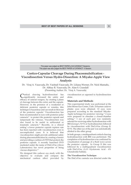
BEST OF BEST PAPERS OF ALL SESSIONS3This paper was judged as BEST PAPER of All CATARACT Sessions.This paper was also judged the BEST PAPER of CATARACT- II Session.Cortico-Capsular Cleavage During Phacoemulsification -Viscodissection Versus Hydro-Dissection: A <strong>Miyake</strong>-<strong>Apple</strong> <strong>View</strong><strong>Analysis</strong>Dr. Viraj A. Vasavada, Dr. Vaishali Vasavada, Dr. Liliana Werner, Dr. Nick Mamalis,Dr. Abhay R. Vasavada, Dr. Alan S. Crandall(Presenting Author: Dr. Viraj A. Vasavada)Cortical cleaving hydrodissection 1 has viscodissection as opposed to hydrodissectionsignificantly increased the safety and alone.efficacy of cataract surgery, by creating a planeMaterials and Methodsof cleavage between the cortex and the capsule.However, in the presence of a weakened or This experimental study was performed at thedeficient posterior capsule or zonules, this John Moran Eye Center, Utah. 14 human cadavertechnique is hazardous and can result in blowout phakic eyes were obtained. 12 eyes wereof the posterior capsule. prepared according to the standard <strong>Miyake</strong>-2 Viscodissection hasbeen recommended in eyes with posterior polar <strong>Apple</strong> technique for posterior viewing. 10 2 eyescataractswere prepared to simulate a closed-chamber2,4 to protect the posterior capsule andtamponade the vitreous. Viscodissection wassetting. 11 1 eye of each pair was randomlyalso found to be useful in subluxated orselected for receiving either hydrodissection withtraumatic cataracts.BSS (Group I, N=7) or hydrodissection followed5,6 Recently, in a clinicalsettingby viscodissection with DisCoVisc (Group II,7 a lower posterior capsule rupture rateN=7). The other eye of the pair was automaticallyhas been reported with viscodissection even inallotted to the other group.uncomplicated cases. It is believed thatviscodissection might provide additional safety In both groups, a multiquadrant cortical-cleavingduring phacoemulsification by creating a barrier hydrodissection with BSS was performed. Theand providing a cushion of OVD that protects the completion of hydrodissection was confirmed byposterior capsule. A recently launched OVD visualization of passage of a fluid wave acrossmarketed under the name of DisCoVisc (Alcon the posterior capsule. In Group II this wasLaboratories) has novel properties of being followed by a multiquadrant viscodissectionviscous-dispersive. with DisCoVisc (1.6% Hyaluronic Acid - 4%8,9Chondroitin Sulfate).We designed this cadaver eye study with theobjective to evaluate and compare the The Viscodissection Techniquemechanical cushion effect provided by The DisCoVisc cannula was directed under the
4 AIOC 2009 PROCEEDINGSanterior capsule. Small amounts were injectedslowly with a side-to-side swiping motion of thecannula around 360 degrees, injecting a total of0.15-0.2 ml. At this point, with the lens in situ, 1eye from each group (<strong>Miyake</strong>-<strong>Apple</strong> preparation)was sectioned for histopathological evaluation.In the remaining 10 eyes prepared according tothe <strong>Miyake</strong>-<strong>Apple</strong> technique, phacoemulsificationwas performed. In the 2 eyesprepared for the closed chamber setting, the eyewas fixed to a training head and phacoemulsificationwas performed.The space created between the capsule and cortexwas evaluated by 2 observers. At each of thestages, i.e. Following Hydrodissection, FollowingViscodissection, Sculpting, Early FragmentRemoval, and Late Fragment Removal, a semiquantitativescale was devised to grade the spacefrom 0 to 3 (0 = no space/ space equal to thatbefore the start of surgery; 3 = maximum spacedetectable). The surgeon was also asked tosubjectively evaluate the space maintained.ResultsInjecting DisCoVisc was found to be easy and itcould be clearly visualized passing beyond theequator, creating a space between capsule andcortex.Some cortico-capsular separation was visible inall eyes when a wave of BSS passed betweencapsule and cortex. With hydrodissection, mostof this fluid egressed out as phacoemulsificationproceeded. In contrast, after DisCoVisc injection,a larger cortico-capsular separation wasuniformly created. This space was maintainedand some cushion effect could be documenteduntil the removal of last fragments.Table 1 shows space created. Significantly greaterspace was created with viscodissection ascompared to hydrodissection. (p=0.05).Significantly greater score was reported in groupII during fragment removal (p=0.04). Surgeon’ssubjective impression confirmed this finding.Histopathological examination of the 2 eyes withthe lens in situ, revealed greater separation of theposterior capsule and cortex followingviscodissection.DiscussionThe technique of viscodissection waspopularized by Krag 13 and Burton, 14 as an adjunctduring ECCE. Viscodissection has been shown tobe invaluable in complicated cases. 2,4 Recentlyreduced PCR rates following viscodissectioneven in a standard cataract scenario have beenreported. 7 The possible mechanisms for thisbeneficial effect include the creation of a physicalbarrier between cortex and posterior capsule,thus providing greater space for in-the-bagmanipulation and preventing movement of thecapsule towards the phaco tip.We found that following viscodissection aTable-1: Objective (Semi-quantitative) Scoring of Capsulo-Cortical SpaceGroup Eye No. Hydro-dissection Viscodissection Sculpting Nuclear Early Frag- Late Frag-Division ment Removal ment RemovalI 1 2 - 2 1.5 1 0.52 1.5 - 1.5 1 0.5 03 2.5 - 2 1 0.5 04 1.5 (in 1 quadrant) - 1 0.5 0.5 05 2 - 1.5 0.5 0.5 06# 2 - - - - -Mean±SD 1.92±0.38 - 1.6±0.42 0.9±0.42 0.6±0.22 0.1±0.22II 1 1.5 2 2 1 1 02 1.5 2.5 2 1.5 1 0.53 2 2.5 2.5 1.5 1 0.54 2 (in 1 quadrant) 3 3 2 1 0.55 1.5 2.5 2 1.5 1 0.56# 2 3 - - - -Mean+SD 1.58±0.38 2.58±0.38 2.3±0.45 1.5±0.35 1.0±0.0 0.4±0.22P value* P = 0.32 P = 0.05 P = 0.05 P = 0.13 P=0.04 P = 0.17
BEST OF BEST PAPERS OF ALL SESSIONS5significant space was created partitioning thecapsule and cortex in all 4 quadrants. A cushioneffect could still be detected even up to the laterstages of fragment removal.In our study we chose DisCoVisc because it hasnovel properties of being a viscous-dispersiveOVD. 15 Hence it affords excellent spacemaintenance, with superior visualization, andcan be removed easily at the end of surgery.The technique of injecting OVD differs from thatof injecting BSS. With hydrodissection, a singlesite forceful injection of BSS produces a fluidwave. On the other hand viscodissection involvesslow, gentle injection of small amounts of OVDaccompanied by a swiping motion of the cannulaaround 360 degrees, re-inserting the cannula indifferent quadrants as and when required. Acomplete wave was not allowed to pass in any of1. Fine IH. Cortical cleaving hydrodissection. JCataract Refract Surg 1992;18:508-12.2. Allen D, Wood C. Minimizing risk to the capsuleduring surgery for posterior polar cataract. JCataract Refract Surg 2002;28:742-4.3. <strong>Apple</strong> DJ, Peng Q, Visessook N. Eradication ofposterior capsule opacification; documentation of amarked decrease in Nd:YAG laser posteriorcapsulotomy rates noted in an analysis of 5416pseudophakic human eyes obtained postmortem.Ophthalmology 2001;108:505-18.4. Fine IH, Packer M, Hoffman RS. Management ofposterior polar cataract. J Cataract Refract Surg 2003;29:16-9.5. Cionni RJ, Osher RH. Management of profoundzonular dialysis or weakness with a newendocapsular ring designed for scleral fixation. JCataract Refract Surg 1998;24:1299-06.6. Cionni RJ, Osher RH, Marques DM, et al. Modifiedcapsular tension ring for patients with congenitalloss of zonular support. J Cataract Refract Surg 2003;29:1668-73.7. Mackool RJ, Nicolich S, Mackool Jr R. Effect ofviscodissection on posterior capsule rupture duringphacoemulsification. J Cataract Refract Surg 2007;33:553.8. Petroll MW, Jafari M, Lane SS, et al. Quantitativeassessment of ophthalmic viscosurgical deviceretention using in vivo confocal microscopy. JCataract Refract Surg 2005;31:236-68.9. Bissen-Miyajima H. In vitro behavior of ophthalmicviscosurgical devices during phacoemulsification. JCataract Refract Surg 2006; 32:1026-31.Referencesthe quadrants. As DisCoVisc disperses moreslowly it leaves time for the capsular bag togradually stretch without leading to a suddenincrease in fluid pressure.The implication of these results is thatviscodissection partitions the posterior capsulefrom the activity inside the capsular bag, thusminimizing the risk of posterior capsule injury.A potential limitation may be in eyes with bulkynuclei, intumescent cataracts as well as shallowanterior chambers, where injection of even smallamounts of OVD may cause rise in theintracapsular pressure.In conclusion, in this cadaver eye study,viscodissection created a greater mechanicalcushion between the lens and the capsular bagwhen compared with hydrodissection alone.10. <strong>Apple</strong> DJ, Lim Es, Morgan RC. Preparation andstudy of human eyes obtained postmortem with the<strong>Miyake</strong> posterior photographic technique.Ophthalmology 1990;97:810-6.11. Auffarth GU, Wesendahl TA, Solomon K. Amodified preparation technique for closed systemocular surgery of human eyes obtainedpostmortem; an improved teaching tool.Ophthalmology 1996;103:977-82.12. Vasavada AR, Goyal D, Shastri L. Corticocapsularadhesions and their effect during cataract surgery. JCataract Surgery 2003;29:309-14.13. Krag S, Thim K, Corydon D. Strength of the lenscapsule during hydroexpression of the nucleus. JCataract Refract Surg 1993;19:205-8.14. Burton RL, Pickering S. Extracapsular cataractsurgery using capsulorhexis with viscoexpressionvia a limbal section. J Cataract Refract Surg 1995;21:297-301.15. Arshinoff SA, Jafari M. New classification ofophthalmic viscosurgical devices-2005. J CataractRefract Surg 2005; 31:2167-71.16. <strong>Miyake</strong> K, <strong>Miyake</strong> C. Intraoperative posteriorchamber lens haptic fixation in the human cadavericeye. Ophthalmic Surg 1985; 16:230-6.17. Auffarth GU, Holzer MP, Visessook N, et al.Removal times for a dispersive and a cohesiveophthalmic viscosurgical device correlated withintraocular lens material. J Cataract Refract Surg2004;30:2410-4.18. Oshika T, Okamoto F, Kaji Y, et al. Retention andremoval of a new viscous dispersive ophthalmicviscosurgical device during cataract surgery inanimal eyes. Br J Ophthalmol 2006;90:485-7.


