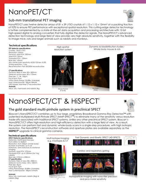
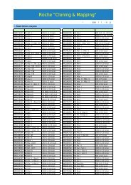
NanoPET/CT ®Sub-mm translational PET imagingNanoPET/CT uses twelve detector arrays of 81 x 39 LYSO crystals of 1.12 x 1.12 x 13mm 3 at a packing fractionof 92% to ensure PET performance with exceptional spatial resolution. This cutting-edge detector technologyis further complemented by a state-of-the-art data acquisition and processing architecture with 12-bithigh-speed digital to analog converters that fully digitize the detector signals. The NanoPET/CT’s advanceddetection technology and large field of view provide very high absolute sensitivity, together with the flexibilityto image mice, rats and larger animals such as rabbits and monkeys.Technical specificationsPET detector specifications:Crystals: 13mm LYSOResolution: 0.8mmDynamic axial FOV: 300mmAxial FOV: 95mmTransaxial FOV: 123mmBore size: 160mmAbs center point sensitivity @250-750 kev: 8.3%Timing resolution: 1.5nsReconstruction: Fast 3DOSEM reconstructionCT specifications:Dynamic field of view: 80 x 300mmDetector active area: 50 x 175mmPixel size: 11, 34, 100µmFocal spot: 9µmX-Ray peak energy: 35-80kv (Variable)Max current/power: 180µA/ 8WattsGPU-based, real-time CT reconstructionAnimals:Mice, rats, marmosets and rabbits 3kgHigh-spatialresolution systemMouse thyroid(F18-NIS)Dynamic & biodistribution studies -Whole body mouse & ratsRat Brain(FDG)NanoSPECT/CT ® & HiSPECT ®The gold standard multi-pinhole system in preclinical SPECTBioscan’s NanoSPECT/CT combines up to four large, proprietary Broadband Gamma Ray Detectors withpatented Multiplexed Multi-Pinhole SPECT (MMP-SPECT) to eliminate many of the sensitivity versus resolutiontrade-offs associated with traditional SPECT systems. Unlike any other preclinical SPECT system, Bioscan’sNanoSPECT/CT offers high-resolution and high-efficiency detection with a large field of view. As a result,the system can perform fast and dynamic whole-body scans in a single-step procedure, with high isotropicresolution. The NanoSPECT’s reconstruction software and aperture plates are available separately as theHiSPECT ® upgrade to clinical gamma cameras.Technical specificationsSPECT Detector specifications:Detectors: Na(Tl)-SpectroscopyFrom 25Kev to 365KeVResolution: from 350µm (whole body)Number of pinholes: up to 64Stationary, semi-stationary & rotationalimagingDynamic Axial FOV: 270mmTransaxial FOV: up to 230mm (for 5 kgrabbits)Bore size: 215mm x 230mmCT specificationDynamic Field of view: 80 x 300mmDetector Active Area: 100 x 100mmPixel Size: 50µmFocal Spot: 9µmX-Ray Peak Energy: 35-80kv (Variable)Max Current/power: 180µA/ 8 WattsMulti-isotope imaginge.g. 4 isotopes & CTFast Dynamic and Kinetic SPECT (4D-SPECT)Cardiac and respiratory gatingAnimals:Mice, rats, marmosets, rabbits (3kg) &rabbits (5kg)5Tc-99m, I-123, In-111and Tl-201, plus X-rayCTNanoparticle imaging with nano-liter precisionand pico-molar sensitivity
MRS 1500 & MRS 3000 Cryogen-free, superconducting MRI at 1.5T & 3.0TOur preclinical MRI systems, the MRS 1500 & MRS 3000, are state-of-the-art, cost-effective translationalMRI instruments with low running costs and no special site requirements. The innovative, cryogen-freesuperconducting magnets virtually have no fringe field, enabling safe use of the system in any facility andby any operator. Designed to complement Bioscan’s other molecular imaging modalities, these MRI systemsdeliver powerful performance and ease-of-use. The systems feature a small footprint and can be placed within2m of another imaging system. MRS instruments are in use worldwide for a wide range of life sciences researchapplications including, functional, molecular, anatomical and multi-modality imaging.Technical specificationsMagnetMagnet type: Cryogen-free, superconductingField strength: 1.5T and 3.0TFOV: 70mm DSVHomogeneity: DSV 30mm +/- 0.10ppmDSV 60mm +/- 1.0ppmClear bore diameter: 160mmExample Pulse SequencesSpin Echo Based SequencesFast SequencesEPI SequencesGradient Echo Based SequencesMagnetic Resonance Angiography2D & 3D Time of Flight sequenceBulk Spectroscopy SequencesIVS SequencesSTEAM spectroscopy sequencePoint Resolved Spectroscopy SequenceFat/water suppression sequenceISIS sequenceAnimals:Mice and RatsMouse Sagittal Image; Spin Echo TE 36ms,Resolution 93 x 93 µ x 1.0mm 1.5T (singleloop surface coil)Mouse Whole Body Image; Spin Echo TR 1200ms,FOV 65mm, THK 1.2mm, 3 averages, Scan Time7m 50s, 130x256, TE = 20 msBioPET/CT-MR Translational multimodality PET/CT/MRI (1.5T/3.0T)Bioscan’s entry into the PET/MR imaging segment combines superior PET performance with translational MRI at1.5T and 3.0T field strengths. Coupling the PET and MRI modalities while retaining superior imaging performanceis a significant technical challenge: As the two modalities are brought into closer proximity, the magnetic fieldinterferes with the PET detectors, creating a trade-off between field strength and PET resolution. To address thechallenge, Bioscan has developed a three-phase approach* to preclinical PET/MRI development, moving fromsequential imaging to integrated inline imaging and finally to simultaneous/dynamic imaging.Phase 1 (available today): Side-By-Side PET/CT and MRISequential imaging with the BioPET/CT positioned next to the MRS 1500 or 3000 system.Sub-mm PET performance, high-resolution, high-contrast MRI at translational fieldstrengths, seamless integration with PET-CT-MRI compatible animal beds andautomated fusion of PET, CT and MRI images with Bioscan’s InVivoScope software. Thisenables both parallel and sequential imaging with the PET, CT and MRI modalities.Phase 2 (Work-In-Progress): Integrated In-line PET/MRIIn-line PET/MRI system similar in configuration to current preclinical PET/CT systems.This phase will combine magnetic field-tolerant PET detection technology with abenchtop cryogen-free superconducting MRI system of minimum 3.0T.Phase 3 (Work-In-Progress): Simultaneous PET/MRINew approach to functional preclinical PET/MRI imaging providing sub-mm PETresolution at fMRI field strengths. Enabled by combining the tDOI technologyfrom Bioscan’s BioPET detectors with Digital Photon Counting (PDPC**) powered PETdetection.* Work supported by IMAPPI (ANR - France)** PDPC= Philips Digital Photon Counting and Digital SiPM Technology (Philips Corporate Technologies, Koninklijke Philips Electronics N.V.)SHAMProtocol Details: 208 g Wistar RatLDA Ligation- chronic infarct SHAM vs. MICardiac Gating: 30 min 18F-FDG uptake25 min acquistion Dose: 1 mCiDigital SiPM: the key todynamic PET/MR imagingMI6