H. G. Brokmeier, B. Weiss, S. B. Yi, W. Ye, K.-D. Liss, T. Lippmann
H. G. Brokmeier, B. Weiss, S. B. Yi, W. Ye, K.-D. Liss, T. Lippmann
H. G. Brokmeier, B. Weiss, S. B. Yi, W. Ye, K.-D. Liss, T. Lippmann

You also want an ePaper? Increase the reach of your titles
YUMPU automatically turns print PDFs into web optimized ePapers that Google loves.
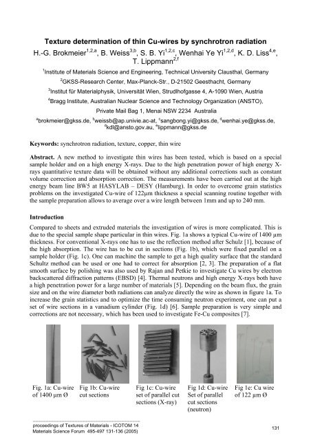
Texture determination of thin Cu-wires by synchrotron radiationH.-G. <strong>Brokmeier</strong> 1,2,a , B. <strong>Weiss</strong> 3,b , S. B. <strong>Yi</strong> 1,2,c , Wenhai <strong>Ye</strong> <strong>Yi</strong> 1,2,d , K. D. <strong>Liss</strong> 4,e ,T. <strong>Lippmann</strong> 2,f1 Institute of Materials Science and Engineering, Technical University Clausthal, Germany2 GKSS-Research Center, Max-Planck-Str., D-21502 Geesthacht, Germany3 Institut für Materialphysik, Universität Wien, Strudlhofgasse 4, A-1090 Wien, Austria4 Bragg Institute, Australian Nuclear Science and Technology Organization (ANSTO),Private Mail Bag 1, Menai NSW 2234 Australiaa brokmeier@gkss.de, b weissb@ap.univie.ac-at, c sangbong.yi@gkss.de, d wenhai.ye@gkss.de,d kdl@ansto.gov.au, e lippmann@gkss.deKeywords: synchrotron radiation, texture, copper, thin wireAbstract. A new method to investigate thin wires has been tested, which is based on a specialsample holder and on a high energy X-rays. Due to the high penetration power of high energy X-rays quantitative texture data will be obtained without any additional corrections such as constantvolume correction and absorption correction. The measurements have been carried out at the highenergy beam line BW5 at HASYLAB – DESY (Hamburg). In order to overcome grain statisticsproblems on the investigated Cu-wire of 122µm thickness a special scanning routine together withthe sample preparation allows to average over a wire length between 1mm and up to 240 mm.IntroductionCompared to sheets and extruded materials the investigation of wires is more complicated. This isdue to the special sample shape particular in thin wires. Fig. 1a shows a typical Cu-wire of 1400 µmthickness. For conventional X-rays one has to use the reflection method after Schulz [1], because ofthe high absorption. The wire has to be cut in sections (Fig. 1b), which were fixed parallel on asample holder (Fig. 1c). One can machine the sample to get a high quality surface that the standardSchultz method can be used or one had to correct for absorption [2, 3]. The preparation of a flatsmooth surface by polishing was also used by Rajan and Petkie to investigate Cu wires by electronbackscattered diffraction patterns (EBSD) [4]. Thermal neutrons and high energy X-rays both havea high penetration power for a large number of materials [5]. Depending on the beam flux, the grainsize and on the wire diameter both radiations can analyze directly the wire as shown in figure 1a. Toincrease the grain statistics and to optimize the time consuming neutron experiment, one can put aset of wire sections in a vanadium cylinder (Fig. 1d) [6]. Sample preparation is very simple andcorrections are not necessary, which has been used to investigate Fe-Cu composites [7].Fig. 1a: Cu-wireof 1400 µm ØFig 1b: Cu-wirecut sections_________________________________________proceedings of Textures of Materials - ICOTOM 14Materials Science Forum 495-497 131-136 (2005)Fig 1c: Cu-wireset of parallel cutsections (X-ray)Fig 1d: Cu-wireSet of parallelcut sections(neutron)Fig 1e: Cu wireof 122 µm Ø131
Figure 1e shows an example of a thin Cu-wire of 122µm wire diameter, which gives someproblems in the sample preparation. The neutron method is no more practicable for thin wires,because one has to collect to many sections in the vanadium cylinder to get reasonable countingtimes. Conventional X-rays show less correction for thin wires because of the wire diameter, but thesample preparation to get a parallel arrangement is complicated. Thus we decide to use a highenergy X-ray beam and to develop a special sample holder fixing thin wires between 10 – 150 µm.Synchrotron texture measurementThe synchrotron radiation is characterized by a wide range of available wavelength with a very highphoton flux and an excellent brilliance. Due to the photon energy, hard X-rays with up to 200 keVshow a high penetration power. In the case of copper the penetration depth to lose 50% of theincoming intensity is 0.015mm for Cu Kα- radiation, 2.0mm for 100 keV X-rays and 8.5mm forthermal neutrons. 2mm penetration depth is sufficient to measure a bundle of thin Cu wires or evena single wire. The X-ray technique at the high energy beam line BW5 follows the principle of apinhole camera (Fig. 2). With its high brilliance one gets a highly parallel beam, which has to bedirected to the sample. A typical beam size for texture measurements given by two automatic drivenincoming slits is 1 x 1 mm² and a typical counting time for one exposure is in the order of someseconds. For complete pole figures one needs a set of individual exposures with different ω-angles.∆ω depends on the sharpness of the texture. Blind areas as known from former film work [8], whichhave been used similar scanning routines, depend on the 2θ – angle of the measured pole figure.Due to the low wavelength of high energy X-rays the 2θ – angle is in the order of some degrees, sothat blind areas don’t occur. That means in the case of round samples one gets a complete polefigure without any corrections.134 5627ω -rotationFig. 2: Beam path at BW5 (1- monochromator hutch, 2- parallel X-ray beam, 3- incident slit,4- diode, 5- incident slit, 6- sample, 7 – MAR345 image plate detector)A special sample holder has been constructed (Fig. 3), which allowsfixing up to ten wire sections parallel. Maximum length of a wiresection is 5 mm. One can see in Fig.1e that thin wires tend to curl up sothat a device for a soft stretching is necessary to get a sufficientcomposite sample. The sample holder can be used for wires between 10– 150 µm. According to the small wire diameter and the curling effectthe preparation needs a magnifying glass. Meanwhile a similar sampleholder is available also for thin foils. Due to the geometry of the sampleholder a restriction in ω-rotation is present, which did not influence theexperiment because of the high texture symmetry._________________________________________proceedings of Textures of Materials - ICOTOM 14Materials Science Forum 495-497 131-136 (2005)Fig. 3: Wire sample holder with ten softly stretched Cu-wires1 32
Position 1Cu-111Pmax = 2.8 mrdPosition 1Cu-200Pmax = 3.6 mrdPosition 2Cu-111Pmax = 2.7 mrdPosition 2Cu-200Pmax = 3.6 mrdPosition 3Cu-111Pmax = 2.9 mrdPosition 3Cu-200Pmax = 3.3 mrdPosition 4Cu-111Pmax = 3.1 mrdPosition 4Cu-200Pmax = 4.6 mrdPosition 5Cu-111Pmax = 3.1 mrdPosition 5Cu-200Pmax = 4.7 mrdPosition 6Cu-111Pmax = 2.8 mrdPosition 6Cu-200Pmax = 3.1 mrdFig. 5: Cu (111) and Cu (200) pole figures for all six positions_________________________________________proceedings of Textures of Materials - ICOTOM 14Materials Science Forum 495-497 131-136 (2005)1 34
Quantitative textureTo get a good statistics the six Cu (111) pole figures and the six Cu (200) pole figures were addedto a Cu (111) sum pole figure and a Cu (200) sum pole figure (Fig. 6a and 6b). As expectedaveraging results in a decrease of the pole density, this is more distinct in Cu (200) than in Cu(111). The quantitative texture (ODF) was calculated with the iterative series expansion method upto a degree of Lmax = 21. Figures 6 c – 6f show the recalculated Cu (111), Cu (200), Cu (200) andCu (112) pole figures. Firstly a good agreement between measured and recalculated pole figures canbe noticed. Secondly a fiber texture already indicated in the measured pole figures (see Fig. 5) isconfirmed by the calculation. Thirdly the fiber axis in wire direction is close to the direction(Fig. 6f).ab111 - Pmax 2.7 mrd 200 - Pmax = 3.2 mrdc d e f111 - Pmax 2.8 mrd 200 - Pmax = 2.6 mrd 220 - Pmax = 2.3 mrd 112 - Pmax = 4.1 mrdFig. 6: Measured (a-b) and recalculated Cu pole figures (c-f) averaged over 240 mm wire lengthCu-rods and Cu-wires show normally the typical double fiber with a stronger texturecomponent and a weaker texture component. Contrary to the standard extrusion texture offcc metals, the highly deformed and annealed 99.9% Cu wire of our experiment shows a fiber whichis close to (see Fig. 7), which has already be described by Wassermann and Grewen for thetension texture of copper [9]. A more detailed description of the quantitative texture will be given atanother paper dealing with a set of Cu wires of different purity, of different annealing and ofdifferent wire diameter._________________________________________proceedings of Textures of Materials - ICOTOM 14Materials Science Forum 495-497 131-136 (2005)Fig. 7: Inverse pole figure in wire direction1 35
SummaryHard X-rays with a high penetration power are an excellent tool to characterize wires intransmission technique. In the case of copper wires of about 150µm or less the investigation on onehand of individual grains and on the other hand over 240 mm wire length was possible. A specialsample holder allows bundling up to 10 wire sections of about 5mm in length so that the study canbe carried without any additional sample treatment like etching or polishing. Moreover, correctionfor constant volume and absorption is not necessary. There are no limitations for other materials.The investigated 99.9% pure Cu was part of a set of Cu samples with different purity, different wirediameter and different annealing. In the present example a fiber texture with a fiber axis close to was observed. The typical double fiber of fcc metals was not obtained.AcknowledgementThis project was supported by the German Ministry of Education and Research under the contactnumber 05KS1MCA/2References[1] L. G. Schulz: J. Appl. Phys. Vol. 20 (1949), p. 1030[2] K.Van Acker, P. Van Houtte and E. Aernoudt: Mater. Sci. Forum Vol. 157-162 (1994), p. 2075[3] T. Montesin and J.J. Heizmann: Textures Microstruc. Vol. 14-18 (1991), p. 573[4] K. Rajan and R. Petkie: Materials Science and Engineering Vol. A 257 (1998), p. 185[5] H.-G. <strong>Brokmeier</strong>, S. B. <strong>Yi</strong>, N. J. Park, J. Homeyer: Mater. Sci. Forum (in press)[6] H.-G. <strong>Brokmeier</strong>: Texturanalyse mittels winkeldispersiver neutronographischer Kernstreuung(GKSS-Report 05/E/9, Germany 1995)[7] R.E. Bolmaro, A. Furty, H.-G. <strong>Brokmeier</strong>: Textures Microstruc. Vol. 33 (1999), p. 125[8] J.F.H. Clusters: Philips Techn. Rundschau Vol. 7 (1942) p13.[9] G. Wassermann and J. Grewen: Texturen metallischer Werkstoffe (Springer Verlag, Germany1962)_________________________________________proceedings of Textures of Materials - ICOTOM 14Materials Science Forum 495-497 131-136 (2005)1 36