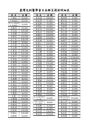
ABSTRACT OF POSTER PRESENTATION (APRIL 17, SATURDAY)
ABSTRACT OF POSTER PRESENTATION (APRIL 17, SATURDAY)
ABSTRACT OF POSTER PRESENTATION (APRIL 17, SATURDAY)

You also want an ePaper? Increase the reach of your titles
YUMPU automatically turns print PDFs into web optimized ePapers that Google loves.
P2-085 Nephrology P2-086 NephrologyGENETIC AND HISTOLOGIC ANALYSIS <strong>OF</strong> AKINDRED WITH AUTOSOMAL RECESSIVEALPORT SYNDROMETHE CELLULAR MECHANISM <strong>OF</strong>CARDIOTOXICITY INDUCED BY PERITONEALDIALYSIS RELATED PERITONITISKenichiro Miura, Takashi Sekine, Kazuhiro Takahashi,Atsuhiro Yanagisawa, Yoshiyuki Namai, Kandai Nozu,Masafumi Oka, Kazumoto Iijima, Ichiro Naito,TAKASHI IGARASHIDepartment of Pediatrics, Tokyo University Graduate School of Medicine,JapanBACKGROUND: Autosomal recessive Alport syndrome(ARAS) is caused by mutations of either genes encodingthe alpha 3 or the alpha 4 chains of type IV collagen. It ischaracterized by the loss of alpha 3, alpha 4, and alpha 5chains in the glomerular basement membrane (GBM) butthe remaining of alpha 5, and alpha 6 chains in theBowman’s capsule. We describe here a girl with ARAS inwhich a renal specimen was analyzed using antibodiesagainst alpha 1 through alpha 6 chains of type IV collagen.Genetic analyses of the family members revealedcompound heterozygous mutations in COL4A3 genes ofthe patient and her brother.PATIENT: Hematuria was noted in a 3-year-old girl by thenational urinary screening test. Proteinuria also developedand the urinary protein-creatinine ratio was 1.5-4.0 g/gCrat the age of 6. Ophthalmologic and otolaryngologicexamination revealed no abnormalities. Her father and herpaternal grandmother, but not her mother, had hematuria.Her younger brother also had hematuria and proteinuria.The light microscopic examination of this patient’s renalspecimen at the age of 6 revealed minor glomerularabnormalities. Immunofluorescence studies forimmunoglobulins and complements were all negative.Electron microscopy showed that there was thinning, butnot splitting and lamellation, of the GBM.Immunofluorescence studies for type IV collagen revealedattenuated staining of the GBM for alpha 4 and alpha 5chains, and preserved staining of the Bowmann’s capsulefor alpha 5 and alpha 6 chains, which was compatible witha diagnosis of ARAS. Immunoreactivity of the GBM foralpha 3 chain was also attenuated, but partially preserved,which was not typical of ARAS. Genetic analyses of thepatient, her parents, and her younger brother wereperformed. The patient and her brother had twoheterozygous mutations in the COL4A3 gene: c.1354G>A(G452R) in exon 22 and c.3822_3823insG in exon 43. Theformer mutation was identified in the father’s gene and thelatter in the mother’s gene.DISCUSSION: Although compound heterozygousmutations in the COL4A3 gene were identified in thepresent patient, immunofluorescence studies showed thatthere was partially preserved staining of the GBM for typeIV collagen alpha 3 chain. Incomplete formation of type IVcollagen alpha 3 chains due to missense mutation in oneallele might be a possible explanation for this observation.[Keywords] autosomal recessive Alport syndrome, type IVcollagen, immunofluorescenceHsin-Hui Wang, 1,2 Ping-Chun Li, 3 Ya-Ling Chiou,4 Tzong-Yann Lee, 5,6 and Ching-Yuang Lin, 4,7,81 Department of Pediatrics, Section of Nephrology, Taipei Veterans GeneralHospital; 2 Department of Pediatrics, Faculty of Medicine, National Yang-MingUniversity, Taipei; 3 Department of Surgery, Division of CardiovascularSurgery,China Medical University and Hospital; 4 Institute of Immunology andMicrobiology, National Yang-Ming University, Taipei; 5 Department of InternalMedicine, Section of Nephrology, En Chu Kong hospital; 6 Department ofIntegrated Diagnostics & Therapeutics, National Taiwan University Hospital,7 College of Medicine, China Medical University; 8 Division of PediatricNephrology, China Medical University Hospital, TaiwanOBJECTIVE: Peritoneal dialysis (PD) is the predominant dialyticmodality for the pediatric patients with end stage renal disease.Peritonitis is the most serious complication and the leading cause ofmortality in patients undergoing PD. In most patients withperitonitis-related mortality, the immediate cause of death was acardiovascular event. The purpose of this study was to examine thecellular mechanism for the clinical observation that PD-relatedperitonitis is accompanied by a high incidence of cardiac mortality.METHODS: The turbid PD effluent (PDE) prior to antibiotictreatment were collected in 8 culture positive peritonitis episodes fromPD patients. The microorganisms were gram-positive bacteria in 5episodes and gram-negative bacteria in 3 episodes. Cultured humancardiomyocytes were treated with PDE during peritonitis, and theeffects of PDE on cultured cardiomyocytes in terms of viability,apoptosis and the expression of several apoptosis-related genes wereassessed. Cell viability was evaluated by MTT assay. Apoptosis wasevaluated by flow cytometry, TUNEL staining with confocalmicroscopy, and Comet assay. The expression of genes was determinedby quantitative real-time RT-PCR.MAIN RESULTS: PDE treatment induced human cardiomyocytedeath in a dose- and time-dependent manner. When cardiomyocyteswere pre-exposed to 12.5, 18, or 25 mg/ml PDE for 24 h, cellviabilities were 70.6±5.7%, 58.7±9.7%, and 41.6±7.8%, respectively(P