FAO Fisheries Technical Paper 402/2 - Library - Network of ...
FAO Fisheries Technical Paper 402/2 - Library - Network of ...
FAO Fisheries Technical Paper 402/2 - Library - Network of ...

Create successful ePaper yourself
Turn your PDF publications into a flip-book with our unique Google optimized e-Paper software.
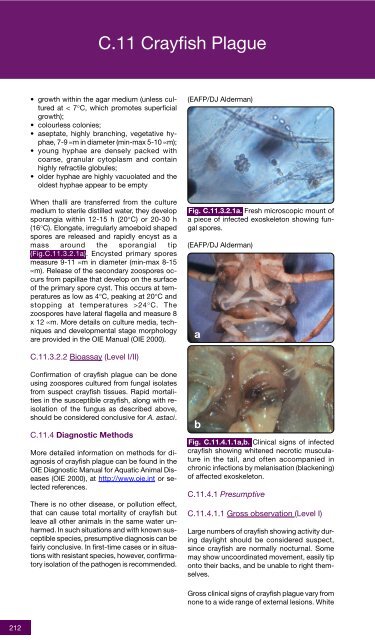
C.11 Crayfish Plague• growth within the agar medium (unless culturedat < 7°C, which promotes superficialgrowth);• colourless colonies;• aseptate, highly branching, vegetative hyphae,7-9 µm in diameter (min-max 5-10 µm);• young hyphae are densely packed withcoarse, granular cytoplasm and containhighly refractile globules;• older hyphae are highly vacuolated and theoldest hyphae appear to be emptyWhen thalli are transferred from the culturemedium to sterile distilled water, they developsporangia within 12-15 h (20°C) or 20-30 h(16°C). Elongate, irregularly amoeboid shapedspores are released and rapidly encyst as amass around the sporangial tip(Fig.C.11.3.2.1a). Encysted primary sporesmeasure 9-11 µm in diameter (min-max 8-15µm). Release <strong>of</strong> the secondary zoospores occursfrom papillae that develop on the surface<strong>of</strong> the primary spore cyst. This occurs at temperaturesas low as 4°C, peaking at 20°C andstopping at temperatures >24°C. Thezoospores have lateral flagella and measure 8x 12 µm. More details on culture media, techniquesand developmental stage morphologyare provided in the OIE Manual (OIE 2000).(EAFP/DJ Alderman)Fig. C.11.3.2.1a. Fresh microscopic mount <strong>of</strong>a piece <strong>of</strong> infected exoskeleton showing fungalspores.(EAFP/DJ Alderman)aC.11.3.2.2 Bioassay (Level I/II)Confirmation <strong>of</strong> crayfish plague can be doneusing zoospores cultured from fungal isolatesfrom suspect crayfish tissues. Rapid mortalitiesin the susceptible crayfish, along with reisolation<strong>of</strong> the fungus as described above,should be considered conclusive for A. astaci.C.11.4 Diagnostic MethodsMore detailed information on methods for diagnosis<strong>of</strong> crayfish plague can be found in theOIE Diagnostic Manual for Aquatic Animal Diseases(OIE 2000), at http://www.oie.int or selectedreferences.There is no other disease, or pollution effect,that can cause total mortality <strong>of</strong> crayfish butleave all other animals in the same water unharmed.In such situations and with known susceptiblespecies, presumptive diagnosis can befairly conclusive. In first-time cases or in situationswith resistant species, however, confirmatoryisolation <strong>of</strong> the pathogen is recommended.bFig. C.11.4.1.1a,b. Clinical signs <strong>of</strong> infectedcrayfish showing whitened necrotic musculaturein the tail, and <strong>of</strong>ten accompanied inchronic infections by melanisation (blackening)<strong>of</strong> affected exoskeleton.C.11.4.1 PresumptiveC.11.4.1.1 Gross observation (Level I)Large numbers <strong>of</strong> crayfish showing activity duringdaylight should be considered suspect,since crayfish are normally nocturnal. Somemay show uncoordinated movement, easily tiponto their backs, and be unable to right themselves.Gross clinical signs <strong>of</strong> crayfish plague vary fromnone to a wide range <strong>of</strong> external lesions. White212