Mohammad Reza Saboktakin, Abel Maharramov, Mohammad Ali ...
Mohammad Reza Saboktakin, Abel Maharramov, Mohammad Ali ...
Mohammad Reza Saboktakin, Abel Maharramov, Mohammad Ali ...
- No tags were found...

Create successful ePaper yourself
Turn your PDF publications into a flip-book with our unique Google optimized e-Paper software.
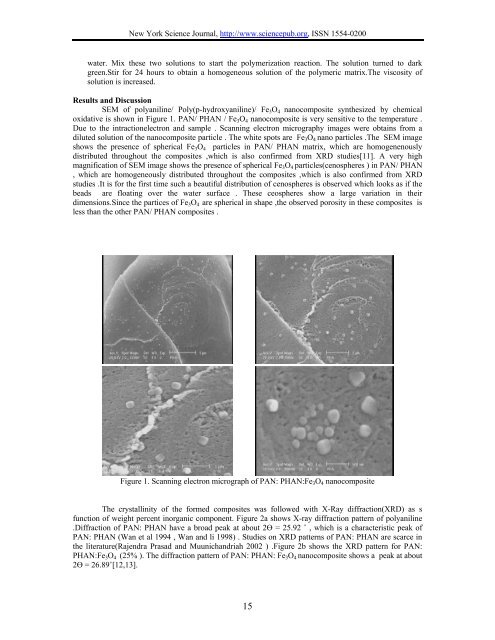
New York Science Journal, http://www.sciencepub.org, ISSN 1554-0200water. Mix these two solutions to start the polymerization reaction. The solution turned to darkgreen.Stir for 24 hours to obtain a homogeneous solution of the polymeric matrix.The viscosity ofsolution is increased.Results and DiscussionSEM of polyaniline/ Poly(p-hydroxyaniline)/ Fe 3 O 4 nanocomposite synthesized by chemicaloxidative is shown in Figure 1. PAN/ PHAN / Fe 3 O 4 nanocomposite is very sensitive to the temperature .Due to the intractionelectron and sample . Scanning electron micrography images were obtains from adiluted solution of the nanocomposite particle . The white spots are Fe 3 O 4 nano particles .The SEM imageshows the presence of spherical Fe 3 O 4 particles in PAN/ PHAN matrix, which are homogenenouslydistributed throughout the composites ,which is also confirmed from XRD studies[11]. A very highmagnification of SEM image shows the presence of spherical Fe 3 O 4 particles(cenospheres ) in PAN/ PHAN, which are homogeneously distributed throughout the composites ,which is also confirmed from XRDstudies .It is for the first time such a beautiful distribution of cenospheres is observed which looks as if thebeads are floating over the water surface . These ceospheres show a large variation in theirdimensions.Since the partices of Fe 3 O 4 are spherical in shape ,the observed porosity in these composites isless than the other PAN/ PHAN composites .Figure 1. Scanning electron micrograph of PAN: PHAN:Fe 3 O 4 nanocompositeThe crystallinity of the formed composites was followed with X-Ray diffraction(XRD) as sfunction of weight percent inorganic component. Figure 2a shows X-ray diffraction pattern of polyaniline.Diffraction of PAN: PHAN have a broad peak at about 2Ө = 25.92 ˚ , which is a characteristic peak ofPAN: PHAN (Wan et al 1994 , Wan and li 1998) . Studies on XRD patterns of PAN: PHAN are scarce inthe literature(Rajendra Prasad and Muunichandriah 2002 ) .Figure 2b shows the XRD pattern for PAN:PHAN:Fe 3 O 4 (25% ). The diffraction pattern of PAN: PHAN: Fe 3 O 4 nanocomposite shows a peak at about2Ө = 26.89˚[12,13].15