
Create successful ePaper yourself
Turn your PDF publications into a flip-book with our unique Google optimized e-Paper software.
DATA SHEET<br />
Understanding Images<br />
<strong>Definiens</strong> <strong>Tissue</strong> <strong>Studio</strong> TM<br />
<strong>Definiens</strong> Oncology Suite<br />
<strong>Definiens</strong> <strong>Tissue</strong> <strong>Studio</strong> TM is the most advanced digital pathology image<br />
analysis solution for biomarker translational research.<br />
<strong>Definiens</strong> <strong>Tissue</strong> <strong>Studio</strong> helps you to solve<br />
biological questions<br />
<strong>Definiens</strong> <strong>Tissue</strong> <strong>Studio</strong> is built on <strong>Definiens</strong><br />
Cognition Network Technology® and is the most advanced<br />
digital pathology image analysis application<br />
for translational biomarker research. It accurately<br />
quantifies biomarkers, revealing the precise attributes<br />
of sub-anatomical objects, cells and sub-cellular<br />
objects, helping to solve your most challenging<br />
biological questions.<br />
<strong>Definiens</strong> <strong>Tissue</strong> <strong>Studio</strong> is easy to use<br />
Every aspect of <strong>Definiens</strong> <strong>Tissue</strong> <strong>Studio</strong> has been<br />
designed to support pathologists and research<br />
scientists who seek further validation and understanding<br />
of tissue-based biomarkers. It takes just<br />
three simple steps to use.<br />
1. Train the application to identify regions of interest<br />
The application can be trained to identify regions<br />
of interest automatically. Simply import sample<br />
slides and highlight the regions of interest with the<br />
”paintbrush“ tool, comparing up to four slides at once.<br />
Then, simply click the ”Learn“ button.<br />
The application uses your images to create a robust<br />
algorithm for identifying the precise regions of interest<br />
(ROI) which you seek. You can manually review the<br />
regions of interest or they can form the basis of a fully<br />
automated batch processing routine.<br />
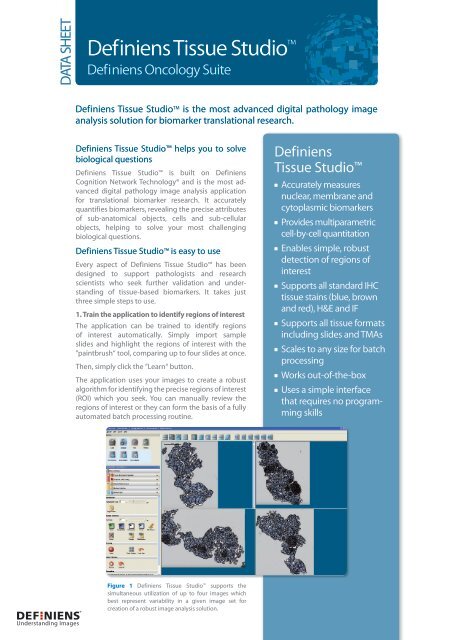
Figure 1 <strong>Definiens</strong> <strong>Tissue</strong> <strong>Studio</strong> supports the<br />
simultaneous utilization of up to four images which<br />
best represent variability in a given image set for<br />
creation of a robust image analysis solution.<br />
<strong>Definiens</strong><br />
<strong>Tissue</strong> <strong>Studio</strong><br />
Accurately measures<br />
nuclear, membrane and<br />
cytoplasmic biomarkers<br />
Provides multiparametric<br />
cell-by-cell quantitation<br />
Enables simple, robust<br />
detection of regions of<br />
interest<br />
Supports all standard IHC<br />
tissue stains (blue, brown<br />
and red), H&E and IF<br />
Supports all tissue formats<br />
including slides and TMAs<br />
Scales to any size for batch<br />
processing<br />
Works out-of-the-box<br />
Uses a simple interface<br />
that requires no programming<br />
skills
2<br />
<strong>Definiens</strong> <strong>Tissue</strong> <strong>Studio</strong><br />
Figure 2 <strong>Definiens</strong> Composer Technology TM<br />
in action: In order to classify the object as a tumor,<br />
the user simply “paints” just a few representative ROIs with the paintbrush tool (green). This<br />
process is then repeated across the other images, and for other ROIs (tissue, lymph node, etc.).<br />
Figure 3 <strong>Definiens</strong> <strong>Tissue</strong> <strong>Studio</strong> TM control panel: The colored icons at the top are used to select<br />
diferent paintbrush colors for sampling ROIs. After using the paintbrush tool, the user simply<br />
clicks the “Learn” button to teach the system to properly identify ROIs. This entire process is<br />
simple, and takes only minutes.<br />
Figure 4 Close-up view of pancreas tissue after training the system how to recognize ROIs<br />
using <strong>Definiens</strong> Composer Technology TM . Islets (both tumor and normal) are depicted in green,<br />
tissue in blue, lymph nodes in dark blue and white space in white.
2. Create unique segmentation and classifcation for sub-cellular biomarker detection<br />
Using the intuitive interface and pre-defned tools, you can create multiparametric segmentation and<br />
classifcation with ease.<br />
<strong>Definiens</strong> <strong>Tissue</strong> <strong>Studio</strong> conducts segmentation and classifcation on a cell-by-cell basis, giving you<br />
unparalleled confdence and understanding of biomarkers that are related to disease progression,<br />
toxicity, or therapeutic response.<br />
Use the new, simplifed graphic user interface to train the application how to recognize the nuclear,<br />
cytoplasmic, and membrane attributes of cells. Even with challenging images, the cell simulation<br />
function will identify nuclear objects and then“grow”the cell out to establish its proper border.<br />
Choose from a complete set of default multiparametric and morphological features you want to<br />
measure – number of positive nuclei, number of negative nuclei, mean cell size, marker area,<br />
staining intensity, and many more. Custom parameters are also available for export.<br />
The solutions you develop with <strong>Definiens</strong> <strong>Tissue</strong> <strong>Studio</strong> will enable you to data mine your tissue<br />
images and uncover underlying correlations related to clinical endpoints of interest.<br />
Figure 5 In this example, a second <strong>Definiens</strong> Composer Technology step was used to further<br />
classify islets into tumor (red) and normal (orange). ROIs (gray boxes) are then selected within<br />
the tumor regions for subsequent cell-by-cell biomarker multiparametric quantifcation.<br />
3. Run the dataset<br />
<strong>Definiens</strong> <strong>Tissue</strong> <strong>Studio</strong> works with all standard image formats – Hamamatsu, Zeiss, Aperio, Applied<br />
Imaging, <strong>Tissue</strong>Gnostics, Bacus – as well as generic formats like .tif and .jpeg. All you need to do<br />
is import the slides and click ”run“.<br />
It is that easy.<br />
<strong>Definiens</strong> <strong>Tissue</strong> <strong>Studio</strong> can batch process locally or send larger jobs to servers. There is no limit to<br />
the number of CPUs that can be added, ensuring that you can handle any project of any size.<br />
<strong>Definiens</strong> <strong>Tissue</strong> <strong>Studio</strong> 3
4<br />
<strong>Definiens</strong> <strong>Tissue</strong> <strong>Studio</strong><br />
Figure 6 Example of nucleus detection in pancreas tumor.<br />
<strong>Definiens</strong> <strong>Tissue</strong> <strong>Studio</strong> is uniquely powerful<br />
<strong>Definiens</strong> <strong>Tissue</strong> <strong>Studio</strong> is built on the powerful <strong>Definiens</strong> Cognition Network Technology®<br />
which does not rely solely on traditional pixel-based, pattern recognition. Instead, it builds<br />
an understanding of images iteratively, identifying objects through their characteristics, their<br />
relationships to other objects and the context in which they occur.<br />
This fundamental diference enables <strong>Definiens</strong> <strong>Tissue</strong> <strong>Studio</strong> to address highly heterogeneous<br />
tissues, identify individual cells and the sub-cellular objects within them, and then<br />
conduct extremely accurate cell-by-cell measurements.<br />
<strong>Definiens</strong> <strong>Tissue</strong> <strong>Studio</strong> is part of <strong>Definiens</strong> Oncology Suite<br />
<strong>Definiens</strong> <strong>Tissue</strong> <strong>Studio</strong> is part <strong>Definiens</strong> Oncology Suite which encompasses applications spanning<br />
cellular assays, histopathology, and non-invasive medical imaging.<br />
Defniens <strong>Tissue</strong> <strong>Studio</strong> incorporates <strong>Definiens</strong> Composer Technology<br />
<strong>Definiens</strong> Composer Technology is a new easy to use machine learning approach that enables<br />
the user to train the application to detect regions of interest using up to four images simultaneously.<br />
The resulting solution can be applied to a complete set of images with a single click for unlimited<br />
parallel batch processing.<br />
<strong>Definiens</strong> <strong>Tissue</strong> <strong>Studio</strong> improves decision making<br />
<strong>Definiens</strong> <strong>Tissue</strong> <strong>Studio</strong> surpasses every other image analysis application for translational research of<br />
tissue-based biomarkers, delivering deeper insights, faster results and better decisions.
<strong>Definiens</strong><br />
<strong>Tissue</strong> <strong>Studio</strong><br />
delivers<br />
DEEPER INSIGHTS by<br />
Quantifing nuclear,<br />
membrane and cytoplasmic<br />
biomarkers on a cell-by-cell<br />
basis<br />
Supporting all standard<br />
IHC tissue stains (blue, brown<br />
and red), H&E and IF<br />
Analyzing all slides and<br />
TMA images from all major<br />
image acquisition devices<br />
FASTER RESULTS by<br />
Identifing regions of<br />
interest automatically<br />
Operating through a simple,<br />
intuitive user interface<br />
Ofering unlimited<br />
throughput with parallel<br />
batch processing<br />
BETTER DECISIONS by<br />
Delivering extremely accurate<br />
and reproducible results<br />
Enabling multiparametric<br />
investigations to identify<br />
underlying correlations<br />
Supporting translational<br />
research through rapid<br />
iterative investigation of<br />
biomarkers<br />
Multiparametric and morphological<br />
features measured<br />
IHC<br />
No. of positive nuclei<br />
No. of negative nuclei<br />
Positive index<br />
Cell and membrane features:<br />
– Mean cell size (μm²)<br />
– Mean IHC marker intensity (cell)<br />
– Mean IHC marker intensity (cytoplasm)<br />
– Mean IHC marker intensity (membrane)<br />
– Mean ratio IHC marker intensity membrane<br />
to cytoplasm<br />
– Marker area (μm²)<br />
No. and % of:<br />
– Nuclei of low intensity<br />
– Nuclei of medium intensity<br />
– Nuclei of high intensity<br />
No. and % of:<br />
– Nuclei with small area<br />
– Nuclei with medium area<br />
– Nuclei with large area<br />
No. and % of:<br />
– Cells of negative intensity (0)<br />
– Cells of low intensity (1+)<br />
– Cells of medium intensity (2+)<br />
– Cells of high intensity (3+)<br />
No. and % of:<br />
– Cells with small area<br />
– Cells with medium area<br />
– Cells with large area<br />
H & E and IF<br />
ROI area/marker area<br />
Custom data export is also supported<br />
<strong>Definiens</strong> <strong>Tissue</strong> <strong>Studio</strong> 5
System requirements<br />
Defniens <strong>Tissue</strong> <strong>Studio</strong> is available with either two or four analysis engines to meet your<br />
processing requirements.<br />
TWO ANALYSIS ENGINES<br />
Minimum:<br />
• Windows XP Professional 64Bit SP2,<br />
Vista-Business/Ultimate 64 Bit SP2<br />
• Intel / AMD Quad Core 64 Bit 1.83Ghz +<br />
• 8 Gb Memory<br />
• 30Gb Disk Space<br />
• 20“ 1600 x 1200 Display<br />
• 100 MB Ethernet network connection<br />
FOUR ANALYSIS ENGINES<br />
Minimum:<br />
• Windows XP Professional 64Bit SP2,<br />
Vista-Business/Ultimate 64 Bit SP2<br />
• Intel / AMD Dual CPU (Socket),<br />
Quad Core 64 Bit 1.83Ghz +<br />
• 12 Gb Memory<br />
• 30Gb Disk Space<br />
• 20“ 1600 x 1200 Display<br />
• 100 MB Ethernet network connection<br />
Recommended:<br />
• Windows XP Professional -64Bit SP2,<br />
Vista-Business/Ultimate 64 Bit SP2<br />
• Intel Xeon/ AMD Quad Core 64 Bit 2.00Ghz+<br />
• 12 Gb Memory (1.33GHz Bus)<br />
• 30Gb Disk Space<br />
• 24“ 1920 x 1200 Display +<br />
• 1Gb Ethernet Connection<br />
Recommended:<br />
• Windows XP Professional 64Bit SP2,<br />
Vista-Business/Ultimate 64 Bit SP2<br />
• Intel Xeon/ AMD Dual CPU (Socket),<br />
Quad Core 64 Bit 2.00Ghz+<br />
• 16 Gb Memory (1.33GHz Bus)<br />
• 30Gb Disk Space<br />
• 24“ 1920 x 1200 Display +<br />
• 1Gb Ethernet Connection<br />
<strong>Definiens</strong> is the number one Enterprise Image Intelligence® company for analyzing and interpreting images<br />
on every scale, from microscopic cell structures to satellite images.<br />
The patented <strong>Definiens</strong> Cognition Network Technology®, developed by Nobel Laureate Prof. Gerd Binnig<br />
and his team, emulates human cognitive processes of perception to extract intelligence from images.<br />
If you are interested in learning more about how <strong>Definiens</strong> could address the challenges you face, please visit<br />
our website.<br />
www.definiens.com<br />
Corporate Headquarters Americas Headquarters<br />
<strong>Definiens</strong> AG <strong>Definiens</strong> Inc.<br />
Trappentreustrasse 1 1719 State Route 10, Suite 118<br />
80339 Munich Parsippany, New Jersey 07054<br />
Germany USA<br />
Tel. +49 (0)89 231180-0 Tel. +1-973-457-7150 DSL-0004-02-31080<br />
Copyright © 2009 <strong>Definiens</strong> AG. <strong>Definiens</strong>, <strong>Definiens</strong> Cellenger, <strong>Definiens</strong> Cognition Network Technology, <strong>Definiens</strong> eCognition, <strong>Definiens</strong> Enterprise Image Intelligence<br />
and Understanding Images are trademarks or registered trademarks of <strong>Definiens</strong> in the United States, the European Community, or certain other jurisdictions.<br />
All registered trademarks, pending trademarks, or service marks are property of their respective owners. The information in this document is subject to change<br />
without notice and should not be construed as a commitment by <strong>Definiens</strong> AG. <strong>Definiens</strong> AG assumes no responsibility for any errors that may appear in this document.