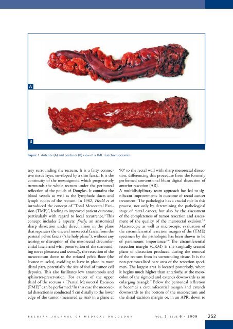
ABFigure 1. Anterior (A) and posterior (B) view of a TME resection specimen.tery surrounding the rectum. It is a fatty connectivetissue layer, enveloped by a thin fascia. It is thecontinuity of the mesosigmoid which progressivelysurrounds the whole rectum under the peritonealreflection of the pouch of Douglas. It contains theblood vessels as well as the lymphatic ducts andlymph nodes of the rectum. In 1982, Heald et alintroduced the concept of “Total Mesorectal Excision(TME)”, leading to improved patient outcome,particularly with regard to local recurrence. 3 Thisconcept includes 2 aspects: firstly, an anatomicalsharp dissection under direct vision in the planethat separates the visceral mesorectal fascia from theparietal pelvic fascia (“the holy plane”), without anytearing or disruption of the mesorectal circumferentialfascia and with preservation of the surroundingnerve plexuses; and secondly, the resection of themesorectum down to the striated pelvic floor (thelevator muscles), avoiding to leave in place its mostdistal part, potentially the site of foci of tumor celldeposits. This also facilitates low anastomosis andsphincter-preservation. For cancer of the upperthird of the rectum a “Partial Mesorectal Excision(PME)” can be performed. 4 In this case the mesorectaldissection is conducted 5 cm distally to the loweredge of the tumor (measured in situ) in a plane at90° to the rectal wall with sharp mesorectal dissection,differencing this procedure from the formerlyperformed conventional blunt digital dissection ofanterior resection (AR).A multidisciplinary team approach has led to significantimprovements in outcome of rectal cancertreatment. 1 The pathologist has a crucial role in thisprocess, not only by determining the pathologicalstage of rectal cancer, but also by the assessmentof the completeness of tumor resection and assessmentof the quality of the mesorectal excision. 5,6Macroscopic as well as microscopic evaluation ofthe circumferential resection margin of the (TME)specimen by the pathologist has been shown to beof paramount importance. 5,6 The circumferentialresection margin (CRM) is the surgically-createdplane of dissection produced during the removalof the rectum from its surrounding tissue. It is thenon-peritonealised bare area of the resection specimen.The largest area is located posteriorly, whereit begins much higher than anteriorly, at the mesocolonof the sigmoid and extends downwards as anenlarging triangle. 7 Below the peritoneal reflectionit becomes a circumferential margin and extendsdownwards to the bottom of the mesorectum andthe distal excision margin or, in an APR, down toB E L G I A N J O U R N A L O F M E D I C A L O N C O L O G Y v o l . 3 i s s u e 6 - 2 0 0 9 252
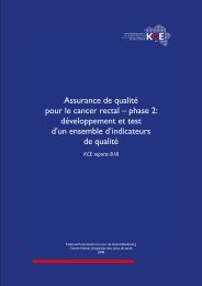
P R A C T I C E G U I D E L I N E SFigure 2. Severely irregular mesorectum in an APR specimen: the muscle layer is visible about 1 cm below the peritoneal reflection.The description of the quality of the mesorectal surface in an APR specimen is limited to the description of the rectumabove the sphincters.the anal skin (Figure 1). 7 Tumor involvement of theCRM is the single most important factor for predictingthe risk of local recurrence in rectal cancerpatients. 6,8-10 It is also important in the predictionof distant metastasis and overall survival. 10 Tumorswithin 1 mm of the surgically created margin havea greatly increased risk of recurrence. 8,9 One studystill showed a high incidence of recurrence at 2 mm,but this finding could not be confirmed in subsequentstudies. 1,11,12For very low primary rectal tumors an abdominoperinealresection (APR) is required. Mesorectal excisionin this case will remove lymphatic, vascular,and neural pathways of metastasis, but there will oftenbe involvement of the surgical resection marginat the level of the sphincters. 13,14 The tapering of themesorectum towards the levators emphasizes thatthere is less tissue for the carcinoma to transverse beforeinvolving the surgical plane of resection in thelow mesorectum and anal canal. In addition, goodvisualization and access are limited with the classicalapproach of APR and it was shown that the planeof resection lies within the sphincter muscle, thesubmucosa, or lumen in more than one third of theAPR cases. In the remainder, the plane of resectionlies on the sphincteric muscles. 14 This predisposes tocircumferential resection margin (CRM) involvement,except in the very early stages. In addition, ahigh intra-operative perforation rate was observed. 14Neoadjuvant therapy has an important place here indownsizing the tumor, enabling complete resection.Moreover, a more radical operation with a predominantlyperineal surgical approach, creating a CRMoutside the levators and giving wider clearance is beingconsidered for low rectal tumors. 13,14Gross external appearance of the surgicallyresected specimen: macroscopic inspectionThe first task of the pathologist when receiving thesurgical excision specimen following TME is thevisual inspection of the completeness of resectionof the mesorectum. 5-8,15 Preferentially, the resectionspecimen should be examined in the fresh, unfixedstate. More importantly however, is that it shouldbe delivered unopened to the pathologist. The externalsurface of the TME should be carefully inspectedand the quality of the mesorectal excisionshould be assessed and can be graded (complete,nearly complete, incomplete). 5,6 The mesorectalsurface of a good resection should be smoothwith a good bulk of mesorectum. There should beno coning near the distal margin. Defects at thesurface should be less than 5 mm deep. In case ofperforation of the resection specimen or in case themuscular layer is visible exteriorly, the resectionis classified as incomplete (Figure 2). In the PRO-CARE protocol the following terminology is used:smooth and regular, mildly irregular or severely irregular(Table 1). 2 This slightly adapted terminologywas introduced to avoid misinterpretation ofthe notion of “incomplete” resection, as in case ofadvanced tumor growth it is not always possible toavoid margin involvement. A Dutch study demonstratedthat despite extensive surgical training only57% of operations were judged to be complete excisionsand nearly one quarter (24%) were classifiedas incomplete excisions. 5 In resection specimens reportedto be incomplete, there was a significantlyhigher rate of circumferential margin involvementand a higher rate of overall recurrence and local tumorrecurrence. 5 In patients with a positive CRM,253v o l . 3 i s s u e 6 - 2 0 0 9B E L G I A N J O U R N A L O F M E D I C A L O N C O L O G Y