Table 1. Grading of the quality of mesorectal excision in TME specimens as proposed in the PROCARE guidelines.Both the specimen as a whole (fresh) and transverse slices (after fixation) should be examined in orderto allow adequate evaluation of the mesorectal excision.Smooth, regular - intact mesorectum with only minor irregularities of a smooth mesorectal surface- no defect deeper than 5 mm- no coning toward the distal margin of the specimen*- a smooth circumferential resection margin on slicingMildly irregular - moderate bulk to the mesorectum, but irregularity of the mesorectal surface- moderate coning of the specimen allowed*- the muscularis propria invisible at every site, with the exception of the insertion of thelevator musclesSeverely irregular - little bulk to the mesorectum with defects down onto the muscularis propria and/orvery irregular circumferential resection margin on slicing* Coning refers to the tendency for the surgeon to cut towards the rectum wall during distal dissection, rather thanstaying outside of the visceral mesorectal fascia; this gives a conical appearance to the surgical resection specimen.assessment of the quality of surgery added nothingto the prediction of local recurrence above CRMinvolvement alone. However, in patients with anegative CRM and incomplete resection, the overallrecurrence rate was doubled from 15 to 29% andsurvival decreased from 91 to 77%. 5 No prognosticdifference was observed between patients witha complete mesorectum compared to those with anearly complete mesorectum. 5It is important to realize that there is less mesorectaltissue anteriorly and laterally than posteriorly.7 In addition the size of the mesorectum varieswidely between individuals and is related to severalfactors such as to body mass, gender, and degree ofcachexia. 16In an APR specimen, the description of the qualityof the mesorectal surface is limited to the descriptionof the rectum above the sphincter.Handling the TME specimenThe relation of the tumor to the serosal surfaceshould be determined, i.e. above, at, or belowthe peritoneal reflection. After examination ofthe external surface, the resection margin shouldbe painted with india ink. The resection specimenis opened anteriorly above the peritonealreflection starting at its proximal end withoutextension into the tumor. This allows effectiveevaluation of the anterior CRM that would otherwisebe destroyed by the opening process. Ideally,the resection specimen should be pinnedout on a corkboard to avoid shrinkage and leftfloating with the cork upwards in formalin fixativefor at least 48 hours. 18 Placement of gauze orpaper tissue wick soaked in formalin within thelumen of the intact bowel segment is necessaryto enhance fixation. After fixation, the resectionspecimen should be sectioned in parallel cuts of3-4 mm intervals, perpendicularly to the lengthof the bowel. The long fixation time is required tomake the tissue firmer and facilitates serial crosssectionalslicing of the specimen. This allows theassessment of the deepest point of tumor invasionand to measure the distance to the place where thetumor is closest to the CRM. The number of tissueblocks to be taken from the tumor is 3 at minimum.2 At least one tissue block should include thetransition from the surrounding normal mucosa tothe tumor and at least one tissue block should betaken of the deepest point of invasion. 2,19 The latterpermits microscopic confirmation and refinementof gross observations at the area of greatest macroscopicconcern. It is known that, in particular afterradiotherapy, the presence of fibrosis may makemacroscopic assessment of the tumor inaccurate. Itis often impossible to distinguish therapy-inducedfibrosis from tumor invasion. 20 In this case, sufficienttissue blocks should be taken from all macroscopicallysuspected areas in order not to miss thedeepest point of invasion. No distinction should bemade between various modes of involvement of theCRM, continuous spread, discontinuous tumordeposits, or involved lymph nodes. The orientationof grossly suspicious nodes that are closely relatedto the CRM should thus be preserved in sections.Tissue slices can be embedded as large-area (giant)blocks or as conventional small blocks. Formalinfixation will allow additional molecular pathologicalexamination.B E L G I A N J O U R N A L O F M E D I C A L O N C O L O G Y v o l . 3 i s s u e 6 - 2 0 0 9 254
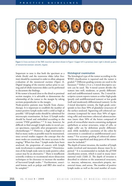
P R A C T I C E G U I D E L I N E SFigure 3. Cross-sections of the TME resection specimen shown in Figure 1 (upper left is proximal, lower right is distal); qualityof mesorectal excision: smooth, regular.Important to note is that both the specimen as awhole (fresh) and the transverse slides (after fixation)should be examined in order to allow adequateevaluation of the mesorectal excision (Figure 3).Digital imaging of the exterior surface prior to cuttingand of whole transverse slides can be performedto document the findings.If the tumor is located close to the distal or proximalsection margins, it is advisable to demonstrate therelationship of the tumor to the margin by takingsections perpendicular to the margin.Node-positive patients may benefit from chemotherapy.It is important to establish the number oftumor-positive lymph nodes with a yield as high aspossible. 6 All lymph nodes should be submitted formicroscopic examination. At least 12 lymph nodesshould be found and embedded according to thecurrent TNM guidelines. 21-23 It may, however, bedifficult to find enough lymph nodes in rectal cancerspecimens, especially after preoperative radiochemotherapy.24,25 However, a high motivation tofind as many nodes as possible must be maintained,since several studies support the concept that themore nodes are examined, the more accurate is thestaging. 6 When less than 7 lymph nodes have beenanalysed, the proportion of cancers with lymphnode involvement is underestimated. 26 Determinationof the lymph node ratio in node-positive coloncancer may be an alternative. 27 There is insufficientscientific evidence to recommend micro-dissectiontechniques or fat clearance to increase the numberof harvested lymph nodes. 21 Furthermore, associatedlesions such as polyps and IBD also need tobe sampled. 21Histological examinationThe histological type of the tumor according to theWHO classification is reported and the tumor isgraded. 28 Different grading systems are used in theliterature. 28 Either a 4 or a 2-tiered descriptive systemcan be used. The 4-tiered system divides thetumors into well, moderate, or poorly differentiatedand undifferentiated tumors. The 2-tiered descriptivesystem reports tumors as either high grade(poorly and undifferentiated tumors) or low grade(well and moderately differentiated tumors). In the2-tiered descriptive system, the high grade correspondsto less than 50% of glandular structures ofthe surface analysed. Signetring cell colorectal carcinomas(composed for more than 50% of signetringcells) and mucinous colorectal adenocarcinomas(more than 50% of the lesion composed ofpools of extracellular mucin-containing malignantepithelium as acinar structures, strips of cells, orsingle cells) are by definition poorly differentiated,while medullary carcinoma of the colon byconvention is considered as undifferentiated carcinoma.28 For accurate grading of colorectal adenocarcinomassuperficial and deep parts of the tumormust be included. 19The depth of tumor invasion, the number of lymphnodes involved and metastatic disease must be reported.21 It is recommended to include the pTNMclassification system, which is used in many internationaltrials (Table 2). The depth of invasion isdescribed in relation to the anatomical structures,i.e. mucosa, submucosa, muscularis propria, mesorectaltissue, and serosa. The number of positivelymph nodes as well as the total number of exam-255v o l . 3 i s s u e 6 - 2 0 0 9B E L G I A N J O U R N A L O F M E D I C A L O N C O L O G Y