Download - Belgian Cancer Registry
Download - Belgian Cancer Registry
Download - Belgian Cancer Registry
- No tags were found...

You also want an ePaper? Increase the reach of your titles
YUMPU automatically turns print PDFs into web optimized ePapers that Google loves.
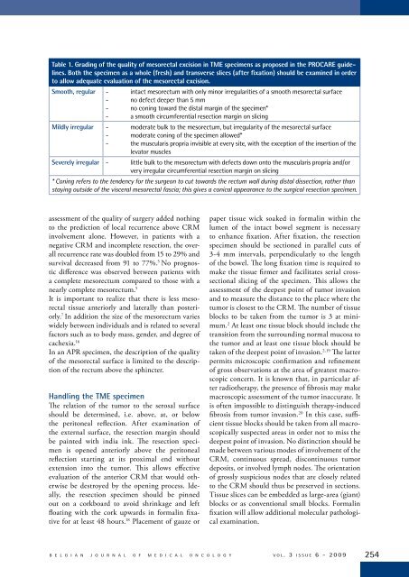
Table 1. Grading of the quality of mesorectal excision in TME specimens as proposed in the PROCARE guidelines.Both the specimen as a whole (fresh) and transverse slices (after fixation) should be examined in orderto allow adequate evaluation of the mesorectal excision.Smooth, regular - intact mesorectum with only minor irregularities of a smooth mesorectal surface- no defect deeper than 5 mm- no coning toward the distal margin of the specimen*- a smooth circumferential resection margin on slicingMildly irregular - moderate bulk to the mesorectum, but irregularity of the mesorectal surface- moderate coning of the specimen allowed*- the muscularis propria invisible at every site, with the exception of the insertion of thelevator musclesSeverely irregular - little bulk to the mesorectum with defects down onto the muscularis propria and/orvery irregular circumferential resection margin on slicing* Coning refers to the tendency for the surgeon to cut towards the rectum wall during distal dissection, rather thanstaying outside of the visceral mesorectal fascia; this gives a conical appearance to the surgical resection specimen.assessment of the quality of surgery added nothingto the prediction of local recurrence above CRMinvolvement alone. However, in patients with anegative CRM and incomplete resection, the overallrecurrence rate was doubled from 15 to 29% andsurvival decreased from 91 to 77%. 5 No prognosticdifference was observed between patients witha complete mesorectum compared to those with anearly complete mesorectum. 5It is important to realize that there is less mesorectaltissue anteriorly and laterally than posteriorly.7 In addition the size of the mesorectum varieswidely between individuals and is related to severalfactors such as to body mass, gender, and degree ofcachexia. 16In an APR specimen, the description of the qualityof the mesorectal surface is limited to the descriptionof the rectum above the sphincter.Handling the TME specimenThe relation of the tumor to the serosal surfaceshould be determined, i.e. above, at, or belowthe peritoneal reflection. After examination ofthe external surface, the resection margin shouldbe painted with india ink. The resection specimenis opened anteriorly above the peritonealreflection starting at its proximal end withoutextension into the tumor. This allows effectiveevaluation of the anterior CRM that would otherwisebe destroyed by the opening process. Ideally,the resection specimen should be pinnedout on a corkboard to avoid shrinkage and leftfloating with the cork upwards in formalin fixativefor at least 48 hours. 18 Placement of gauze orpaper tissue wick soaked in formalin within thelumen of the intact bowel segment is necessaryto enhance fixation. After fixation, the resectionspecimen should be sectioned in parallel cuts of3-4 mm intervals, perpendicularly to the lengthof the bowel. The long fixation time is required tomake the tissue firmer and facilitates serial crosssectionalslicing of the specimen. This allows theassessment of the deepest point of tumor invasionand to measure the distance to the place where thetumor is closest to the CRM. The number of tissueblocks to be taken from the tumor is 3 at minimum.2 At least one tissue block should include thetransition from the surrounding normal mucosa tothe tumor and at least one tissue block should betaken of the deepest point of invasion. 2,19 The latterpermits microscopic confirmation and refinementof gross observations at the area of greatest macroscopicconcern. It is known that, in particular afterradiotherapy, the presence of fibrosis may makemacroscopic assessment of the tumor inaccurate. Itis often impossible to distinguish therapy-inducedfibrosis from tumor invasion. 20 In this case, sufficienttissue blocks should be taken from all macroscopicallysuspected areas in order not to miss thedeepest point of invasion. No distinction should bemade between various modes of involvement of theCRM, continuous spread, discontinuous tumordeposits, or involved lymph nodes. The orientationof grossly suspicious nodes that are closely relatedto the CRM should thus be preserved in sections.Tissue slices can be embedded as large-area (giant)blocks or as conventional small blocks. Formalinfixation will allow additional molecular pathologicalexamination.B E L G I A N J O U R N A L O F M E D I C A L O N C O L O G Y v o l . 3 i s s u e 6 - 2 0 0 9 254