Morphological characterization and molecular ... - THE BIOSCAN
Morphological characterization and molecular ... - THE BIOSCAN
Morphological characterization and molecular ... - THE BIOSCAN
- No tags were found...

You also want an ePaper? Increase the reach of your titles
YUMPU automatically turns print PDFs into web optimized ePapers that Google loves.
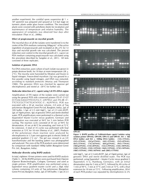
MORPHOLOGICAL CHARACTERIZATION AND MOLECULAR PHYLOGENYanother experiment, the conidial spore suspension @ 1 ×10 6 spore/ml was prepared <strong>and</strong> sprayed at 3-4 leaf stage onturmeric plants under glass house condition. The inoculatedplants were covered with polythene sheets for incubation <strong>and</strong>maintenance of temperature <strong>and</strong> relative humidity. Theappearance of symptoms was observed four days afterinoculation (Than et al., 2008a).Effect of propiconazole on mycelial growthThe mycelial discs of all the isolates were transferred in to thecenter of the PDA medium containing 500μg mL -1 of the activeingredient of propiconazole <strong>and</strong> incubated at 28±2 o C for 12days <strong>and</strong> mycelial growth rate was calculated. The percentreduction over control in the mycelial growth of C. capsici onPDA medium containing propiconazole was calculated usingthe procedure described by Sangdee et al., 2011. All testsconsisted of three replicates.Isolation of genomic DNAFor DNA extraction, pure culture of each isolate was grown inpotato dextrose broth, for 10 days at room temperature (28 ±2°C). The mycelia were harvested by filtration <strong>and</strong> frozen inliquid nitrogen. Freeze-dried mycelium (1g) was ground to afine powder using liquid nitrogen, <strong>and</strong> DNA was extracted,according to st<strong>and</strong>ard protocols (Murray <strong>and</strong> Thompson1980). The genomic DNA was checked by agarose gelelectrophoresis <strong>and</strong> stored at –20°C for further use.Molecular detection of C. capsici using 5.8 ITS rDNA regionAmplifications of ITS region of the isolates were carried outusing the general PCR with conserved primers ITS-1F (5'-GTCCTAACAAGGTTTCCGTA-3'; J297952) <strong>and</strong> ITS-4R (5'-TTCTCCGCTTATTGATATGC-3'; AJ297953). PCR wasexecuted with a 20 μL reaction volume, 2.0 units of Taqpolymerase (Bangalore Genei Pvt Ltd, Banglore, India), 2μL of10X buffer, 1.5μL of 2.5 mM MgCl 2, 1μL of 2.5 mM dNTP,2μL of 10μM primer, 4μL of genomic DNA <strong>and</strong> sterile distilledwater. PCR amplifications were performed in a thermal cycler(Eppendorf Master Cycler nexus gradient, German) <strong>and</strong>denaturation was executed at 94 o C for 5 min before PCRcycling. The reaction cycle consisted of 45 sec at 94 o C fordenaturation, 45 sec at 46 o C for annealing, <strong>and</strong> 1min at 72 o Cfor extension. A total of 35 cycles was performed with finalextension at 72 o C for 10 min (Shenoy et al., 2007). Productsof the polymerase chain reaction were analyzed byelectrophoresis in 1.5 per cent agarose gels in electric fields ofpotential gradient 2 V cm -1 . The gel was viewed in an UVtransilluminator <strong>and</strong> the b<strong>and</strong>ing pattern was photographed<strong>and</strong> analyzed. The sizes of the PCR products were determinedby comparison with st<strong>and</strong>ard 100bp ladder (Bangalore GeneiPvt. Ltd., Bangalore, India).Molecular diversity using RAPD analysisIn total, eighteen 10-mer primers were used for RAPD analysis(Table 1). All the RAPD primers were purchased from Operon(Operon Biotechnologies, Cologne, Germany) <strong>and</strong> used assingle primers. PCR amplification was performed using aEppendorf nexus gradient master cycler <strong>and</strong> a 20μL totalvolume containing 2.0 units of Taq polymerase (BangaloreGenei Pvt Ltd, Banglore, India), 2μL of 10X buffer, 1.5μL of 2.5OPA-02OPA-09OPA-13OPA-01Figure 1: RAPD profiles of Colletotrichum capsici isolates usingr<strong>and</strong>om primer OPA-02, OPA-09, OPA-13 <strong>and</strong> OPF-01. M1 = 1kbDNA ladder <strong>and</strong> M2 = 100bp DNA ladder. (1)Cc1TNAU,(2)Cc2TNAU, (3)Cg1TNAU, (4)Cc3TNAU, (5)Cc4TNAU,(6)Cc5TNAU, (7)Cc6TNAU, (8)Cg2TNAU, (9)Cc7TNAU,(10)Cg3TNAU, (11)Cc8TNAU, (12).Cc9TNAU, (13)Cc10TNAU,(14)Cc11TNAU, (15)Cc12TNAU (16)Cc13TNAU, (17)Cc14TNAU,(18)Cc15TNAU, (19)Cc16TNAU <strong>and</strong> (20)Cc17TNAUmM MgCl 2, 1μL of 2.5 mM dNTP, 2μL of 10μM primer, 4μL ofgenomic DNA <strong>and</strong> sterile distilled water. The PCR wasperformed, using Eppendorf – Master Cycler nexus gradient S(Eppendorf, A G, Hamburg, Germany), with an initialdenaturation step for 5 min at 94°C, followed by 40 cycles of1 min at 94°C, 1 min at 37°C <strong>and</strong> 2 min at 72°C, with a finalextension for 10 min at 72°C. Following amplification, 20¼lof each PCR product was separated by electrophoresis in 2%333