IMAGING & ONCOLOGY - Society of Radiographers
IMAGING & ONCOLOGY - Society of Radiographers
IMAGING & ONCOLOGY - Society of Radiographers
- No tags were found...

You also want an ePaper? Increase the reach of your titles
YUMPU automatically turns print PDFs into web optimized ePapers that Google loves.
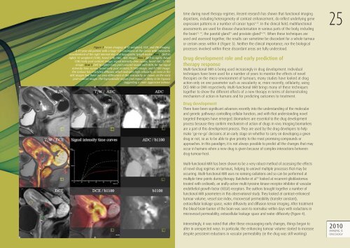
Figure 3 Fusion imaging <strong>of</strong> T2 weighted, DCE, and DW Imaging.A 31-year-old patient with a large T2b carcinoma <strong>of</strong> the cervix with metastaticenvolvement <strong>of</strong> the right internal iliac and hypogastric lymph nodes. Row 1 (left toright): T2 weighted (T2W), Fused T2W/ADC, ADC image. Row 2 (left to right): FusedT2W/early post-contrast image, signal intensity-time curves, fused ADC/b1100image. Row 3 (left to right): Early post-contrast image with ROIs for the signalintensity-time curves, fused early post contrast/b1100 image, and b1100 image.The tumour has restricted diffusion which indicates high cellularity as seen in theADC images but there are area differences in the vascularity as shown on the earlypost-contrast image. The hypovascular region (red region) is likely to be hypoxicsuggesting a more aggressive tumour.time during novel therapy regimes. Recent research has shown that functional imagingdepictions, including heterogeneity <strong>of</strong> contrast enhancement, do refl ect underlying geneexpression patterns in a number <strong>of</strong> cancer types 6-12 . In the clinical fi eld, multifunctionalassessments are used for disease characterisation in various parts <strong>of</strong> the body, includingthe brain 13, 14 , the parotid gland 15 and prostate gland 16-18 . When these techniques areused and assessed together, the results can sometime be discordant for a whole tumouror certain areas within it (Figure 3). Neither the clinical importance, nor the biologicalprocesses involved within these discordant areas are fully understood.Drug development role and early prediction <strong>of</strong>therapy responseMulti-functional MRI is being used increasingly in drug development. Individualtechniques have been used for a number <strong>of</strong> years to monitor the effects <strong>of</strong> noveltherapies on the micro-environment <strong>of</strong> tumours; many studies have looked at drugaction only on one parameter such as vascularity or, more recently, cellularity, usingDCE-MRI or DWI respectively. Multi-functional MRI brings many <strong>of</strong> these techniquestogether to show the different effects <strong>of</strong> a new therapy in terms <strong>of</strong> demonstratingmechanism <strong>of</strong> action in humans and for predicting outcomes to treatment.Drug developmentThere have been significant advances recently into the understanding <strong>of</strong> the molecularand genetic pathways controlling cellular function, and with that understanding noveltargeted therapies have emerged. Biomarkers are essential to the drug developmentprocess because they confirm mechanism <strong>of</strong> action <strong>of</strong> drugs in vivo. Imaging biomarkersare a part <strong>of</strong> this development process. They are used by the drug developers to helpmake ‘go-no-go’ decisions at an early stage on whether to carry on developing a givendrug or not, so as to be able to give priority to the most promising compounds orapproaches. In this paradigm, it is not always possible to predict all the changes that mayoccur in humans when a new drug is given because <strong>of</strong> complex interactions betweendrug-tumour-host.Multi-functional MRI has been shown to be a very robust method <strong>of</strong> accessing the effects<strong>of</strong> novel drug regimes on tumours, helping to unravel multiple processes that may beoccurring. Multi-functional MRI uses no ionising radiations and so can be performed atmultiple time points during therapy. Batchelor et al 19 looked at recurrent glioblastomastreated with cediranib, an orally-active multi-tyrosine kinase receptor inhibitor <strong>of</strong> vascularendothelial growth factor (VEGF) receptors. The authors brought together a number <strong>of</strong>functional MRI parameters in this observational study. They looked at contrast-enhancedtumour volume, vessel size index, microvessel permeability (transfer constant),extracellular leakage space, water diffusivity and diffusion tensor imaging. After treatmentthe blood-brain-barrier <strong>of</strong> the brain was seen to normalise within days with reductions inmicrovessel permeability, extracellular leakage space and water diffusivity (Figure 4).25Interestingly, it was noted that after these encouraging early changes, things began toalter in unexpected ways. In particular, the enhancing tumour volume started to increasedespite persistent reductions in vascular permeability (ie the drug was still working)2010<strong>IMAGING</strong> &<strong>ONCOLOGY</strong>