Conformation Sensitive Capillary Electrophoresis Conformation ...
Conformation Sensitive Capillary Electrophoresis Conformation ...
Conformation Sensitive Capillary Electrophoresis Conformation ...
- No tags were found...

You also want an ePaper? Increase the reach of your titles
YUMPU automatically turns print PDFs into web optimized ePapers that Google loves.
<strong>Conformation</strong> <strong>Sensitive</strong> <strong>Capillary</strong> <strong>Electrophoresis</strong>Daniel WardHigh Throughput Screening Facility (WessexSalisbury)
CSCE examples (BRCA1)wtwtwtwt1186 A>G1445 T>A1048 del A1623 del TTAAA
Batch exampleFrag 1 Frag 2Plate 1FFrag 5Frag 1Frag 3Frag 3 Frag 4Pool 1Plate 2VPool 2Frag 5 Frag 6Plate 3NFrag 4Frag 2 Frag 6• All PCRs to work at one annealing temperature (61ºC)• Any fragment can be labeled in any colour
Universally tagged PCRM13F = tgt aaa acg acg gcc agtM13R = cag gaa aca gct atg accM13F tagGS1GS2M13R tagPCRTemplate DNAProductM13FM13RSequencingreaction
Assay Design: Standardised primeroptimisation and design specificationM13F tagFM13FFGS1M13F = tgt aaa acg acg gcc agtM13R = cag gaa aca gct atg accGS2M13RM13R tagPCRTemplate DNAProductF50bp buffer50bp buffer
Assay Design: Standardised primeroptimisation and design specificationInsertM13RApproximate quantities in PCRPlasmid 50,000 copiesPrimers 24,000 copies eachCut siteM13FM13RM13F
Assay Design: Standardised primeroptimisation and design specificationUS1 tagFUS1FGS1US1 = gta gcg cga cgg cca gtUS2 = cag ggc gca gcg atg acGS2US2US2 tagPCRTemplate DNAProductF50bp buffer50bp bufferStep°CTimeCycleTaq activation9510minDenature950secGS annealing6130secPrimer extension7230secx40 cyclesFinal extension725minHold15∞
Assay Design: Standardised primeroptimisation and design specification35bp 50bp 75bp 100bp32bpFUS1F5’G T A G C G C G A C G G C C A G TC A T C G C T G C G C C C T GC A T C G C G C T G C C G G TC A G T A G C G A C G C G G G A C5’US2
Assay Design: Standardised primeroptimisation and design specificationUS1 tagFUS1FGS1US1 = gta gcg cga cgg cca gtUS2 = cag ggc gca gcg atg acGS2US2US2 tagPCRTemplate DNAProductF50bp buffer50bp bufferStep°CTimeCycleTaq activation9510minDenature950secGS annealing6130secPrimer extension7230secx40 cyclesFinal extension725minHold15∞
Primer optimisationAim:clean trace with single peak within analysis window (1000 to25000 RFU for 3730)• Primary optimisation [US1F]:[GS1]:[GS2]• Determine fixed [US1F] (15 fmol/μl l reaction)• Determine titration ranges for individual optimisations[GS1] 3, 9, 27, 81 fmol/μl l reaction[GS2] 40, 80, 160, 320 fmol/μl l reactionUS1FGS1GS2• Individual fragment optimisation [GS1]:[GS2][
US1FBRCA2 Ex03GS1GS2[GS2] fmol/µl rxn40 80 160 3203[GS1] fmol/µl rxn92781
US1FBRCA2 Ex23GS1GS2[GS2] fmol/µl rxn40 80 160 3203[GS1] fmol/µl rxn92781
US1FBRCA2 Ex11EGS1GS2[GS2] fmol/µl rxn40 80 160 3203[GS1] fmol/µl rxn92781
Optimisation•Experience gained over 2yrs used to refine process•Automated protocolDiseaseGeneFragmentsDesignedOptimised(%)1°(%)2°(%)3°(%)Breast cancerBRCA1333310064297Breast cancerBRCA2464610084151MarfansFBN161619292•FBN1 (61 fragments)•Design– 1 week•Wait for primers!•Optimisation 2 days•1° Redesign requirement – 5 fragments (8%)
Controls• Per fragment• Mutation positive plasmid control• Mutation negative plasmid control• Polymorphism control• Water control60.1 G>A• Per run• 60.1 G>A reference control (x1)• Per week• 60.1 G>A reference control (every capillary)
Post PCR processing• Dilution ~1:50 in 0.05mM EDTA• Mix products by colour (and/or size)• Currently 3plex 3by colour (FAM, VIC, NED)• Potential up to 20 analyses per capillary (4x colour, 5x size)• Add size standard (0.02μl l LIZ 500 per load)• Loading volume 10 μl• Wax overlay
Mixed productsBC112BC111LBC210BBC118BC103BC108
Special cases Compound heterozygositywt2196 G>A2201 C>T +2430 T>C2196 G>A +2201 C>T +2430 T>C
Special cases Homozygotes• May give mobility shift (esp. insdels)BRCA2 3396 A/ABRCA2 3396 A/GBRCA2 3396 G/G
Sequential loading
Dilution of productsDilution in waterDilution in 0.05 mM EDTA
Loss of resolutionPrimary injectionn th injection•Break down of the dynamic coating of the internal walls ofthe capillary•Resolution can be recovered by leaving the instrument in anidle state (currently four hours)
Validation• CSCE setup using Sanger protocol (Davies et al 2006)which gives general conditions for CSCE• Generic Mutation Detection controls (GMD controls)• four different amplicons (20%, 40%, 60% and 80% GC rich)• At 3 different positions in each ampilcon four different mutationshave been introduced• 48 mutant controls and 4 wild type (WT) controls• GMD set passed through the system and scoredmanually
Validation•Scoring system for CSCE tracesWT and 0 1 23 4 5
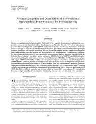
Validation•Testing the Sanger protocol using the GMD controls gives asensitivity of 98% (47/48); the only mutation not detected was80.1G>A•Two main factors affecting detection capability are the GC contentof the fragment and the position of the mutation in the fragment
Conclusions• We have developed an ampilfication system that allows:• Single PCR annealing temperature• Flexible fragment labelling• Reduced primer cost• Simple and informative optimisation• BRCA1 & 2 screen set up and operational (79 fragments)• Marfans screen (FBN1) in optimisation (~60 fragments)• Next targets HNPCC and NOTCH1
AcknowledgementsCSCE• Chris Mattocks NGRL (Wessex)• Helen Davies Cancer Genome group, Sanger institute• Nick Owen NGRL (Wessex)Generic and disease specific mutation controls• Helen White NGRL (Wessex)• Vicky Hall NGRL (Wessex)• Gemma Potts NGRL (Wessex)HTSF• Julie Sillibourne• Tracey Merrifield• AnneMarie Coupe• Alison Skinner• Stacey Sandell