dental biometrics used in forensic science - Call for papers= Asian ...
dental biometrics used in forensic science - Call for papers= Asian ...
dental biometrics used in forensic science - Call for papers= Asian ...
Create successful ePaper yourself
Turn your PDF publications into a flip-book with our unique Google optimized e-Paper software.
JERS/Vol.III/ Issue I/January-March, 2012/26-29<br />
Journal of Eng<strong>in</strong>eer<strong>in</strong>g Research and Studies E-ISSN0976-7916<br />
Research Article<br />
DENTAL BIOMETRICS USED IN FORENSIC SCIENCE<br />
Shubhangi Jadhav 1 , Revati Shriram 2<br />
Address <strong>for</strong> Correspondence<br />
1 Dept. of Instrumentation and Control, D Y Patil College of Eng<strong>in</strong>eer<strong>in</strong>g, Pimpri, Pune, INDIA<br />
2 Cumm<strong>in</strong>s College of Eng<strong>in</strong>eer<strong>in</strong>g <strong>for</strong> Women, Karvenagar, Pune, Maharashtra, INDIA<br />
ABSTRACT<br />
Dental <strong>biometrics</strong> <strong>used</strong> <strong>in</strong> <strong><strong>for</strong>ensic</strong> <strong>science</strong> <strong>for</strong> human identification. It utilizes <strong>dental</strong> radiographs. This radiograph provides<br />
<strong>in</strong><strong>for</strong>mation related to teeth shape, teeth contour and relative position of neighbor<strong>in</strong>g teeth, also it gives shapes of <strong>dental</strong><br />
work like crowns, fill<strong>in</strong>g & bridges etc. This paper <strong>in</strong>cludes different method <strong>used</strong> <strong>for</strong> <strong>dental</strong> <strong>biometrics</strong> and related<br />
<strong>in</strong><strong>for</strong>mation. Dental <strong>biometrics</strong> requires antemortem (AM) and postmortem (PM) radiographs <strong>for</strong> f<strong>in</strong>d<strong>in</strong>g unidentified<br />
subject. Dental <strong>biometrics</strong> hav<strong>in</strong>g three stages: Preprocess<strong>in</strong>g and segmentation of radiographs, Contour extraction or <strong>dental</strong><br />
work extraction, Atlas registration and match<strong>in</strong>g. Segmentation can be done by various methods that are mentioned <strong>in</strong> this<br />
paper. Contour or shape of teeth and <strong>dental</strong> work can be extracted by us<strong>in</strong>g active contour model (ACM) or active shape<br />
model (ASM) methods. Atlas registration is the method <strong>used</strong> <strong>for</strong> label<strong>in</strong>g to teeth, which will help <strong>in</strong> the match<strong>in</strong>g stage.<br />
Match<strong>in</strong>g of AM radiograph with PM radiograph can be done by us<strong>in</strong>g algorithms.<br />
KEYWORDS Biometrics, Dental Radiograph, Forensic Science, Teeth contour, Dental work.<br />
1. INTRODUCTION<br />
Determ<strong>in</strong><strong>in</strong>g identity of an <strong>in</strong>dividual is becom<strong>in</strong>g<br />
very important now days. For this purpose biometric<br />
system is <strong>used</strong>. A biometric is a measurable physical<br />
characteristic which are reliable than a password.<br />
Many biometric systems are employed which work<br />
on the basis of image analysis. “Biometrics” is a<br />
general term <strong>used</strong> alternatively to describe a<br />
characteristic or process.<br />
The biometric systems are divided <strong>in</strong>to two<br />
categories as: behavioral <strong>biometrics</strong> and<br />
physiological <strong>biometrics</strong>. In behavioral biometric<br />
systems a person is identified based on how he<br />
per<strong>for</strong>ms someth<strong>in</strong>g. The most popular technique<br />
under this category is the voice verification<br />
technique. Here user has to actively participate <strong>in</strong> the<br />
process of identification i.e. he needs to per<strong>for</strong>m<br />
some action <strong>in</strong> front of the mach<strong>in</strong>e. Other examples<br />
of physiological biometric characteristic are mak<strong>in</strong>g a<br />
signature, walk<strong>in</strong>g, typ<strong>in</strong>g on a keyboard. In<br />
physiological biometric system a person is identified<br />
based on a unique characteristic of some body organ<br />
of that person. It’s a passive process. All the user has<br />
to do is stand <strong>in</strong> front of the system or at max touch<br />
the sensor of the system with a f<strong>in</strong>ger tip (f<strong>in</strong>ger<br />
biometry) and wait till identification process is<br />
completed. The typical examples of physiological<br />
biometric system are Iris biometry, face biometry,<br />
f<strong>in</strong>ger biometry, ear biometry, <strong>dental</strong> biometry etc.<br />
Dental biometry <strong>used</strong> <strong>in</strong> <strong><strong>for</strong>ensic</strong> identification. This<br />
technique requires ante mortem and postmortem<br />
radiographs. In this both radiographs are segmented<br />
and matched <strong>for</strong> identification of unidentified victim.<br />
There are some various techniques of <strong>dental</strong><br />
biometric that are proposed by different authors as <strong>in</strong><br />
below section.<br />
1.1. Forensic Identification<br />
Forensic identification is <strong>used</strong> <strong>for</strong> suspect<br />
identification and victim identification. Victim<br />
identification is done by physical <strong>biometrics</strong>. Dental<br />
radiograph can be <strong>used</strong> <strong>for</strong> victim identification based<br />
on <strong>dental</strong> evidences. [1]<br />
1.2. Types of Dental Radiographs<br />
There are three types of <strong>dental</strong> radiograph (X-ray):<br />
• Bitew<strong>in</strong>g X-ray- Bitew<strong>in</strong>g x-ray is taken at<br />
rout<strong>in</strong>e check-ups.<br />
• Periapical x-ray- It shows entire tooth, <strong>in</strong>clud<strong>in</strong>g<br />
crown, root and bone.<br />
• Panoramic x-ray- It gives broader overview of<br />
entire dentition. It shows not only teeth also<br />
s<strong>in</strong>us, upper and lower jawbone.<br />
1.3. Dental Biometry<br />
Forensic <strong>dental</strong> <strong>biometrics</strong> <strong>used</strong> to identify<br />
unidentified victims. Automatic <strong><strong>for</strong>ensic</strong><br />
identification utilizes <strong>dental</strong> radiographs [1].<br />
Biometrics can be classified <strong>in</strong> two category based on<br />
characteristic like behavioral and physical. Physical<br />
biometric represents iris, f<strong>in</strong>gerpr<strong>in</strong>t, face recognition<br />
etc. Behavioral biometric represent voice, gait,<br />
signature and all behavioral traits of <strong>in</strong>dividual. [1]<br />
Evaluation of <strong>biometrics</strong> features requires<br />
characteristics such as universality, uniqueness,<br />
permanence, per<strong>for</strong>mance, collectability and<br />
acceptability. [1]<br />
2. DENTAL IDENTIFICATION SYSTEM<br />
The components of Dental identification system<br />
are:<br />
1) Dental Radiograph<br />
2) Radiograph preprocess<strong>in</strong>g and segmentation<br />
3) Contour extraction<br />
4) Atlas registration<br />
5) Match<strong>in</strong>g of Radiograph<br />
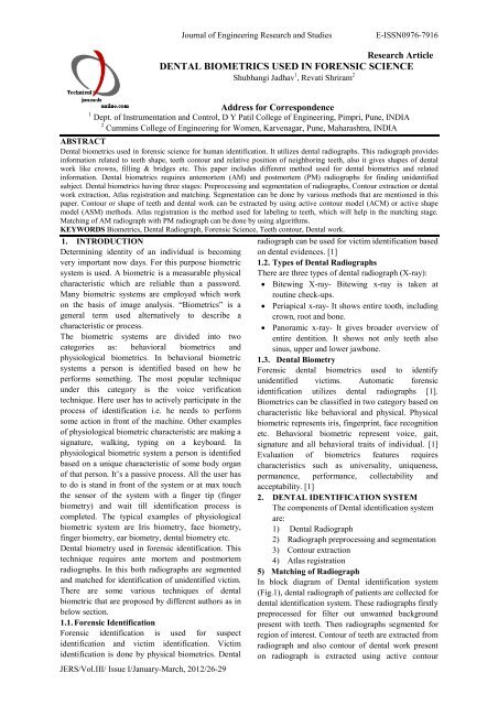
In block diagram of Dental identification system<br />
(Fig.1), <strong>dental</strong> radiograph of patients are collected <strong>for</strong><br />
<strong>dental</strong> identification system. These radiographs firstly<br />
preprocessed <strong>for</strong> filter out unwanted background<br />
present with teeth. Then radiographs segmented <strong>for</strong><br />
region of <strong>in</strong>terest. Contour of teeth are extracted from<br />
radiograph and also contour of <strong>dental</strong> work present<br />
on radiograph is extracted us<strong>in</strong>g active contour
model. Atlas registration is <strong>used</strong> to give label<strong>in</strong>g or<br />
number<strong>in</strong>g to the teeth present <strong>in</strong> jaw, it will help <strong>in</strong><br />
match<strong>in</strong>g stage. Radiograph from database selected<br />
and this radiograph are preprocessed, segmented and<br />
contour extracted from it. Also registration of teeth is<br />
given as per atlas registration. And last stage of this<br />
system is match<strong>in</strong>g, <strong>in</strong> this feature extracted from<br />
these two radiograph matched with each other us<strong>in</strong>g<br />
algorithm and f<strong>in</strong>al result is identification of person<br />
based on match<strong>in</strong>g distance between radiographs[1]<br />
[5].<br />
Radiograph Collection<br />
Radiograph segmentation<br />
Match<strong>in</strong>g tooth<br />
contour<br />
Contour Extraction<br />
Dental work Extraction<br />
Atlas Registration<br />
Match<strong>in</strong>g of Radiograph<br />
Fusion<br />
Subject Identification<br />
Fig.1 Block diagram of Dental<br />
Identification System<br />
3. HUMAN IDENTIFIACTION BASED ON<br />
DENTAL RESTORATIONS<br />
Preprocess<strong>in</strong>g and Segmentation:<br />
Dental radiograph <strong>in</strong>itially converted <strong>in</strong>to gray scale<br />
image. Region of <strong>in</strong>terest (ROI) are decided on<br />
radiograph. Algorithm is <strong>used</strong> to determ<strong>in</strong>ed gray<br />
threshold value of ROI. Then histogram is prepared<br />
<strong>for</strong> the grayscale threshold. To smooth the histogram<br />
filter is <strong>used</strong> to filter out unwanted <strong>in</strong>tensities from<br />
image .After this gray image converted <strong>in</strong>to b<strong>in</strong>ary<br />
image. Active contour model (Snake method) is <strong>used</strong><br />
<strong>for</strong> segmentation of image [6] [15].<br />
Dental Code:<br />
Dental code is <strong>used</strong> to locate teeth, size of teeth and<br />
distance between DR or teeth.<br />
Dental Atlas registration:<br />
Method or code is developed to locate teeth this is<br />
known as <strong>dental</strong> atlas registration. In this we can give<br />
number<strong>in</strong>g to teeth from left to right of jaw and also<br />
we can differentiate between upper jaw and lower<br />
jaw. As per [2], HMM and Markov model was <strong>used</strong><br />
JERS/Vol.III/ Issue I/January-March, 2012/26-29<br />
Journal of Eng<strong>in</strong>eer<strong>in</strong>g Research and Studies E-ISSN0976-7916<br />
Match<strong>in</strong>g <strong>dental</strong><br />
work<br />
<strong>for</strong> this registration purpose. This registration will<br />
help <strong>in</strong> match<strong>in</strong>g stage.<br />
Size of DR:<br />
The method uses to resize DR <strong>in</strong> same size. It means<br />
amount of pixels should be same <strong>for</strong> all DR. So it<br />
will help <strong>in</strong> match<strong>in</strong>g.<br />
Distance between DR:<br />
This is more useful <strong>in</strong> match<strong>in</strong>g stage to create more<br />
sensitive algorithm of match<strong>in</strong>g. In this distance of<br />
neighbor<strong>in</strong>g teeth is calculated.<br />
Match<strong>in</strong>g:<br />
After creation of <strong>dental</strong> code (DC) ,this DC will<br />
match to the DC of database radiograph and distance<br />
between this two DCs calculated. So f<strong>in</strong>ally we<br />
received match<strong>in</strong>g percentage between the AM and<br />
PM radiograph. Based on result subject get identified<br />
[6] [15].<br />
2.1 Nomir & Adlab-Mottaleb<br />
In [2], Nomir & Adlab-Mottaleb <strong>in</strong>troduce a fully<br />
automated approach <strong>for</strong> <strong>dental</strong> x-ray images. The<br />
technique depends on apply<strong>in</strong>g the follow<strong>in</strong>g stages:<br />
iterative threshold to divide the image <strong>in</strong>to two parts<br />
teeth & background, adaptive threshold <strong>in</strong> order to<br />
<strong>in</strong>crease the accuracy and remove teeth <strong>in</strong>terfer<strong>in</strong>g,<br />
horizontal <strong>in</strong>tegral part <strong>in</strong> order to separate the upper<br />
jaw and lower jaw, f<strong>in</strong>al, vertical <strong>in</strong>tegral projection<br />
<strong>in</strong> order to separate <strong>in</strong>dividual tooth.<br />
3.2 Eyad Haj Said, Diaa Eld<strong>in</strong> M Nassar, Gamal<br />
Fabry & Hany Ammar<br />
In [3], Eyad Haj Said, Diaa Eld<strong>in</strong> M Nassar, Gamal<br />
Fabry & Hany Ammar presented method of teeth<br />
segmentation us<strong>in</strong>g mathematical morphology<br />
approach offers fully automated teeth segmentation<br />
and reduces segmentation error due to <strong>in</strong>herent to that<br />
improve the def<strong>in</strong>ition of teeth versus background<br />
and segmentation per<strong>for</strong>mance.<br />
3.3 Supaporn Kiaths<strong>in</strong>, Adison Leelasantitham,<br />
Kos<strong>in</strong> Chamnogthai & Kohji Higuchi<br />
A match of x-ray teeth films us<strong>in</strong>g image process<strong>in</strong>g<br />
based on special features of teeth by Supaporn<br />
Kiaths<strong>in</strong>, Adison Leelasantitham, Kos<strong>in</strong><br />
Chamnogthai & Kohji Higuchi [4]. This method can<br />
help dentist to match simply pair of teeth us<strong>in</strong>g<br />
special features present on teeth film. This presents<br />
some steps: teeth’s picture is scanned and adjusted by<br />
scanner & computer, converted <strong>in</strong>to b<strong>in</strong>ary code, and<br />
decoded <strong>in</strong> cha<strong>in</strong> code (Direction code)<br />
3.4 Hong Chen & A.K. Ja<strong>in</strong><br />
Dental biometry is <strong>used</strong> to identify human. In [5],<br />
Hong Chen & A.K. Ja<strong>in</strong> <strong>in</strong>troduced <strong>dental</strong> <strong>biometrics</strong><br />
us<strong>in</strong>g active contour extraction model (ACM). In this<br />
they proposed a new dynamic energy term i.e.<br />
directional snake to extract contours of teeth. As per<br />
this paper traditional snake cannot able to<br />
discrim<strong>in</strong>ate edges of multiple adjacent objects. So<br />
there can be presence of overlapp<strong>in</strong>g images. To<br />
remove this problem Hong Chen & A.K. Ja<strong>in</strong> utilized<br />
direction gradients. The contour extraction process<br />
hav<strong>in</strong>g three steps: <strong>in</strong>itialization- In this gum l<strong>in</strong>e is
<strong>used</strong> to separate the crown and roots of teeth <strong>for</strong> the<br />
snake <strong>in</strong>itialization, convergence of Gradient, f<strong>in</strong>e<br />
adjustment<br />
Hong Chen & A.K. Ja<strong>in</strong> [6] presented Dental<br />
Biometrics: Alignment and Match<strong>in</strong>g of Dental<br />
Radiographs. This proposed system has ma<strong>in</strong> two<br />
stages: feature extraction, match<strong>in</strong>g.<br />
In this to extract contours of <strong>dental</strong> work the <strong>in</strong>tensity<br />
histogram of the tooth image is automated with the<br />
mixture of Gaussian model.<br />
In the match<strong>in</strong>g stage three steps given: Tooth level<br />
match<strong>in</strong>g, tooth contours are matched us<strong>in</strong>g a shape<br />
registration method, and the <strong>dental</strong> work is matched<br />
on overlapp<strong>in</strong>g areas.<br />
Distance between postmortem and ante mortem<br />
radiographs provide candidates identities to estimate<br />
subject identification.<br />
Hofer M & Marana<br />
In [6], Hofer M & Marana AN presented Dental<br />
<strong>biometrics</strong> <strong>for</strong> human identification based on <strong>dental</strong><br />
work. Dental <strong>biometrics</strong> <strong>used</strong> <strong>for</strong> <strong><strong>for</strong>ensic</strong><br />
identification, <strong>in</strong> this <strong>dental</strong> work <strong>in</strong><strong>for</strong>mation is <strong>used</strong><br />
to identify human. This paper three process<strong>in</strong>g steps<br />
were given as per follow<strong>in</strong>g: Segmentation (Feature<br />
Extraction), creation of <strong>dental</strong> code, match<strong>in</strong>g<br />
Primarily segmentation is done by detect<strong>in</strong>g<br />
threshold and f<strong>in</strong>ally snake (active contour model)<br />
algorithm <strong>used</strong>. Dental code is prepared as per the<br />
position, size of <strong>dental</strong> work and neighbor<strong>in</strong>g <strong>dental</strong><br />
work. Match<strong>in</strong>g done by edit<strong>in</strong>g distance between<br />
<strong>dental</strong> works.<br />
4. DISCUSSION<br />
Dental biometry is <strong>used</strong> <strong>in</strong> <strong><strong>for</strong>ensic</strong> identification.<br />
Some other methods are also present <strong>for</strong> <strong><strong>for</strong>ensic</strong><br />
identification. But it is more useful when there is no<br />
any other identity like iris [11], face, f<strong>in</strong>gerpr<strong>in</strong>ts [16]<br />
etc of victim rema<strong>in</strong><strong>in</strong>g. Dental atlas can rema<strong>in</strong> <strong>in</strong><br />
well condition <strong>for</strong> some time period after the death of<br />
person so by tak<strong>in</strong>g radiograph at the time of PM and<br />
match<strong>in</strong>g with it to database hav<strong>in</strong>g with <strong><strong>for</strong>ensic</strong><br />
department it can give you identification of victim or<br />
subject. Also <strong>dental</strong> biometry can be <strong>used</strong> <strong>in</strong> human<br />
identification system. Also match<strong>in</strong>g of x-ray films<br />
which is the part of <strong>dental</strong> biometry can help dentist<br />
to their cl<strong>in</strong>ical diagnosis [1] [5]. Dental biometry<br />
requires AM and PM radiograph only <strong>for</strong><br />
identification. Dental biometry cannot require<br />
additional <strong>in</strong><strong>for</strong>mation. DNA requires additional<br />
<strong>in</strong><strong>for</strong>mation of subject <strong>for</strong> identification. Dental<br />
biometry comes under physical biometry so it is<br />
easier to measure quantitatively as compared to<br />
behavioral biometry. Hand ve<strong>in</strong> pattern and ret<strong>in</strong>a<br />
scan are physical biometry but it hav<strong>in</strong>g low<br />
acceptability because it may reveal person’s health<br />
status and it is <strong>in</strong>vasive method. In disasters, victim<br />
or subject’s physical characteristics like iris, face,<br />
f<strong>in</strong>gerpr<strong>in</strong>ts not available that time biometry related<br />
to these characteristic not worked. So there is then<br />
need of <strong>dental</strong> biometry because <strong>dental</strong> jaw<br />
JERS/Vol.III/ Issue I/January-March, 2012/26-29<br />
Journal of Eng<strong>in</strong>eer<strong>in</strong>g Research and Studies E-ISSN0976-7916<br />
rema<strong>in</strong><strong>in</strong>g <strong>in</strong> well condition <strong>in</strong> massive disasters.<br />
Dental biometry is <strong>used</strong> <strong>for</strong> <strong><strong>for</strong>ensic</strong> identification.<br />
Also match<strong>in</strong>g of <strong>dental</strong> radiograph can be <strong>used</strong> <strong>in</strong><br />
cl<strong>in</strong>ical diagnosis <strong><strong>for</strong>ensic</strong> medic<strong>in</strong>e (Forensic<br />
Dentistry) [1].<br />
Dental biometry hav<strong>in</strong>g some limitation based on<br />
below mentioned po<strong>in</strong>ts so <strong>in</strong> some cases it is not<br />
good choice <strong>for</strong> identification:<br />
• Positive identification: AM and PM radiographs<br />
should have sufficient data to match, with no any<br />
discrepancy like miss<strong>in</strong>g tooth.<br />
• Possible identification: If consistent features are<br />
present <strong>in</strong> AM or PM radiograph but if there is<br />
problem <strong>in</strong> quality of either AM or PM<br />
radiograph then this may create no positive<br />
identification.<br />
• Insufficient evidence: Sufficient <strong>in</strong><strong>for</strong>mation is<br />
not available <strong>for</strong> the basis of conclusion.<br />
• Exclusion: The radiograph <strong>in</strong><strong>for</strong>mation is<br />
<strong>in</strong>consistent [1].<br />
5. CONCLUSION<br />
Segmentation of Radiographs: A fast march<strong>in</strong>g based<br />
algorithm is <strong>used</strong> to segment radiographs <strong>in</strong>to<br />
homogeneous regions so that each region conta<strong>in</strong>s<br />
only one tooth [9]. This method utilizes the<br />
characteristics of <strong>dental</strong> radiographs <strong>in</strong> that soft tissue<br />
and bone have different pixel <strong>in</strong>tensity. This method<br />
can be extended to analyze other types of<br />
radiographs. ASM-Based Tooth Contour Extraction:<br />
An active shape model (ASM) is <strong>used</strong> to extract tooth<br />
contours. The traditional ASMs have some problems<br />
that are solved by new ASM. Extraction of Contours<br />
of Dental Work: An anisotropic diffusion <strong>used</strong> to<br />
enhance the images and extract the contours of <strong>dental</strong><br />
work with a reasonable assumption that pixel values<br />
of <strong>dental</strong> restoration follow a Gaussian distribution<br />
[1] [2] [3] [5] [10] [17]. Hybrid HMM/SVM Model<br />
<strong>for</strong> Representation of Dental Atlas: Tooth states <strong>in</strong><br />
the HMM comb<strong>in</strong>ed with SVMs model the shape of<br />
teeth, and distance states <strong>in</strong> the HMM model the<br />
<strong>in</strong>ter-teeth distance ca<strong>used</strong> by miss<strong>in</strong>g teeth.<br />
Construction of Observation sequences: An<br />
observation sequence is an alternat<strong>in</strong>g sequence of<br />
tooth shapes and distances between successive teeth.<br />
To compensate <strong>for</strong> the difference between image<br />
resolutions of AM and PM images, the tooth shapes<br />
are normalized to affixed width, and the distances<br />
between teeth are regularized with the width of<br />
neighbor<strong>in</strong>g teeth [1] [7] [17]. Location of DW<br />
(Dental Work) can be f<strong>in</strong>d<strong>in</strong>g out by implement<strong>in</strong>g<br />
algorithm. In this all DWs <strong>in</strong> the DWM from left to<br />
right based on the center of mass po<strong>in</strong>t of each<br />
<strong>in</strong>dividual DW sorted. This is requir<strong>in</strong>g creat<strong>in</strong>g<br />
<strong>dental</strong> code. The upper jaw and lower jaw is<br />
identified by the valley location with respect to upper<br />
jaw and lower jaw [6] [15].<br />
Match<strong>in</strong>g of tooth contours: Tooth contours are open<br />
curves. To match tooth contours, correspondence<br />
between the tooth contours has to be established. An
iterative procedure is present to align tooth contours<br />
and compute distances based on the correspond<strong>in</strong>g<br />
segments of the two curves. Algorithm is presented to<br />
align <strong>dental</strong> work and by us<strong>in</strong>g we can compute the<br />
distance between two images us<strong>in</strong>g a metric of<br />
overlapp<strong>in</strong>g pixels. Dental restorations are not always<br />
present <strong>in</strong> the <strong>dental</strong> radiographs. To fuse the<br />
match<strong>in</strong>g distances between tooth contours and<br />
<strong>dental</strong> restoration contours, a scheme presented based<br />
on posterior probability. A scheme <strong>used</strong> to compute<br />
the distance between an image and a subject and the<br />
distance between two subjects [1].<br />
REFERENCES<br />
1. Hong Chen, “Automatic Forensic Identification<br />
based on <strong>dental</strong> radiographs”, Michigan State<br />
University, 2007.<br />
2. O. Nomir and M. Abdel-Mottaleb, “Human<br />
Identification from Dental X-Ray Images Based<br />
on the Shape and Appearance of the Teeth”,<br />
IEEE Transactions on In<strong>for</strong>mation Forensics and<br />
Security, vol. 2, Issue 2, pp. 188 – 197, 2007.<br />
3. E.H. Said; D.E.M. Nassar; G. Fahmy and H.H.<br />
Ammar, “ Teeth segmentation <strong>in</strong> digitized <strong>dental</strong><br />
X-ray films us<strong>in</strong>g mathematical morphology”,<br />
IEEE Transactions on In<strong>for</strong>mation Forensics and<br />
Security, vol. 1, Issue 2, pp. 178 – 189, 2006<br />
4. Supaporn Kiattism, Adisorn Leelasantitham,<br />
Kos<strong>in</strong> Chamnongthai and Kohji Higuchi, “ A<br />
match of x-ray teeth films us<strong>in</strong>g image<br />
process<strong>in</strong>g based on special features of teeth”,<br />
SICE Annual conference 2008, August 20-<br />
22,2008, The University Electro-<br />
Communications, Japan.<br />
5. Hong Chen and A.K. Ja<strong>in</strong>, “Dental <strong>biometrics</strong>:<br />
alignment and match<strong>in</strong>g of <strong>dental</strong> radiographs”,<br />
IEEE Transactions on Pattern Analysis and<br />
Mach<strong>in</strong>e Intelligence, vol. 27, Issue 8, pp. 1319 –<br />
1326, 2005.<br />
6. Hofer M and Marana AN, “Dental <strong>biometrics</strong> :<br />
Human identification based on <strong>dental</strong> work<br />
<strong>in</strong><strong>for</strong>mation” eHealth2008 – Medical In<strong>for</strong>matics<br />
meets eHealth. Tagungsband der eHealth2008 –<br />
Wien, 29.-30. Mai 2008<br />
7. Andrea Castellani, Debora Botturi, Manuele<br />
Bicago, and Paolo Fior<strong>in</strong>i “ Hybrid HMM/SVM<br />
model <strong>for</strong> the analysis and segmentation of<br />
teleoperation tasks”, In Proc. IEEE International<br />
Conference on Robotics & Automation, page<br />
2918-2923,New Orleans, LA, April 2004.<br />
8. G. Fahmy, D Nassar, E. Haj-Said, H. Chen, O.<br />
Nomir, J. Zhou, R. Howell, H.H. Ammar, M.<br />
Abdel-Mottaleb and A. K. Ja<strong>in</strong> “Towards an<br />
automated <strong>dental</strong> identification system (ADIS).In<br />
Proc. ICBA(International Conference on<br />
Biometric Authentication),volume LNCS 3072,<br />
pages 789–796, Hong Kong, July 2004.<br />
9. Hong Chen and Carol Novak, “ Segmentation of<br />
hand radiographs us<strong>in</strong>g fast march<strong>in</strong>g methods”,<br />
In Proc. SPIE conference on Medical Imag<strong>in</strong>g,<br />
volume 6144, pages 68-76,San Diego, Cali<strong>for</strong>nia,<br />
2006.<br />
10. Hao Huang and Jughua Wang, “ Anistropic<br />
diffusion <strong>for</strong> object segmentation”, In Proc.<br />
IEEE International conference on systems, Man<br />
and Cybernetics, volume3 pages 1563–1567,<br />
Nashville, TN, October 2000.<br />
11. John Daughman, “How Iris Recognition Works”,<br />
IEEE Transactions on circuits and systems <strong>for</strong><br />
video technology, Vol.14, January 2004.<br />
JERS/Vol.III/ Issue I/January-March, 2012/26-29<br />
Journal of Eng<strong>in</strong>eer<strong>in</strong>g Research and Studies E-ISSN0976-7916<br />
12. A.K. Ja<strong>in</strong> and H. Chen, “Match<strong>in</strong>g of Dental Xray<br />
Images <strong>for</strong> Human Identification”, Pattern<br />
Recognition, vol37, no.7, pp.1519-1532, 2004.<br />
13. H. Chen and A.K. Ja<strong>in</strong>, “Tooth Contour<br />
Extraction <strong>for</strong> Match<strong>in</strong>g Dental Radiographs”,<br />
Proc. 17 th <strong>in</strong>ternational conference. Pattern<br />
Recognition, vol.III, pp.522-525, Aug2004.<br />
14. P.Perona and J.Malik, Scale-Space and Edge<br />
Detection Us<strong>in</strong>g Anistropic Diffusion, IEEE<br />
Trans.Pattern Analysis and Mach<strong>in</strong>e Intelligence,<br />
Vol.12, no.7, pp. 629-639, July 1990.<br />
15. Michael Hofer, Aparecido Nilceu, “ Marana2<br />
Dental Biometrics: Human Identification Based<br />
on Dental Work In<strong>for</strong>mation”, Car<strong>in</strong>thia<br />
University of Applied Sciences, Klagenfurt,<br />
Austria UNESP, Faculdade de C<strong>in</strong>cias,<br />
Departmento de Computao, Bauru SP, Brazil<br />
16. R.Khanna and W. Shen, “Automated f<strong>in</strong>gerpr<strong>in</strong>t<br />
identification system (AFIS) benchmark<strong>in</strong>g us<strong>in</strong>g<br />
the National Institute of Standards and<br />
Technology (NIST) special database4”, <strong>in</strong> Proc.<br />
Inst. Elect.Electron. Eng. 28 th Annu. Int.<br />
Carnahan Conf., Albuquerque, NM, Oct.12-14,<br />
1994, pp. 188–194.<br />
17. Juri Lember, Kristi Kuljus and Alexe<br />
Koloydenco, “Theory of segmentation”,<br />
University Tartu, Swedish University of<br />
Agricultural Sciences, Royal Holloway,<br />
University of London, Estonia, Sweden, UK.<br />
18. Anil K. Ja<strong>in</strong>, Hong Chen and Silviu M<strong>in</strong>ut,<br />
“Dental Biometrics: Human Identification us<strong>in</strong>g<br />
<strong>dental</strong> radiographs”, Department of Computer<br />
Science, Michigan State University, East<br />
Lans<strong>in</strong>g, MI 48824.<br />
19. Marcelo Cavalcanti, Axel Ruprecht, William<br />
Johnson, Southard Thomas, Jane Jakobsen, and<br />
Sao Paulo, “Oral and maxillofacial radiology”,<br />
Oral Surg Oral Med Oral Pathol, Volume 88:<br />
pages 353.357, 1999.<br />
20. Greth Jonasson and Gudrun Bankvall,<br />
“Estimation of skeletal bone m<strong>in</strong>eral density by<br />
means of the trabecular pattern of the alveolar<br />
bone, its <strong>in</strong>ter<strong>dental</strong> thickness, and the bone mass<br />
of the mandible”, Oral Surg Oral Med Oral<br />
Pathol Oral Radiol Endod, Volume92:pages 346-<br />
352 Ba2001.


