Prevalence of oral lesions among Saudi dental ... - BioMedSearch
Prevalence of oral lesions among Saudi dental ... - BioMedSearch
Prevalence of oral lesions among Saudi dental ... - BioMedSearch

Create successful ePaper yourself
Turn your PDF publications into a flip-book with our unique Google optimized e-Paper software.
original article<br />
<strong>Prevalence</strong> <strong>of</strong> <strong>oral</strong> <strong>lesions</strong> <strong>among</strong> <strong>Saudi</strong> <strong>dental</strong><br />
patients<br />
Azizah Al-Mobeeriek, Abdullah M. AlDosari<br />
From the Department <strong>of</strong> Maxill<strong>of</strong>acial Surgery and Diagnostic Sciences, College <strong>of</strong> Dentistry, King Saud University, Riyadh, <strong>Saudi</strong> Arabia<br />
Correspondence and reprints: Azizah F. Al-Mobeeriek, MD •Department <strong>of</strong> Maxill<strong>of</strong>acial Surgery and Diagnostic Sciences, College <strong>of</strong> Dentistry,<br />
King Saud University, PO Box 231189, Riyadh 11321, <strong>Saudi</strong> Arabia • T: +966-1-478-4524 ext 307 F: +966-1-467-8719 • azizafm2001@yahoo.<br />
com • Accepted for publication August 2008<br />
Ann <strong>Saudi</strong> Med 2009; 29(5): 365-368<br />
BACKGROUND AND OBJECTIVES: Few studies have been conducted in the <strong>Saudi</strong> population on <strong>oral</strong> mucosal<br />
<strong>lesions</strong>. The purpose <strong>of</strong> this study was to evaluate the type and extent <strong>of</strong> <strong>oral</strong> <strong>lesions</strong> in a study <strong>among</strong> <strong>dental</strong><br />
patients at a college <strong>of</strong> dentistry in <strong>Saudi</strong> Arabia.<br />
PATIENTS AND METHODS: Over a 3-year period, 2552 <strong>dental</strong> outpatients were interviewed and investigated<br />
clinically for the presence <strong>of</strong> <strong>oral</strong> mucosal conditions. A thorough <strong>oral</strong> clinical examination was performed, including<br />
a radiographic examination. The diagnosis was confirmed histopathologically when necessary.<br />
RESULTS: Of 383 (15.0%) patients found to have <strong>oral</strong> mucosal <strong>lesions</strong>, females constituted 57.7% (n=221) and<br />
males 42.3% (n=162). The age range <strong>of</strong> the patients was between 15 to 73 years with a mean age <strong>of</strong> 38.2 years.<br />
The most commonly affected age group was 31 to 40 years, which comprised 21.4% (n=82) <strong>of</strong> all affected individuals.<br />
The least affected age group were individuals older than 61 years. The most common lesion was Fordyce<br />
granules (3.8%; n=98), followed by leukoedema (3.4%; n=86) and traumatic <strong>lesions</strong> (ulcer, erosion) in 1.9%<br />
(n=48). Tongue abnormalities were present in 4.0% (n=101) <strong>of</strong> all <strong>oral</strong> conditions observed, ranging from 1.4%<br />
(n=36) for fissured tongue to 0.1% (n=2) for bifid tongue. Other findings detected were torous platinus (1.3%;<br />
n=34), mandibular tori (0.1%; n=2) aphthous ulcer (0.4%; n=10), herpes simplex (0.3%; n=7), frictional hyperkeratosis<br />
(0.9%; n=23), melanosis (0.6%; n=14), lichen planus (0.3%; n=9) and nicotinic stomatitis (0.5%; n=13).<br />
CONCLUSION: The findings <strong>of</strong> this study provide information on the types and prevalence <strong>of</strong> <strong>oral</strong> <strong>lesions</strong><br />
<strong>among</strong> <strong>Saudi</strong> <strong>dental</strong> patients. This provides baseline data for future studies about the prevalence <strong>of</strong> <strong>oral</strong> <strong>lesions</strong><br />
in the general population.<br />
Few studies have been conducted in the <strong>Saudi</strong><br />
population on <strong>oral</strong> mucosal <strong>lesions</strong>. An 8-year<br />
retrospective study using biopsied specimens<br />
found that focal fibrous hyperplasia (FFH), pyogenic<br />
granuloma (PG), peripheral giant cell granuloma<br />
(PGCG) and peripheral ossifying fibroma (POF)<br />
constitute 9% <strong>of</strong> all biopsied <strong>lesions</strong>. 1 A 5-year (1985-<br />
1990) survey in <strong>Saudi</strong> Arabia indicated that 64.6% <strong>of</strong><br />
biopsied <strong>lesions</strong> were benign tumors, hyperplasia and<br />
granulomas, 20.3% were cysts and cyst-like <strong>lesions</strong><br />
and 4.8% were other mucous membrane <strong>lesions</strong>. 2<br />
The prevalence <strong>of</strong> leukoplakia was studied in relationship<br />
to tobacco habits and was found to be 11.4%<br />
in the Gizan area <strong>of</strong> <strong>Saudi</strong> Arabia. 3,4 The <strong>oral</strong> cavity<br />
was ranked as one <strong>of</strong> the most common ten malignancies<br />
from 1975-2006. 5 The tongue was the most<br />
affected site (30.1%), mostly on the lateral borders <strong>of</strong><br />
the anterior two-thirds, followed by the oropharynx,<br />
lips, cheeks, gingiva and floor <strong>of</strong> the mouth, palate<br />
and salivary gland. 6 Oral health survey data are essential<br />
for proper health planning programs. In addition,<br />
most <strong>of</strong> the conducted studies were <strong>of</strong> tumors<br />
or ulcers and did not include all <strong>oral</strong> <strong>lesions</strong> in adult<br />
populations. The pattern <strong>of</strong> disease is also changing<br />
due to increasing awareness, changes in lifestyle and<br />
increasing interest in <strong>oral</strong> health. In <strong>Saudi</strong> Arabia<br />
there is a great need for clinical studies to establish<br />
baseline data on the prevalence <strong>of</strong> <strong>oral</strong> <strong>lesions</strong>. The<br />
aim <strong>of</strong> this study was to determine the type and the<br />
prevalence <strong>of</strong> <strong>oral</strong> <strong>lesions</strong> <strong>among</strong> patients seeking<br />
<strong>dental</strong> care.<br />
PATIENTS AND METHODS<br />
This study was carried out on adult <strong>Saudi</strong> <strong>dental</strong> outpatients<br />
who attended the Oral Diagnostic Clinic at<br />
the College <strong>of</strong> Dentistry, King Saud University, Riyadh,<br />
Ann <strong>Saudi</strong> Med 29(5) September-October 2009 www.saudiannals.net 365
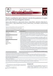
Percentage (%)<br />
366<br />
25<br />
20<br />
15<br />
10<br />
5<br />
0<br />
original article<br />
<strong>Saudi</strong> Arabia, for an <strong>oral</strong> examination and a <strong>dental</strong><br />
treatment plan. The study sample included adult subjects<br />
who were older than 15 years <strong>of</strong> age. The college<br />
is an open public facility and referral hospital. A total<br />
<strong>of</strong> 2552 patients were interviewed and clinically investigated<br />
for the presence <strong>of</strong> <strong>oral</strong> <strong>lesions</strong> from June 2002-<br />
to December 2005. History taking and a thorough <strong>oral</strong><br />
clinical examination was performed, including a radiographic<br />
examination. The type and distribution <strong>of</strong> the<br />
<strong>oral</strong> mucosal conditions was recorded. An interview<br />
was conducted to collect information. The examination<br />
was performed by a single examiner. Data were statistically<br />
analyzed using SPSS (SPSS, Inc. Chicago, IL).<br />
RESULTS<br />
Among the 2552 patients, only 383 patients (15.0%)<br />
had <strong>oral</strong> <strong>lesions</strong>. Females constituted 57.7% (n=221)<br />
and males 42.3% (n=162). The age range <strong>of</strong> the patients<br />
was between 15 to 73 years. The mean age <strong>of</strong> the sample<br />
group was 38.2 years. The mean age for females was<br />
33.0 years and 47.1 years for males. Of the total sample,<br />
the most commonly affected age was between 31 to<br />
40 years (21.4%), followed by 15 to 20 years (18.5%),<br />
and 21 to 30 years (17.8%) (Figure 1). Twenty-four patients<br />
(0.9%) admitted smoking habits and 196 patients<br />
(7.7%) had a systemic disease, but there were no differences<br />
between smokers and non-smokers, or between<br />
healthy people and those with systemic diseases.<br />
Oral <strong>lesions</strong> were more prevalent <strong>among</strong> females<br />
than males (Table 1). Fordyce granules were observed<br />
in 3.84% (n=98) on the buccal and labial mucosa, and<br />
were significantly more common in females. Females<br />
15-20 21-30 31-40 41-50 51-60 61-70 >70 Missing<br />
Age (years)<br />
data<br />
Figure 1. <strong>Prevalence</strong> <strong>of</strong> <strong>oral</strong> conditions by age range and sex (n=383).<br />
Male<br />
Female<br />
Total<br />
ORAL LESIONS AMONG SAUDIS<br />
showed a higher prevalence than males, accounting for<br />
two-thirds <strong>of</strong> the affected individuals (n=24), but the<br />
difference was not statistically significant. Both torous<br />
platinus and mandibular tori wre more common in females,<br />
but the difference was not statistically significant.<br />
Other <strong>lesions</strong> were significantly more common <strong>among</strong><br />
females: aphthous ulcer and herpes simplex. On the<br />
other hand, frictional hyperkeratosis was more common<br />
<strong>among</strong> males than females, but the difference was<br />
not statistically significant. Other pathologies detected<br />
were excessive melanin pigmentation (melanosis).<br />
Females showed a higher incidence than males, but the<br />
difference was not statistically significant. Both lichen<br />
planis and nicotinic stomatitis were significantly higher<br />
<strong>among</strong> males.<br />
Tongue abnormalities were present in 3.96%<br />
(n=101) <strong>of</strong> total sample and in 26.4% <strong>of</strong> all <strong>oral</strong> conditions<br />
observed. The prevalence <strong>of</strong> tongue <strong>lesions</strong><br />
was higher <strong>among</strong> females than males, but the difference<br />
was not statistically significant. the most common<br />
tongue condition was fissured tongue, constituting<br />
about 35.6% <strong>of</strong> all tongue conditions. it is also ranked<br />
fourth most common lesion in our population. Hairy<br />
tongue was present in 0.55% <strong>of</strong> the sample (n=14) and<br />
was significantly more common in males than in females.<br />
Tongue tie was also significanlty more common<br />
in females than males. Other tongue <strong>lesions</strong> were lingual<br />
varcosis. Females showed a higher incidence than<br />
males, but the difference was no statistically significant.<br />
Both bifid tonge and glossitis had a low occurrence and<br />
were seen manily <strong>among</strong> females.<br />
DISCUSSION<br />
Oral s<strong>of</strong>t tissue <strong>lesions</strong> present a significant health problem<br />
with a considerable morbidity. Despite its importance,<br />
there are few reports on its prevalence <strong>among</strong> the<br />
<strong>Saudi</strong> population and its association with <strong>oral</strong> habits,<br />
when compared to <strong>dental</strong> caries and periodontal diseases.<br />
In accordance with others, 1,2 our results showed<br />
a higher prevalence <strong>of</strong> <strong>oral</strong> mucosal <strong>lesions</strong> <strong>among</strong> females<br />
(57.7%) and young adults (31-40 years) (21.4%).<br />
Other reports, however, indicated that <strong>oral</strong> <strong>lesions</strong> tend<br />
to increase with age in association with tobacco consumption<br />
and denture use. 7,8 The age <strong>of</strong> the patient is<br />
crucial in patient assessment, treatment planning and<br />
health education.<br />
The present study suggests the distribution pattern<br />
<strong>of</strong> <strong>oral</strong> diseases in <strong>Saudi</strong> Arabia is similar to other countries.<br />
Benign <strong>lesions</strong> are more common in the young and<br />
females while cancerous and precancerous <strong>lesions</strong> are<br />
more common in the elderly. 9,10 Along with other investigations,<br />
1-6,11 it may also shed some light on the pattern<br />
Ann <strong>Saudi</strong> Med 29(5) September-October 2009 www.kfshrc.edu.sa/annals
ORAL LESIONS AMONG SAUDIS<br />
Table 1. Distribution <strong>of</strong> <strong>oral</strong> conditions in <strong>Saudi</strong> <strong>dental</strong> patients (n=2552).<br />
Type<br />
No.<br />
original article<br />
Males Females Total<br />
% <strong>of</strong> total<br />
sample<br />
No.<br />
% <strong>of</strong> total<br />
sample<br />
Ann <strong>Saudi</strong> Med 29(5) September-October 2009 www.saudiannals.net 367<br />
No.<br />
% <strong>of</strong> total<br />
sample<br />
Fordyce granules a 30 1.18 68 2.67 98 3.84<br />
Leukoedema b 53 2.08 33 1.29 86 3.37<br />
Traumatic <strong>lesions</strong><br />
(ulcers, erosions)<br />
14 0.55 34 1.33* 48 1.88<br />
Torous platinus 0 0 34 1.33 34 1.33<br />
Frictional<br />
hyperkeratosis<br />
14 0.55 9 0.35 23 0.90<br />
Melanosis 5 0.195 9 0.35 14 0.55<br />
Nicotinic stomatitis a 11 0.43 2 0.08 13 0.51<br />
Aphthae a 0 0 10 0.39 10 0.39<br />
Lichen planus 7 0.27 2 0.08 9 0.35<br />
Herpes simplex c 0 0 7 0.27 7 0.27<br />
Mandibular tori 0 0 2 0.08 2 0.08<br />
Non-nicotinic stomatitis 2 0.08 2 0.08 4 0.16<br />
Focal fibrous hyperplasia 2 0.08 3 0.12 5 0.195<br />
Tongue <strong>lesions</strong><br />
Fissured tongue 12 0.47 24 0.94 36 1.41<br />
Bifid tongue 0 0 2 0.08 2 0.08<br />
Glossitis 0 0 3 0.12 3 0.12<br />
Scalloped tongue 0 0 8 0.31 8 0.31<br />
Lingual varcosis 2 0.08 8 0.31 10 0.39<br />
Geographic tongue 7 0.27 6 0.24 13 0.51<br />
Hairy tongue c 10 0.39 4 0.16 14 0.55<br />
Tongue tie 2 0.08 12 0.47 15* 0.59<br />
Total tongue <strong>lesions</strong> 33 1.3 67 2.6 101 3.96<br />
Total 171 282 454<br />
*One case missing sex; a P
368<br />
original article ORAL LESIONS AMONG SAUDIS<br />
Traumatic ulcer was the third most common s<strong>of</strong>t<br />
tissue lesion. Females were affected more than males.<br />
It was also found to be one <strong>of</strong> the common s<strong>of</strong>t tissue<br />
<strong>lesions</strong> in Spain, Italy, and Chile elderly and in the institutionalized<br />
elderly in Denmark. 21-24 The main reason<br />
<strong>among</strong> the elderly was poorly constructed dentures. 23-25<br />
As in the to Kenya 13 and in elderly Malaysians, 26 frictional<br />
hyperkeratosis ranked as one <strong>of</strong> the most common<br />
<strong>oral</strong> mucosal <strong>lesions</strong>. This finding supports the results<br />
from the United States <strong>of</strong> America, 27 China 28 and<br />
Bangladesh, 29 where keratotic <strong>lesions</strong> are a common clinical<br />
presentation possibly due to population differences,<br />
particularly in term <strong>of</strong> tobacco habits.<br />
Consistent with data from China, 28 tongue conditions<br />
REFERENCES<br />
1. Narty N, Masadomi H, Al-Gilani M, Al-Mobeerik<br />
A. Localized inflammatory hyperplasia <strong>of</strong> the <strong>oral</strong><br />
cavity: clinico-pathological study <strong>of</strong> 164 cases.<br />
<strong>Saudi</strong> Dent J. 1994 Sep;6(3).<br />
2. Masadomi H, Algilani M, Narty N, Alsaif N. Tumors,<br />
cyst, cyst-like and allied <strong>lesions</strong> <strong>of</strong> the jaws<br />
and <strong>oral</strong> mucosa in Riyadh, KSA. <strong>Saudi</strong> Dent J.<br />
1992 Jan;4(S1). Salem G, Jul R, Schiodt T. Actaodontol-scand.<br />
1984 Feb;42(1):41-5.<br />
3. Salem G. Leukoplakia and tobacco habits in<br />
Gizan, <strong>Saudi</strong> Arabia. <strong>Saudi</strong> Dent J. 1992 May;4(2).<br />
4. Rabadi S. Cancer at Dhahran health center,<br />
<strong>Saudi</strong> Arabia. Ann <strong>Saudi</strong> Med. 1987;7(4).<br />
5. AlDosari AM. Preliminary study <strong>of</strong> <strong>oral</strong> cancer<br />
in <strong>Saudi</strong> Arabia. <strong>Saudi</strong> Med J. 1987;8(5):476-480.<br />
6. Al-Mobeeriek AF. Oral and upper aero-digestive<br />
tract malignancy. A review <strong>of</strong> a five-year experience.<br />
Ann <strong>of</strong> <strong>Saudi</strong> Med. 1998;257-259.<br />
7. Shulman JD, Beach MM, Rivera-Hidalgo F. The<br />
prevalence <strong>of</strong> <strong>oral</strong> mucosal<strong>lesions</strong> in US adults:<br />
data from the Third National Health and Nutrition<br />
Examination Survey, 1988-1994. J Am Dent Assoc.<br />
2004 Sep;135(9):1279-86.<br />
8. Kleinman DV, Swango PA, Pindborg JJ. Epidemiology<br />
<strong>of</strong> <strong>oral</strong> mucosal <strong>lesions</strong> in United States<br />
schoolchildren: 1986-87. Community Dent Oral Epidemiol.<br />
1994 Aug;22(4):243-53.<br />
9. Bouquot JE. Common <strong>oral</strong> <strong>lesions</strong> found during<br />
a mass screening examination. Am Dent Assoc.<br />
1986 Jan;112(1):50-7.<br />
10. Kovac-Kovacic M, Skaleric U. The prevalence<br />
<strong>of</strong> <strong>oral</strong> mucosal <strong>lesions</strong> in a population<br />
in Ljubljana, Slovenia. J Oral Pathol Med. 2000<br />
Aug;29(7):331-5.<br />
11. Mani NJ. Preliminary report on prevalence<br />
<strong>of</strong> <strong>oral</strong> cancer and precancerous <strong>lesions</strong> <strong>among</strong><br />
<strong>dental</strong> patients in <strong>Saudi</strong> Arabia. Community Dent<br />
Oral Epidemiol. 1985;13:247-248.<br />
12. dos Santos PJ, Bessa CF, de Aguiar MC, do<br />
Carmo MA. Cross-sectional study <strong>of</strong> <strong>oral</strong> muco-<br />
sal conditions <strong>among</strong> a central Amazonian Indian<br />
community, Brazil. J Oral Pathol Med. 2004<br />
Jan;33(1):7-12.<br />
13. Macigo FG, Mwaniki DL, Guthua SW. <strong>Prevalence</strong><br />
<strong>of</strong> <strong>oral</strong> mucosal <strong>lesions</strong> in a Kenyan population<br />
with special reference to <strong>oral</strong> leukoplakia.<br />
East Afr Med J. 1995 Dec;72(12):778-82.<br />
14. Axell T. A prevalence study <strong>of</strong> <strong>oral</strong> mucosal<br />
<strong>lesions</strong> in an adult Swedish population. Odontol<br />
Revy. 1976;27(36):1-103.<br />
15. Van Wyk CW, Farman AG, Staz J. Oral health<br />
status <strong>of</strong> institutionalized elderly Cape Coloreds<br />
from the Cape Peninsula <strong>of</strong> South Africa. Community<br />
Dent Oral Epidemiol. 1977 Jul;5(4):179-84.<br />
16. Corbet EF, Holmgren CJ, Phillipsen HP. Oral<br />
mucosal <strong>lesions</strong> in 65-74- year-old Hong Kong<br />
Chinese. Community Dent Oral Epidemiol. 1994<br />
Oct;22(5 Pt 2):392-5.<br />
17. Axell T, Henricsson V. Leukoedema-an epidemiologic<br />
study with special reference to the<br />
influence <strong>of</strong> tobacco habits. Community Dent Oral<br />
Epidemiol. 1981 Jun;9(3):142-6.<br />
18. Zain RB, Razak IA. Association between cigarette<br />
smoking and prevalence <strong>of</strong> <strong>oral</strong> mucosal<br />
<strong>lesions</strong> <strong>among</strong> Malaysian army personnel. Community<br />
Dent Oral Epidemiol. 1989 Jun;17(3):148-9.<br />
19. Gupta PC, Murti PR, Bhonsle RB, Mehta FS,<br />
Pindborg JJ. Effect <strong>of</strong> cessation <strong>of</strong> tobacco use<br />
on the incidence <strong>of</strong> <strong>oral</strong> mucosal <strong>lesions</strong> in a 10yr<br />
follow-up study <strong>of</strong> 12,212 users. Oral Dis. 1995<br />
Mar;1(1):54-8.<br />
20. Reichart PA, Schmidtberg W, Samaranayake<br />
LP, Scheifele C. Betel quid associated <strong>oral</strong><br />
<strong>lesions</strong> and <strong>oral</strong> Candida species in a female<br />
Cambodian cohort. J Oral Pathol Med. 2002<br />
Sep;31(8):468-72.<br />
21. Garcia-Pola Vallejo MJ, Martinez Diaz-Canel<br />
AI, Garcia Martin JM, Gonzalez Garcia M. Risk<br />
factors for <strong>oral</strong> s<strong>of</strong>t tissue <strong>lesions</strong> in an adult Spanish<br />
population. Community Dent Oral Epidemiol.<br />
were a frequent observation in our study, comprising almost<br />
one-third <strong>of</strong> the mucosal <strong>lesions</strong>. Fissured tongue<br />
ranked fourth <strong>among</strong> <strong>oral</strong> abnormalities and was the<br />
most common tongue conditions. Worldwide, fissured<br />
tongue occurrence varies, but remains a common tongue<br />
condition ranging from 28% <strong>among</strong> elderly Thai, 30 27.3%<br />
<strong>among</strong> Amazonians, 12 21.1% <strong>among</strong> Slovenians 10 and<br />
5.2% in Turkey. 31 Fissured tongue was also one <strong>of</strong> the common<br />
tongue conditions, constituting 45.7% in Jordan. 32<br />
The findings <strong>of</strong> this study possibly provide important<br />
and missing information about the types and prevalence<br />
<strong>of</strong> <strong>oral</strong> <strong>lesions</strong> <strong>among</strong> <strong>Saudi</strong> <strong>dental</strong> patients and can serve<br />
as baseline data for future studies on the prevalence <strong>of</strong><br />
different <strong>oral</strong> <strong>lesions</strong> in the general population.<br />
2002 Aug;30(4):277-85.<br />
22. Campisi G, Margiotta V. Oral mucosal <strong>lesions</strong><br />
and risk habits <strong>among</strong> men in an Italian study<br />
population. J Oral Pathol Med. 2001 Jan;30(1):22-8.<br />
23. Espinoza I, Rojas R, Aranda W, Gamonal J.<br />
<strong>Prevalence</strong> <strong>of</strong> <strong>oral</strong> mucosal <strong>lesions</strong> in elderly<br />
people in Santiago, Chile. J Oral Pathol Med. 2003<br />
Nov;32(10):571-5.<br />
24. Vigild M. Oral mucosal <strong>lesions</strong> <strong>among</strong> institutionalized<br />
elderly in Denmark. Community Dent<br />
Oral Epidemiol. 1987 Dec;15(6):309-13.<br />
25. Moskona D, Kaplan I. Oral health and treatment<br />
needs in a non-institutionalized elderly population:<br />
experience <strong>of</strong> a <strong>dental</strong> school associated geriatric<br />
clinic. Gerodontology. 1995 Dec;12(12):95-8.<br />
26. Taiyeb Ali TB, Razak IA, Raja Latifah RJ, Zain<br />
RB. An epidemiological survey <strong>of</strong> <strong>oral</strong> mucosal <strong>lesions</strong><br />
<strong>among</strong> elderly Malaysians. Gerodontology.<br />
1995 Jul;12(1):37-40.<br />
27. Bouquot JE, Gorlin RJ. Leukoplakia, lichen planus,<br />
and other <strong>oral</strong> keratoses in 23,616 white Americans<br />
over the age <strong>of</strong> 35 years. Oral Surg Oral Med<br />
Oral Pathol. 1986 Apr;61(4): 373-81.<br />
28. Lin HC, Corbet EF, Lo EC. Oral mucosal <strong>lesions</strong> in<br />
adult Chinese. J Dent Res. 2001 May;80(5):1486-90.<br />
29. Pearson N, Croucher R, Marcenes W, O’Farrell<br />
M. <strong>Prevalence</strong> <strong>of</strong> <strong>oral</strong> <strong>lesions</strong> <strong>among</strong> a sample <strong>of</strong><br />
Bangladeshi medical users aged 40 years and<br />
over living in Tower Hamlets, UK. Int Dent J. 2001<br />
Feb; 51(1):30-4.<br />
30. Jainkittivong A, Aneksuk V, Langlais RP. Oral<br />
mucosal conditions in elderly <strong>dental</strong> patients. Oral<br />
Dis. 2002 Jul;8(4):218-23.<br />
31. Mumcu G, Cimilli H, Sur H, Hayran O, Atalay<br />
T. <strong>Prevalence</strong> and distribution <strong>of</strong> <strong>oral</strong> <strong>lesions</strong>: a<br />
cross-sectional study in Turkey. Oral Dis. 2005<br />
Mar;11(2):81-7.<br />
32. Darwazeh AM, Pillai K. <strong>Prevalence</strong> <strong>of</strong> tongue<br />
<strong>lesions</strong> in 1013 Jordanian <strong>dental</strong> outpatients. Community<br />
Dent Oral Epidemiol. 1993 Oct;21(5):323-4.<br />
Ann <strong>Saudi</strong> Med 29(5) September-October 2009 www.kfshrc.edu.sa/annals