Platelet count/bipolar spleen diameter ratio for the ... - BioMedSearch
Platelet count/bipolar spleen diameter ratio for the ... - BioMedSearch
Platelet count/bipolar spleen diameter ratio for the ... - BioMedSearch

Create successful ePaper yourself
Turn your PDF publications into a flip-book with our unique Google optimized e-Paper software.
KOWSAR<br />
Hepat Mon. 2011;11(4):278-284<br />
Journal home page: www.HepatMon.ir<br />
<strong>Platelet</strong> <strong>count</strong>/<strong>bipolar</strong> <strong>spleen</strong> <strong>diameter</strong> <strong>ratio</strong> <strong>for</strong> <strong>the</strong> prediction of esophageal<br />
varices: The special Egyptian situation<br />
Mona A. Abu El Makarem 1* , Mohamed E. Shatat 1 , Yehya Shaker 1 , Ahmad A. Abdel Aleem 1 ,<br />
Ali M. El Sherif 1 , Maysa Abdel Moaty 1 , Hosny S. Abdel Ghany 2 , Atef Elakad 1 , Amal M. Kamal<br />
Eldeen 3<br />
1<br />
Department of Internal Medicine, Minia University, Minya, Egypt<br />
2<br />
Department of Radiology, Minia University, Menia, Egypt<br />
3<br />
Department of Clinical pathology, Minia University, Minya, Egypt<br />
ARTICLE INFO<br />
Article Type:<br />
Original article<br />
Article history:<br />
Received: 14 Sep 2010<br />
Revised: 04 Dec 2010<br />
Accepted: 11 Dec 2010<br />
Keywords:<br />
Cirrhosis<br />
<strong>Platelet</strong> <strong>count</strong><br />
Bipolar disorders<br />
Spleen<br />
ABSTRACT<br />
Background: Esophageal variceal hemorrhage is a devastating complication of portal<br />
hypertension that occurs in approximately one-third of cirrhotic patients.<br />
Objectives: We assessed <strong>the</strong> value of <strong>the</strong> platelet <strong>count</strong>/ <strong>bipolar</strong> <strong>spleen</strong> <strong>diameter</strong> <strong>ratio</strong><br />
as a noninvasive parameter <strong>for</strong> <strong>the</strong> prediction of esophageal varices (EVs) in Egyptian<br />
cirrhotic patients.<br />
Patients and Methods: Laboratory and ultrasonographic and imaging variables were<br />
prospectively evaluated in 175 patients with liver cirrhosis. All patients underwent upper<br />
gastrointestinal endoscopy. Patients with active gastrointestinal bleeding at <strong>the</strong><br />
time of admission were excluded.<br />
Results: The platelet <strong>count</strong>/ <strong>bipolar</strong> <strong>spleen</strong> <strong>diameter</strong> <strong>ratio</strong> in patients with EVs was significantly<br />
lower than in patients without EVs. In an analysis of <strong>the</strong> receiver operating<br />
characteristic curves (ROCs), we calculated an optimal cutoff value of 939.7 <strong>for</strong> this <strong>ratio</strong>,<br />
which gave 100% sensitivity and negative predictive values, 86.3% specificity, a 95.6%<br />
positive predictive value, and an area under <strong>the</strong> ROC curve of 0.94 ± 0.02, reflecting<br />
its overall diagnostic accuracy. These findings were extended to a subset analysis of<br />
compensated cirrhotic patients.<br />
Conclusions: The platelet <strong>count</strong>/ <strong>bipolar</strong> <strong>spleen</strong> <strong>diameter</strong> <strong>ratio</strong> has excellent accuracy in<br />
<strong>the</strong> noninvasive assessment of EVs in patients with compensated or decompensated<br />
liver cirrhosis. It is easy to calculate and can lower <strong>the</strong> financial and sanitary burdens<br />
of endoscopy units, especially in developing <strong>count</strong>ries.<br />
c 2011 Kowsar M.P.Co. All rights reserved.<br />
Implication <strong>for</strong> health policy/practice/research/medical education:<br />
Exploring well-validated non invasive tools <strong>for</strong> <strong>the</strong> prediction of esophageal varicesmay be useful as afirst line tool to identify<br />
cirrhotic patients in which <strong>the</strong> risk ofclinically relevant OV is trivial, and to reduce <strong>the</strong> number ofupper endoscopies. Noninvasive<br />
methods play an important role in diagnosis of severity in liver diseases. Reading this article is recommended to all internists,<br />
gastroenterologists and hepatologists.<br />
Please cite this paper as:<br />
Abu El Makarem MA, Shatat ME, Shaker Y, Abdel Aleem AA, El Sherif AM, Abdel Moaty M, et al. <strong>Platelet</strong> <strong>count</strong>/<strong>bipolar</strong> <strong>spleen</strong> <strong>diameter</strong><br />
<strong>ratio</strong> <strong>for</strong> <strong>the</strong> prediction of esophageal varices: The special Egyptian situation. Hepat Mon.2011;11(4):278-84.<br />
* Corresponding author at: Mona Abu El Makarem, Internal Medicine<br />
Department, Minia University, Minia 61111, Egypt. Tel: +20-862366553, Fax:<br />
+20-86242813.<br />
E-mail: mona.makarem@yahoo.com<br />
c 2011 Kowsar M.P.Co. All rights reserved.<br />
Background<br />
Portal hypertension and esophageal varices (EVs) are<br />
common major complications of liver cirrhosis, occurring<br />
in approximately 24% to 80% of cases, with an
Noninvasive prediction of esophageal varices Abu El Makarem MA et al.<br />
extremely high mortality rate (1-3). There<strong>for</strong>e, <strong>the</strong> prevention<br />
of variceal bleeding is an important goal in management<br />
patients with liver cirrhosis. Universal endoscopic<br />
screening of EVs is recommended in conjunction<br />
with primary prophylaxis in patients who are at high<br />
risk of variceal bleeding (4, 5). But, this screen is invasive,<br />
and many patients will not have varices, rendering this<br />
method cost-ineffective. Thus, noninvasive diagnosis of<br />
portal hypertension may be useful (2). Recently, several<br />
studies have attempted to identify <strong>the</strong> variables that can<br />
predict <strong>the</strong> presence of EVs—even large EVs—noninvasively,<br />
examining various biochemical, clinical, and ultrasonographic<br />
parameters alone or in combination, with<br />
promising results overall (6-9).<br />
Most such variables, however, have several limitations,<br />
which has hindered <strong>the</strong> wide application of <strong>the</strong>se results.<br />
Early studies were retrospective and were per<strong>for</strong>med in a<br />
specific subgroup of patients—those who were going to<br />
be placed on a wait list <strong>for</strong> liver transplantation (8, 10-14);<br />
thus, <strong>the</strong> study groups lacked homogeneity and <strong>the</strong> wide<br />
representation of <strong>the</strong> cirrhotic population that is seen in<br />
clinical practice. Fur<strong>the</strong>r, in patients with chronic liver<br />
disease, although <strong>the</strong> presence of thrombocytopenia is<br />
due primarily to portal hypertension (15), thrombocytopenia<br />
can depend on o<strong>the</strong>r factors, such as shortened<br />
mean platelet lifetime, decreased thrombopoetin production,<br />
and <strong>the</strong> myelotoxic effects of alcohol or hepatitis<br />
viruses (16); thus, <strong>the</strong> autoimmune profile and bone<br />
marrow aspirate should be assessed <strong>for</strong> greater accuracy<br />
be<strong>for</strong>e conclusions can be made on <strong>the</strong> final data that<br />
thrombocytopenia is owing to liver cirrhosis.<br />
Finally, <strong>the</strong>re has been a lack in uni<strong>for</strong>mity in <strong>the</strong> classification<br />
and diagnosis of EVs in previous studies (8, 10-14),<br />
in which EVs were not categorized by a single endoscopist<br />
or in <strong>the</strong> same endoscopy unit. Moreover, <strong>the</strong>ir focus<br />
on patients with large EVs might have led to <strong>the</strong> omission<br />
of an important subset of patients who required medical<br />
counseling; thus, <strong>the</strong> analysis of <strong>the</strong> presence or absence<br />
of EVs might prevent data from being misinterpreted<br />
and allow results to be generalized (15). The platelet<br />
<strong>count</strong>:<strong>spleen</strong> <strong>diameter</strong> <strong>ratio</strong>, proposed by Giannini et al.<br />
(15), appears to be one of <strong>the</strong> best noninvasive predictors<br />
of EVs that have emerged (17).<br />
Objectives<br />
In this prospective study, we evaluated <strong>the</strong> platelet<br />
<strong>count</strong>:<strong>spleen</strong> <strong>diameter</strong> <strong>ratio</strong> in 175 consecutive unselected<br />
Egyptian cirrhotic patients—with varying ethnicities<br />
and clinical presentations and poor nutritional status,<br />
many of whom had a viral etiology—in predicting EVs.<br />
Patients and Methods<br />
Eligible patients<br />
This study included 175 consecutive patients with liver<br />
cirrhosis due to hepatitis C virus. Liver cirrhosis was diagnosed<br />
by physical, laboratory, and radiological evalua-<br />
Hepat Mon. 2011;11(4):278-284<br />
279<br />
tions. Diagnosis was confirmed by histological examination<br />
of Tru-cut needle-isolated liver biopsy <strong>for</strong> patients<br />
with Child-Pugh class A. After written consent was obtained,<br />
<strong>the</strong> participants underwent upper GIT endoscopy<br />
in our endoscopy unit, Minia University Hospital, between<br />
April 2008 and March 2010. The exclusion criteria<br />
were: active variceal bleeding at admission, a history of<br />
endoscopic variceal sclero<strong>the</strong>rapy or band ligation, transjugular<br />
intrahepatic portosystemic stent shunt placement,<br />
a history of surgery <strong>for</strong> portal hypertension, medication<br />
use <strong>for</strong> primary prophylaxis of variceal bleeding,<br />
alcohol abuse, and thrombocytopenia due to causes<br />
o<strong>the</strong>r than hypersplenism. O<strong>the</strong>r complications of liver<br />
cirrhosis were not exclusion criteria, although <strong>the</strong>y were<br />
recorded.<br />
In<strong>for</strong>med consent<br />
The study protocol was approved by <strong>the</strong> Institutional<br />
Ethics Committee of <strong>the</strong> School of Medicine, Minia University,<br />
Egypt, and all patients gave <strong>the</strong>ir in<strong>for</strong>med consent<br />
to participate. The study was conducted in accordance<br />
with <strong>the</strong> ethical guidelines of <strong>the</strong> 1975 Decla<strong>ratio</strong>n<br />
of Helsinki and <strong>the</strong> International Conference on Harmonization<br />
Guidelines <strong>for</strong> Good Clinical Practice.<br />
Clinical and Laboratory Assessment<br />
At <strong>the</strong> time of <strong>the</strong> upper endoscopy, a history was taken<br />
and a clinical examination was per<strong>for</strong>med, with special<br />
emphasis on <strong>the</strong> stigmata of chronic liver disease and<br />
a careful abdominal examination. After hospital admission,<br />
venous blood was drawn to determine <strong>the</strong> complete<br />
blood <strong>count</strong>, prothrombin time and concent<strong>ratio</strong>n, liver<br />
function, and renal function. In thrombocytopenic patients<br />
(platelets < 140,000/cmm), anti-nuclear antibody<br />
(ANA), anti-smooth muscle antibody (ASMA), and gamma-globulin<br />
levels were measured and bone marrow aspi<strong>ratio</strong>n<br />
was per<strong>for</strong>med to exclude causes of thrombocytopenia<br />
o<strong>the</strong>r than portal hypertension.<br />
Imaging Study<br />
Abdominal ultrasonography: Cirrhosis was diagnosed<br />
per <strong>the</strong> criteria of Tchelepi et al. (18). Maximum <strong>bipolar</strong><br />
<strong>spleen</strong> <strong>diameter</strong> was measured by ultrasonography and<br />
expressed in millimeters per Lamb et al. (19). The platelet<br />
<strong>count</strong>/ <strong>bipolar</strong> splenic <strong>diameter</strong> <strong>ratio</strong> was calculated <strong>for</strong><br />
all patients. Duplex study of <strong>the</strong> portal vein: Mean portal<br />
blood flow velocity (MPBV) was measured in <strong>the</strong> portal<br />
vein trunk per Mori Yasu et al. (20) and expressed in <strong>the</strong><br />
Duplex Doppler system as V mean. After <strong>the</strong>se evaluations<br />
were completed, all cirrhotic patients were classified<br />
per Child-Pugh's criteria (21), and <strong>the</strong>ir scores were<br />
calculated.<br />
Upper Endoscopy<br />
Patients were evaluated <strong>for</strong> <strong>the</strong> presence of EVs, gas-
280 Abu El Makarem MA et al.<br />
Noninvasive prediction of esophageal varices<br />
Table 1. Distribution of patients with and with esophageal varices by Child-Pugh score<br />
Variables Patients with EVs a<br />
tropathy, and o<strong>the</strong>r findings. All endoscopies were per<strong>for</strong>med<br />
by a single expert endoscopist who was blinded<br />
to <strong>the</strong> patient's data.<br />
Statistical analysis<br />
(No. = 131) (74.9%)<br />
Child A 14 (10.7%) 32 (72.7%)<br />
Child B 52 (39.7%) 7 (15.9%)<br />
Child C 65 (49.6%) 5 (11.4 %)<br />
All data were tabulated. SPSS® (USA) version 11 was used<br />
<strong>for</strong> <strong>the</strong> statistical analysis. A descriptive analysis was per<strong>for</strong>med<br />
<strong>for</strong> all data. Numerical data were expressed as<br />
mean ±SD and range, and categorical data were expressed<br />
as number and percentage. T-test was used to compare 2<br />
independent groups of data. Chi-square test was used to<br />
compare categorical groups of data. Multivariate logistic<br />
regression analysis was per<strong>for</strong>med on parameters that<br />
differed significantly in <strong>the</strong> univariate analysis between<br />
patients with no EVs and those with EVs to determine <strong>the</strong><br />
variables that were independently associated with <strong>the</strong><br />
presence of EVs. Receiver operating characteristic (ROC)<br />
curves were generated to determine <strong>the</strong> cutoff values <strong>for</strong><br />
<strong>the</strong> best sensitivity and specificity of <strong>the</strong> variables with<br />
regard to <strong>the</strong> presence of EVs. Also, <strong>the</strong> ROC curves were<br />
used to identify <strong>the</strong> cutoff prevalence-adjusted negative<br />
and positive predictive values <strong>for</strong> <strong>the</strong> presence of EVs. The<br />
validity of <strong>the</strong> model was determined using <strong>the</strong> concor-<br />
Hepat Mon. 2011;11(4):278-284<br />
dance (c) statistic (equivalent to <strong>the</strong> area under <strong>the</strong> ROC<br />
curve). A model with a c-value above 0.7 is considered to<br />
be useful, and a c-value between 0.8 and 0.9 indicates excellent<br />
diagnostic accuracy. A P-value was considered to<br />
be nonsignificant if > 0.05 and significant if ≤ 0.05.<br />
Results<br />
Patients without EVs a<br />
(No. = 44) (25.1%)<br />
a EVs = esophageal varices. Data are expressed as numbers and percentages and compared by chi-square test.<br />
Table 2. Demographic, clinical, and laboratory characteristics of cirrhotics with and without esophageal varices<br />
Variables Cirrhotics with EVs a<br />
(No. = 131)<br />
p-value<br />
0.001<br />
Frequency of EVs and Correlation with Clinical, Laboratory,<br />
and Radiology Findings<br />
One hundred fifteen men and 60 women were included<br />
in <strong>the</strong> study. The mean age was 48 years (range 36–60<br />
years). Forty-six patients were Child-Pugh class A (26.3%),<br />
59 were class B (33.7%), and 70 were class C (40%). EVs<br />
were detected in 131 patients (74.9%). Fur<strong>the</strong>r, EVs were<br />
observed in 14 (30.4%) in <strong>the</strong> 46 patients with compensated<br />
cirrhosis. The presence of EVs correlated significantly<br />
with <strong>the</strong> severity of liver cirrhosis (p = 0.001), as<br />
measured by Child-Pugh score (Table 1). Patients with<br />
EVs were older; had lower platelet <strong>count</strong>s; higher Child-<br />
Pugh scores; developed hepatic encephalopathy, ascites,<br />
and jaundice more frequently; and had lower prothrombin<br />
concent<strong>ratio</strong>ns, lower platelet <strong>count</strong>/<strong>bipolar</strong> <strong>spleen</strong><br />
Cirrhotics without EVs a<br />
(No. = 44)<br />
Age ( years) 51.09 ± 5.1 46.8 ± 7.9 0.001<br />
Sex<br />
Male (%)<br />
Female (%)<br />
83 (63.4 %)<br />
48 (36.6 %)<br />
32 (72.7 %)<br />
12 (27.3%)<br />
Splenomegaly (%) 131 (100%) 44 (100 %) _<br />
Child-Pugh score 9.8 ± 2.8 6.8 ± 2.3 0.001<br />
<strong>Platelet</strong> <strong>count</strong> (No/cmm) 119480.9 ± 38725.7 213000 ± 69232.6 0.001<br />
a EVs = esophageal varices<br />
Table 3. Characteristics of imaging study in cirrhotics with and without esophageal varices<br />
Variables Cirrhotics with EVs c<br />
(No. =131)<br />
Cirrhotics without EVs c<br />
(No. = 44)<br />
PVD a (mm) 13.04 ± 1.9 12.4 ± 2.0 0.07<br />
p-value<br />
Bipolar <strong>spleen</strong> <strong>diameter</strong> (mm) 159.4 ± 24.2 140.5 ± 20.7 0.001<br />
<strong>Platelet</strong> <strong>count</strong>/<strong>bipolar</strong> <strong>spleen</strong> <strong>diameter</strong><br />
<strong>ratio</strong><br />
747.6 ± 197.6 1588.8 ± 744.9 0.001<br />
MPBV b 12.8 ± 2.2 12.8 ± 2.8 0.9<br />
a PVD = portal vein <strong>diameter</strong><br />
b MPBV = mean portal blood velocity<br />
c EVs = esophageal varices<br />
0.2<br />
p-value
Noninvasive prediction of esophageal varices Abu El Makarem MA et al.<br />
<strong>diameter</strong> <strong>ratio</strong>s, and higher <strong>bipolar</strong> <strong>spleen</strong> <strong>diameter</strong>s<br />
(Tables 1, 2, 3). Although patients with EVs had higher portal<br />
vein <strong>diameter</strong> PVDs and mean portal blood velocity,<br />
<strong>the</strong>se findings were not significant (p = 0.07, p = 0.9; respectively)<br />
(Table 3).<br />
Factors Associated with EVS by Multivariable Analysis<br />
In <strong>the</strong> multivariate ordinal regression analysis, <strong>the</strong><br />
presence of EVs was associated significantly with platelet<br />
<strong>count</strong>: <strong>spleen</strong> <strong>diameter</strong> <strong>ratio</strong> (odds <strong>ratio</strong>; 1.028, 95%<br />
Table 4. Best-fitting multiple logistic regression predictors of esophageal varices<br />
Predictor Regression coefficient Odds <strong>ratio</strong> 95% CI p-value<br />
<strong>Platelet</strong> <strong>count</strong>/<strong>bipolar</strong> <strong>spleen</strong> <strong>diameter</strong> <strong>ratio</strong> 0.633 1.028 1.016-1.041 0.001<br />
Age 0.150 1.205 1.043-1.392 0.01<br />
Table 5. Predictive accuracy of <strong>the</strong> best cutoff value of platelet <strong>count</strong>/ <strong>bipolar</strong> <strong>spleen</strong> <strong>diameter</strong> <strong>ratio</strong> in <strong>the</strong> diagnosis of esophageal varices<br />
Cutoff value Sensitivity Specificity NPV a PPV b Accuracy<br />
939.7 100% 86.3% 100% 95.6% 96.5%<br />
a NPV = negative predictive value<br />
b PPV = positive predictive value<br />
CI: 1.016-1.04, P = 0.001) and age (odds <strong>ratio</strong>; 1.205, 95%<br />
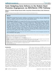
CI: 1.043-1.392, P = 0.01) (Table 4). The area under <strong>the</strong> ROC<br />
curve <strong>for</strong> <strong>the</strong> platelet <strong>count</strong>/ <strong>bipolar</strong> <strong>spleen</strong> <strong>diameter</strong> <strong>ratio</strong><br />
was 0.94 ± 0.02 (Figure 1), which represents <strong>the</strong> overall<br />
diagnostic accuracy of <strong>the</strong> <strong>ratio</strong> in predicting EVs. Thus,<br />
on random selection of an individual with EVs versus no<br />
EVs, <strong>the</strong> score of <strong>the</strong> <strong>for</strong>mer will be lower 94% of <strong>the</strong> time.<br />
Table 5 shows <strong>the</strong> sensitivity, specificity, PPV, and NPV of<br />
<strong>the</strong> <strong>ratio</strong> at various cutoff levels. At a score of 939.7, <strong>the</strong><br />
<strong>ratio</strong> has high sensitivity (100%), robust negative predictive<br />
value NPV (100%), and high specificity (86.3%) and<br />
positive predictive value PPV (95.6%).<br />
In a separate analysis, <strong>the</strong> platelet <strong>count</strong>/<strong>bipolar</strong> <strong>spleen</strong><br />
<strong>diameter</strong> <strong>ratio</strong> was significantly higher in compensated<br />
cirrhotics (Child-Pugh class A) without EVs compared<br />
with those with EVs (1802 ± 987 and 770 ± 146.6, respectively;<br />
p = 0.0001). In this analysis, <strong>the</strong> <strong>ratio</strong> maintained high<br />
sensitivity (100 %), robust NPV (100%), and high specificity<br />
(81.8%) and PPV (97%), with an overall diagnostic accuracy<br />
of 87.2% (Table 6). Notably, none of <strong>the</strong> thrombocytopenic<br />
patients showed any evidence of thrombocytopenia <strong>for</strong><br />
etiologies o<strong>the</strong>r than hypersplenism, as evidenced by <strong>the</strong><br />
normal results <strong>for</strong> ANA, ASMA, gamma-globulin levels<br />
and bone marrow aspi<strong>ratio</strong>n (data not shown).<br />
Discussion<br />
Management of EVs is an everyday challenge. Current<br />
guidelines recommend that all patients should undergo<br />
endoscopic screening <strong>for</strong> varices when cirrhosis is diagnosed,<br />
after which patients with medium and large varices<br />
should be treated to prevent bleeding. For all o<strong>the</strong>r<br />
patients, regular periodic evaluation is required (22). In<br />
Egypt, however, <strong>the</strong> management of patients with liver<br />
Hepat Mon. 2011;11(4):278-284<br />
281<br />
cirrhosis complicated by <strong>the</strong> interplay between clinical,<br />
economic, social, and cultural factors and <strong>the</strong> generally<br />
poor compliance to both follow-up and treatment strategies.<br />
Endoscopic follow-up, which is recommended by<br />
international guidelines, is not feasible in most patients<br />
<strong>for</strong> many reasons. Liver diseases are common in Egypt<br />
due to <strong>the</strong> higher prevalence of viral hepatitis (23, 24)<br />
and increased incidence of schistosomiasis (25); moreover,<br />
most patients present in <strong>the</strong> late phase of liver disease.<br />
Fur<strong>the</strong>r, <strong>the</strong> majority of patients is uninsured and<br />
must pay <strong>for</strong> expenses out of pocket, unaware of <strong>the</strong> risk<br />
Table 6. Validation of cutoff value of platelet <strong>count</strong>/ <strong>bipolar</strong> <strong>spleen</strong> <strong>diameter</strong><br />
<strong>ratio</strong> in <strong>the</strong> diagnosis of esophageal varices in patients with compensated<br />
liver cirrhosis<br />
Sensitivity Specificity NPV<br />
a<br />
NPV = negative predictive value<br />
b<br />
PPV = positive predictive value<br />
a PPV b Accuracy<br />
100% 81.8 % 100 % 70 % 87.2 %<br />
of <strong>the</strong> variceal bleeding, and <strong>the</strong>y are apparently healthy<br />
asymptomatic compensated patients. Compliance can<br />
also be limited, because it requires that patients who are<br />
asymptomatic undergo a procedure repeatedly that is<br />
perceived to be unpleasant.<br />
Endoscopic follow-up is also impractical due to <strong>the</strong> fear<br />
of infection; despite ef<strong>for</strong>ts to ensure sterilization, <strong>the</strong>re<br />
remains a risk of re-infection by ano<strong>the</strong>r subtype of <strong>the</strong><br />
same virus infect <strong>the</strong> patient or ano<strong>the</strong>r viral hepatitis<br />
with increased risk of decompensation in compensated<br />
patients (26). Finally, endoscopy units are not available<br />
in all hospitals, particularly in rural areas, necessitating<br />
o<strong>the</strong>r easier modalities <strong>for</strong> <strong>the</strong> diagnosis and monitoring<br />
of portal hypertension. Thus, a method of predicting<br />
<strong>the</strong> presence of EVs noninvasively is in great demand to<br />
avoid unnecessary endoscopy and improve <strong>the</strong> costeffectiveness<br />
of management; <strong>the</strong> latter is a particularly<br />
important conside<strong>ratio</strong>n in many African and Middle<br />
Eastern <strong>count</strong>ries, including Egypt, where liver cirrhosis<br />
is highly prevalent. Ideally, a method <strong>for</strong> identifying<br />
patients with varices should be simple, noninvasive, inexpensive,<br />
reproducible, accurate, and readily available;<br />
have high sensitivity and specificity; follow <strong>the</strong> natural<br />
history; reflect <strong>the</strong> effect of <strong>the</strong> treatment accurately;<br />
and indicate <strong>the</strong> prognosis and possibility of <strong>the</strong> success<br />
of a treatment. Several noninvasive and minimally
282 Abu El Makarem MA et al.<br />
Noninvasive prediction of esophageal varices<br />
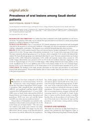
Sensitivity<br />
1-Sepecificity<br />
Figure 1. Receiver operating characteristic curve (roc) of platelet <strong>count</strong>/ <strong>bipolar</strong><br />
<strong>spleen</strong> <strong>diameter</strong> <strong>ratio</strong> and age<br />
AUC = area under <strong>the</strong> curve<br />
AUC <strong>for</strong> <strong>ratio</strong> = 0.94 ± 0.02, AUC <strong>for</strong> age = 0.33 ± 0.05<br />
invasive methods have emerged in recent years, assessing<br />
<strong>the</strong> potential of various laboratory, clinical, and ultrasonographic<br />
parameters, linked directly or indirectly<br />
to portal hypertension, such as splenomegaly, decreased<br />
platelet <strong>count</strong>, and portal vein <strong>diameter</strong> (8-10, 22, 27-31).<br />
Three such methods have been examined extensively:<br />
platelet <strong>count</strong>:<strong>spleen</strong> <strong>diameter</strong> <strong>ratio</strong> (15), Fibrotest (32),<br />
and Fibroscan (33). Fibrotest appears to be insufficiently<br />
precise, and Fibroscan requires fur<strong>the</strong>r evaluation; nei<strong>the</strong>r<br />
test is widely available in Egypt due to financial and<br />
technical conside<strong>ratio</strong>ns. In a seminal trial, Giannini and<br />
colleagues used <strong>the</strong> platelet <strong>count</strong>:<strong>spleen</strong> <strong>diameter</strong> <strong>ratio</strong><br />
as a parameter, linking thrombocytopenia to <strong>spleen</strong> size<br />
to predict portal hypertension (15). Although <strong>the</strong>se international<br />
trials; <strong>the</strong> differences in <strong>the</strong> populations characters<br />
and <strong>the</strong> need to a well-designed repeated multiple<br />
large multi-central trials, be<strong>for</strong>e any final recommendation<br />
can be concluded, lead to that <strong>the</strong> conclusions of <strong>the</strong><br />
Baveno IV consensus workshop on portal hypertension<br />
were that, endoscopic screening has been <strong>the</strong> optimal<br />
method of detecting varices (4, 5). For <strong>the</strong>se reasons, we<br />
planned <strong>the</strong> current study to evaluate <strong>the</strong> use of a similar<br />
<strong>ratio</strong> in Egyptian cirrhotic patients who typically present<br />
late and have varying ethnicities, poorer nutritional status,<br />
and significant viral etiology to overcome <strong>the</strong> drawbacks<br />
of previous studies. (We think that this paragraph<br />
doesn’t need modification).<br />
In our study, <strong>the</strong> prevalence of EVs was 74.9%, which<br />
might be attributed to <strong>the</strong> late presentation of our patients<br />
and <strong>the</strong> increased incidence of schistosomiasis<br />
in our region (25). In addition to age, serum bilirubin,<br />
prothrombin activity, platelet <strong>count</strong>, <strong>spleen</strong> <strong>diameter</strong>,<br />
and Child-Pugh scores, <strong>the</strong> platelet <strong>count</strong>:<strong>spleen</strong> <strong>diameter</strong><br />
<strong>ratio</strong> was associated with portal hypertension.<br />
These results were confirmed by multivariate logistic<br />
regression, which demonstrated that <strong>the</strong> platelet <strong>count</strong>:<br />
<strong>spleen</strong> <strong>diameter</strong> <strong>ratio</strong> (odds <strong>ratio</strong>: 1.028, 95% CI: 1.016-1.04,<br />
Hepat Mon. 2011;11(4):278-284<br />
P = 0.001) and age were independent predictive factors<br />
<strong>for</strong> EVs (odds <strong>ratio</strong>: 1.205, 95% CI: 1.043-1.392, P = 0.01). Although<br />
portal vein <strong>diameter</strong>, as assessed by ultrasonography,<br />
was higher in cirrhotic patients with EVs, this<br />
correlation failed to reach statistical significance (P =<br />
0.07), as did mean portal blood flow velocity; <strong>the</strong>se data<br />
are consistent with results of o<strong>the</strong>r large trials (8, 10-14).<br />
Moreover, per<strong>for</strong>ming Doppler sonographic examination<br />
requires sophisticated skills and equipment, limiting<br />
its value in <strong>the</strong> identification of patients with cirrhosis<br />
who are at risk of variceal bleeding (34). Our analysis<br />
of <strong>the</strong> area under <strong>the</strong> ROC curve (AUROC) revealed that<br />
<strong>the</strong> cutoff of <strong>the</strong> platelet <strong>count</strong>:<strong>spleen</strong> <strong>diameter</strong> <strong>ratio</strong><br />
(939.7) was <strong>the</strong> optimal value <strong>for</strong> accurate prediction of<br />
EVs with an AUC of 0.95; this value corresponded to positive<br />
and negative predictive values of 96% and 100%, respectively.<br />
It has been reported that a negative predictive<br />
value (NPV) of 100% is desirable <strong>for</strong> screens to minimize<br />
<strong>the</strong> oversight of individuals who are at risk (13, 17). This<br />
finding is consistent with <strong>the</strong> initial study by Giannini<br />
et al. (15), who used a cutoff of 909 with an AUROC curve<br />
of 0.981, corresponding to positive and negative predictive<br />
values of 95.6% and 100%, respectively, <strong>for</strong> <strong>the</strong> presence<br />
of varices. The same <strong>ratio</strong> has also been examined by<br />
many groups in many <strong>count</strong>ries in patient populations<br />
that differed from <strong>the</strong> group in which it was developed,<br />
generating consistent results—suggesting that <strong>the</strong> <strong>ratio</strong><br />
is generalizable (35-38). We believe that this <strong>ratio</strong> is valuable<br />
and unique—a hypo<strong>the</strong>sis that is supported by many<br />
clinical, financial, and statistical findings. Clinically, <strong>the</strong><br />
increase in <strong>spleen</strong> size in patients with chronic liver disease<br />
is nearly always a manifestation of portal hypertension<br />
(39, 40); conversely, although thrombocytopenia<br />
can result from immune-mediated mechanisms or lower<br />
thrombopoietin syn<strong>the</strong>sis (16, 41), in most cases it is usually<br />
caused by splenic pooling of platelets due to portal<br />
hypertension (15, 42). This model is supported by our<br />
results, in which thrombocytopenia was attributed to<br />
hypersplensim in all patients. Integrating platelet <strong>count</strong><br />
and <strong>spleen</strong> size in a <strong>ratio</strong> allowed us to determine <strong>the</strong> extent<br />
of thrombocytopenia that most likely resulted from<br />
hypersplenism. Financially, <strong>the</strong> <strong>ratio</strong> is easy to calculate<br />
and can be used at <strong>the</strong> bedside, Biannual calculation of<br />
<strong>the</strong> <strong>ratio</strong> will not generate additional costs in <strong>the</strong> management<br />
of cirrhotic patients, because platelet <strong>count</strong> is<br />
assessed routinely and abdominal ultrasonography is<br />
usually per<strong>for</strong>med at least semiannually to monitor hepatocellular<br />
carcinoma (43). In fact, <strong>spleen</strong> <strong>bipolar</strong> measurements<br />
consistently show high reproducibility and<br />
low intra- and interobserver variability (44, 45).<br />
Statistically, <strong>the</strong> platelet <strong>count</strong>:<strong>spleen</strong> <strong>diameter</strong> <strong>ratio</strong><br />
and age were <strong>the</strong> only parameters that were independently<br />
associated with <strong>the</strong> presence of EVs in <strong>the</strong> multivariate<br />
analysis. The AUROC curves <strong>for</strong> age and platelet<br />
<strong>count</strong>:<strong>spleen</strong> <strong>diameter</strong> <strong>ratio</strong> were 0.31 and 0.95, respectively,<br />
indicating that <strong>the</strong> <strong>ratio</strong> can be used as <strong>the</strong> sole<br />
predictive factor <strong>for</strong> EVs. Notably, in a subset analysis,
Noninvasive prediction of esophageal varices Abu El Makarem MA et al.<br />
this <strong>ratio</strong> remained valuable in <strong>the</strong> prediction of <strong>the</strong><br />
presence of EVs in patients with no signs of decompensation,<br />
with 100% sensitivity and NPV. This property might<br />
be particularly useful; its clinical importance has recently<br />
been emphasized (3, 10, 46, 47). Although our results<br />
were based on a subset analysis and in smaller sample<br />
sizes, routine periodic endoscopy, as recommended<br />
by <strong>the</strong> Ministry of National Health in Egypt, should be<br />
considered in <strong>the</strong> follow-up of cirrhotic patients; based<br />
on limitations in financial resources, this sample <strong>ratio</strong>;<br />
platelet <strong>count</strong>:<strong>spleen</strong> <strong>diameter</strong> <strong>ratio</strong> should be applied.<br />
In conclusion, <strong>the</strong> platelet <strong>count</strong>:<strong>spleen</strong> <strong>diameter</strong> <strong>ratio</strong><br />
is an accurate noninvasive method of assessing EVs in<br />
Egyptian patients with compensated or decompensated<br />
liver cirrhosis. It is easy to calculate and can reduce <strong>the</strong><br />
financial and sanitary burdens of endoscopy units, particularly<br />
in developing <strong>count</strong>ries. Additional large multicenter<br />
studies on this <strong>ratio</strong> should be per<strong>for</strong>med.<br />
Financial support<br />
This study was supported by <strong>the</strong> Department of Internal<br />
Medicine, Department of Clinical Pathology, University<br />
of Minia, Minia City, Egypt, and Minia University<br />
Hospital Research Foundation. The University of Minia,<br />
Minia University Hospital Research Foundation had no<br />
role in <strong>the</strong> design or conduct of <strong>the</strong> study; <strong>the</strong> collection,<br />
analysis, or interpretation of <strong>the</strong> data; or <strong>the</strong> prepa<strong>ratio</strong>n<br />
or review of <strong>the</strong> manuscript.<br />
Authors contribution<br />
Specific author contributions: Dr. Mona Abu El Makarem<br />
had full access to all of <strong>the</strong> data in <strong>the</strong> study and accuracy<br />
of <strong>the</strong> data analysis, study concept, and design.<br />
Yehia Shaker, Mohammed Shatat, Maysa Abd-elmoty, Atef<br />
Elakad : acquisition of data. Mona Abu El Makarem, Mohammed<br />
Shatat, Amal M.Kamal: drafting of <strong>the</strong> manuscript.<br />
Mohammed Shatat, Ali M. El Sherif: statistical<br />
analysis, Ali. Mona Abu El Makarem, Mohammed Shatat,<br />
Ahmed Ali, Atef Elakad: administrative technical or material<br />
support. Hosny S. Abdel Ghany: radiological assessment.<br />
Conflict of Interest<br />
None of <strong>the</strong> authors has an affiliation or conflict of interest.<br />
References<br />
1. Schiedermaier P. Splanchnic hemodynamics: cirrhotic versus<br />
non-cirrhotic portal hypertension. J Gastroenterol Hepatol<br />
2004;19(s7):S150-S4.<br />
2. Hong WD, Zhu QH, Huang ZM, Chen XR, Jiang ZC, Xu SH, et al.<br />
Predictors of esophageal varices in patients with HBV-related<br />
cirrhosis: a retrospective study. BMC Gastroenterol. 2009;9:11.<br />
3. Jensen DM. Endoscopic screening <strong>for</strong> varices in cirrhosis:<br />
findings, implications, and outcomes. Gastroenterology.<br />
2002;122(6):1620-30.<br />
4. de Franchis R. Updating consensus in portal hypertension:<br />
Hepat Mon. 2011;11(4):278-284<br />
283<br />
report of <strong>the</strong> Baveno III Consensus Workshop on definitions,<br />
methodology and <strong>the</strong>rapeutic strategies in portal hypertension.<br />
J Hepatol. 2000;33(5):846-52.<br />
5. de Franchis R. Evolving consensus in portal hypertension.<br />
Report of <strong>the</strong> Baveno IV consensus workshop on methodology<br />
of diagnosis and <strong>the</strong>rapy in portal hypertension. J Hepatol.<br />
2005;43(1):167-76.<br />
6. Gorka W, al Mulla A, al Sebayel M, Altraif I, Gorka TS. Qualitative<br />
hepatic venous Doppler sonography versus portal flowmetry in<br />
predicting <strong>the</strong> severity of esophageal varices in hepatitis C cirrhosis.<br />
AJR Am J Roentgenol. 1997;169(2):511-5.<br />
7. Lavergne J, Molina E, Reddy KR, Jeffers L, Leon R, Nader AK, et al.<br />
Ascites predicts <strong>the</strong> presence of high grade varices by screening<br />
gastroscopy. Gastrointest Endosc. 1997;45(4):AB187.<br />
8. Pilette C, Oberti F, Aube C, Rousselet MC, Bedossa P, Gallois Y, et<br />
al. Non-invasive diagnosis of esophageal varices in chronic liver<br />
diseases. J Hepatol. 1999;31(5):867-73.<br />
9. Madhotra R, Mulcahy HE, Willner I, Reuben A. Prediction of<br />
esophageal varices in patients with cirrhosis. J Clin Gastroenterol.<br />
2002;34(1):81-5.<br />
10. Schepis F, Camma C, Nice<strong>for</strong>o D, Magnano A, Pallio S, Cinquegrani<br />
M, et al. Which patients with cirrhosis should undergo endoscopic<br />
screening <strong>for</strong> esophageal varices detection? Hepatology.<br />
2001;33(2):333-8.<br />
11. Chalasani N, Imperiale TF, Ismail A, Sood G, Carey M, Wilcox CM,<br />
et al. Predictors of large esophageal varices in patients with cirrhosis.<br />
Am J Gastroenterol. 1999;94(11):3285-91.<br />
12. Ng FH, Wong SY, Loo CK, Lam KM, Lai CW, Cheng CS. Prediction<br />
of oesophagogastric varices in patients with liver cirrhosis. J<br />
Gastroenterol Hepatol. 1999;14(8):785-90.<br />
13. Zaman A, Hapke R, Flora K, Rosen HR, Benner K. Factors predicting<br />
<strong>the</strong> presence of esophageal or gastric varices in patients with<br />
advanced liver disease. Am J Gastroenterol. 1999;94(11):3292-6.<br />
14. Zaman A, Becker T, Lapidus J, Benner K. Risk factors <strong>for</strong> <strong>the</strong> presence<br />
of varices in cirrhotic patients without a history of variceal<br />
hemorrhage. Arch Intern Med. 2001;161(21):2564-70.<br />
15. Giannini E, Botta F, Borro P, Risso D, Romagnoli P, Fasoli A, et al.<br />
<strong>Platelet</strong> <strong>count</strong>/<strong>spleen</strong> <strong>diameter</strong> <strong>ratio</strong>: proposal and validation of<br />
a non-invasive parameter to predict <strong>the</strong> presence of oesophageal<br />
varices in patients with liver cirrhosis. Gut. 2003;52(8):1200-5.<br />
16. Peck-Radosavljevic M. Thrombocytopenia in liver disease. Can J<br />
Gastroenterol. 2000;14(Suppl D):60D-6D.<br />
17. de Franchis R. Noninvasive diagnosis of esophageal varices: is it<br />
feasible? Am J Gastroenterol. 2006;101(11):2520-2.<br />
18. Tchelepi H, Ralls P, Radin R, Grant E. Sonography of diffuse liver<br />
disease. J Ultrasound Med. 2002;21(9):1023.<br />
19. Lamb PM, Lund A, Kanagasabay RR, Martin A, Webb JA, Reznek<br />
RH. Spleen size: how well do linear ultrasound measurements<br />
correlate with three-dimensional CT volume assessments? Br J<br />
Radiol. 2002;75(895):573-7.<br />
20. Moriyasu F, Ban N, Nishida O, Nakamura T, Miyake T, Uchino<br />
H, et al. Clinical application of an ultrasonic duplex system in<br />
<strong>the</strong> quantitative measurement of portal blood flow. J Clin Ultrasound.<br />
1986;14(8):579-88.<br />
21. Pugh RN, Murray-Lyon IM, Dawson JL, Pietroni MC, Williams R.<br />
Transection of <strong>the</strong> oesophagus <strong>for</strong> bleeding oesophageal varices.<br />
Br J Surg. 1973;60(8):646-9.<br />
22. Garcia-Tsao G, Sanyal AJ, Grace ND, Carey W. Prevention and management<br />
of gastroesophageal varices and variceal hemorrhage<br />
in cirrhosis. Hepatology. 2007;46(3):922-38.<br />
23. EMOH. Egyptian Ministry of Health Annual Report: 2007: Egyptian<br />
Ministry of Health; 2009 March 6.<br />
24. Attia MA. Prevalence of hepatitis B and C in Egypt and Africa.<br />
Antivir Ther. 1998;3(Suppl 3):1-9.<br />
25. El-Zayadi AR. Curse of schistosomiasis on Egyptian liver. World J<br />
Gastroenterol. 2004;10(8):1079-81.<br />
26. Mikhail NN, Lewis DL, Omar N, Taha H, El-Badawy A, Abdel-<br />
Mawgoud N, et al. Prospective study of cross-infection from upper-GI<br />
endoscopy in a hepatitis C-prevalent population. Gastrointest<br />
Endosc. 2007;65(4):584-8.<br />
27. Burton JR, Jr., Liangpunsakul S, Lapidus J, Giannini E, Chalasani<br />
N, Zaman A. Validation of a multivariate model predicting presence<br />
and size of varices. J Clin Gastroenterol. 2007;41(6):609-15.<br />
28. Garcia-Tsao G, Escorsell A, Zakko M, Patch D, Matloff D, Grace
284 Abu El Makarem MA et al.<br />
Noninvasive prediction of esophageal varices<br />
N, et al. Predicting <strong>the</strong> presence of significant portal hypertension<br />
and varices in compensated cirrhotic patients. Hepatology.<br />
1997;26(pt 2):927-30.<br />
29. Thomopoulos KC, Labropoulou-Karatza C, Mimidis KP, Katsakoulis<br />
EC, Iconomou G, Nikolopoulou VN. Non-invasive predictors<br />
of <strong>the</strong> presence of large oesophageal varices in patients with cirrhosis.<br />
Dig Liver Dis. 2003;35(7):473-8.<br />
30. Berzigotti A, Gilabert R, Abraldes JG, Nicolau C, Bru C, Bosch J, et<br />
al. Noninvasive prediction of clinically significant portal hypertension<br />
and esophageal varices in patients with compensated<br />
liver cirrhosis. Am J Gastroenterol. 2008;103(5):1159-67.<br />
31. Pagliaro L, D’Amico G, Pasta L, Politi F, Vizzini G, Traina M, et al.<br />
Portal hypertension in cirrhosis: natural history. In: Bosch J,<br />
Groszmann RJ. Portal Hypertension Pathophysiology and Treatment<br />
Ox<strong>for</strong>d, UK: Blackwell Scientific. 1994. p. 72-92.<br />
32. Thabut D, Trabut JB, Massard J, Rudler M, Muntenau M, Messous<br />
D, et al. Non-invasive diagnosis of large oesophageal varices with<br />
FibroTest in patients with cirrhosis: a preliminary retrospective<br />
study. Liver Int. 2006;26(3):271-8.<br />
33. Kazemi F, Kettaneh A, N'Kontchou G, Pinto E, Ganne-Carrie N,<br />
Trinchet JC, et al. Liver stiffness measurement selects patients<br />
with cirrhosis at risk of bearing large oesophageal varices. J<br />
Hepatol. 2006;45(2):230-5.<br />
34. Li FH, Hao J, Xia JG, Li HL, Fang H. Hemodynamic analysis of<br />
esophageal varices in patients with liver cirrhosis using color<br />
Doppler ultrasound. World J Gastroenterol. 2005;11(29):4560-5.<br />
35. Legasto GMA, Sevilla J, Balay A, Tan JA, Cham LV, Vitug A, et al.<br />
<strong>Platelet</strong> <strong>count</strong>/<strong>spleen</strong> <strong>diameter</strong> <strong>ratio</strong>: a noninvasive parameter<br />
to predict <strong>the</strong> presence of esophageal varices. Phil J Gastroenterol<br />
2006;2:33-8.<br />
36. Alempijevic T, Kovacevic N. Right liver lobe <strong>diameter</strong>:albumin<br />
<strong>ratio</strong>: a new non-invasive parameter <strong>for</strong> prediction of oesophageal<br />
varices in patients with liver cirrhosis (preliminary report).<br />
Gut. 2007;56(8):1166-7; author reply 7.<br />
Hepat Mon. 2011;11(4):278-284<br />
37. Agha A, Anwar E, Bashir K, Savarino V, Giannini EG. External validation<br />
of <strong>the</strong> platelet <strong>count</strong>/<strong>spleen</strong> <strong>diameter</strong> <strong>ratio</strong> <strong>for</strong> <strong>the</strong> diagnosis<br />
of esophageal varices in hepatitis C virus-related cirrhosis.<br />
Dig Dis Sci. 2009;54(3):654-60.<br />
38. Baig WW, Nagaraja MV, Varma M, Prabhu R. <strong>Platelet</strong> <strong>count</strong> to<br />
<strong>spleen</strong> <strong>diameter</strong> <strong>ratio</strong> <strong>for</strong> <strong>the</strong> diagnosis of esophageal varices: Is<br />
it feasible? Can J Gastroenterol. 2008;22(10):825-8.<br />
39. Bolognesi M, Merkel C, Sacerdoti D, Nava V, Gatta A. Role of <strong>spleen</strong><br />
enlargement in cirrhosis with portal hypertension. Dig Liver Dis.<br />
2002;34(2):144-50.<br />
40. Liangpunsakul S, Ulmer BJ, Chalasani N. Predictors and implications<br />
of severe hypersplenism in patients with cirrhosis. Am J<br />
Med Sci. 2003;326(3):111-6.<br />
41. Pockros PJ, Duchini A, McMillan R, Nyberg LM, McHutchison<br />
J, Viernes E. Immune thrombocytopenic purpura in patients<br />
with chronic hepatitis C virus infection. Am J Gastroenterol.<br />
2002;97(8):2040-5.<br />
42. Giannini EG, Zaman A, Kreil A, Floreani A, Dulbecco P, Testa E, et<br />
al. <strong>Platelet</strong> <strong>count</strong>/<strong>spleen</strong> <strong>diameter</strong> <strong>ratio</strong> <strong>for</strong> <strong>the</strong> noninvasive diagnosis<br />
of esophageal varices: results of a multicenter, prospective,<br />
validation study. Am J Gastroenterol. 2006;101(11):2511-9.<br />
43. Bruix J, Sherman M. Management of hepatocellular carcinoma.<br />
Hepatology. 2005;42(5):1208-36.<br />
44. Winkfield B, Aube C, Burtin P, Cales P. Inter-observer and intraobserver<br />
variability in hepatology. Eur J Gastroenterol Hepatol.<br />
2003;15(9):959-66.<br />
45. O'Donohue J, Ng C, Catnach S, Farrant P, Williams R. Diagnostic<br />
value of Doppler assessment of <strong>the</strong> hepatic and portal vessels<br />
and ultrasound of <strong>the</strong> <strong>spleen</strong> in liver disease. Eur J Gastroenterol<br />
Hepatol. 2004;16(2):147-55.<br />
46. Ong J. Clinical predictors of large esophageal varices: how accurate<br />
are <strong>the</strong>y? Am J Gastroenterol. 1999;94(11):3103-5.<br />
47. Rajvanshi P, Kowdley KV. Prediction of varices in patients with<br />
cirrhosis: a high-stakes numbers game? J Clin Gastroenterol.<br />
2002;34(1):4-5.