Download PDF - BioMedSearch
Download PDF - BioMedSearch
Download PDF - BioMedSearch
You also want an ePaper? Increase the reach of your titles
YUMPU automatically turns print PDFs into web optimized ePapers that Google loves.
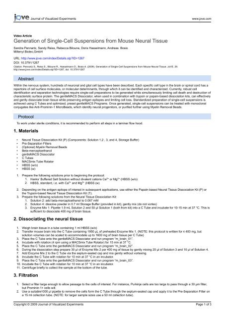
Video Article<br />
Generation of Single-Cell Suspensions from Mouse Neural Tissue<br />
Sandra Pennartz, Sandy Reiss, Rebecca Biloune, Doris Hasselmann, Andreas Bosio<br />
Miltenyi Biotec,GmbH<br />
URL: http://www.jove.com/index/Details.stp?ID=1267<br />
DOI: 10.3791/1267<br />
Citation: Pennartz S., Reiss S., Biloune R., Hasselmann D., Bosio A. (2009). Generation of Single-Cell Suspensions from Mouse Neural Tissue. JoVE. 29.<br />
http://www.jove.com/index/Details.stp?ID=1267, doi: 10.3791/1267<br />
Abstract<br />
Within the nervous system, hundreds of neuronal and glial cell types have been described. Each specific cell type in the brain or spinal cord has a<br />
repertoire of cell surface molecules, or molecular determinants, through which it can be identified and characterized. Currently, robust cell<br />
identification and separation technologies require single-cell preparations to be generated while simultaneously limiting cell death and destruction of<br />
characteristic surface protein. The gentleMACS Dissociator, when used in combination with trypsin or papain-based dissociation kits, can effectively<br />
and gently dissociate brain tissue while preserving antigen epitopes and limiting cell loss. Standardized preparation of single-cell suspensions is<br />
achieved using C Tubes and optimized, preset gentleMACS Programs. Once generated, single-cell suspensions can be treated with monoclonal<br />
conjugates like Anti-Prominin-1 MicroBeads, which identify neural progenitors, or purified further using Myelin Removal Beads.<br />
Protocol<br />
To work under sterile conditions, it is recommended to perform all steps in a laminar flow hood.<br />
1. Materials<br />
2. Dissociating the neural tissue<br />
3. Filtration<br />
Journal of Visualized Experiments www.jove.com<br />
• Neural Tissue Dissociation Kit (P) (Components: Solution 1,2 , 3, and 4, Storage Buffer)<br />
• Pre-Separation Filters<br />
• (Optional) Myelin Removal Beads<br />
• Beta-mercaptoethanol<br />
• gentleMACS Dissociator<br />
• C Tubes<br />
• MACSmix Tube Rotator<br />
• HBSS (w/o)<br />
• HBSS (w)<br />
1. Prepare the following solutions prior to beginning the protocol:<br />
1. Hanks’ Buffered Salt Solution without divalent cations Ca 2+ or Mg 2+ (HBSS (w/o)<br />
2. HBSS, standard, i.e. with Ca 2+ and Mg 2+ (HBSS (w)<br />
2. Depending on the antigen epitope of interest in subsequent applications, use either the Papain-based Neural Tissue Dissociation Kit (P) or<br />
the Trypsin-based Neural Tissue Dissociation Kit (T).<br />
3. Prepare the following solutions from the Neural Tissue Dissociation Kit:<br />
1. Solution 2: add beta-mercaptoethanol to 0.067 mM<br />
2. Solution 4: dissolve powder in 0.7 ml Storage Buffer (provided in kit), gently mix (do not vortex)<br />
3. Enzyme Mix 1: Pipette 1.9 mL Solution 2 and 50 μl Solution 1 (both from kit) into a C Tube and incubate for 10-15 min at 37 °C. This is<br />
sufficient to dissociate 400 mg of brain tissue.<br />
1. Weigh brain tissue in a tube containing 1 ml HBSS (w/o)<br />
2. Transfer mouse brain into the C Tube containing 1950 μL of preheated Enzyme Mix 1. (NOTE: this protocol is written for ≤ 400 mg, but<br />
solution volumes can be scaled to accommodate up to 1600 mg of brain tissue per C Tube)<br />
3. Place the C Tube onto the gentleMACS Dissociator and run program “m_brain_01”.<br />
4. Incubate with rotation (4 rpm using a MACSmix Tube Rotator) for 15 min at 37 °C.<br />
5. Place the C Tube onto the gentleMACS Dissociator and run program “m_brain_02”.<br />
6. During the dissociation step prepare 30 μl of Enzyme Mix 2 per 400 mg of tissue by gently mixing 20 μl of Solution 3 and 10 μl of Solution 4.<br />
7. Add Enzyme Mix 2 to the C Tube via the septum-sealed cap and mix gently without vortexing.<br />
8. Incubate the C Tube with rotation for 10 min at 37 °C in an incubator.<br />
9. Place the C Tube onto the gentleMACS Dissociator and run program “m_brain_03”.<br />
10. Incubate the C Tube with rotation for 10 min at 37 °C in an incubator.<br />
11. Centrifuge briefly to collect the sample at the bottom of the tube.<br />
1. Select a filter large enough to allow passage to the cells of interest. For instance, Purkinje cells are too large to pass through a 30 μm filter,<br />
but Prominin-1+ cells will.<br />
2. Use a suitable1000 μl pipette to remove the cells form the C Tube through the septum-sealed cap and apply it to the Pre-Separation Filter on<br />
a 15 ml collection tube. (NOTE: for larger sample sizes use a 50 ml collection tube).<br />
Copyright © 2009 Journal of Visualized Experiments<br />
Page 1 of 3
Journal of Visualized Experiments www.jove.com<br />
3. Wash the filter with 10 ml of HBSS (w).<br />
4. Centrifuge the filtered cells at 300 x g for 10 min at RT.<br />
5. Resuspend the supernatent in your medium or buffer of choice for subsequent applications.<br />
6. To remove myelin debris, use Myelin Removal Beads.<br />
4. Representative Result: Please See Figures 1-3<br />
Figure. 1 The brain dissociation with the gentleMACS Dissociator resulted in 97% viable cells, as flow cytometric analysis shows.<br />
Figure. 2 Light microscope picture of neurosphere formation after magnetic cell sorting using Anti-Prominin-1 MicroBeads after 7 days of<br />
cultivation in MACS® NeuroMedium supplemented with MACS Supplement B27 PLUS. Cells were prepared from CD1 mouse brain (P3) using<br />
the Neural Tissue Dissociation Kit (P).<br />
Copyright © 2009 Journal of Visualized Experiments<br />
Page 2 of 3
Figure. 3 Myelin debris in single-cell suspensions considerably impairs cell isolation, and removal of myelin debris by Myelin Removal Beads<br />
increases efficiency cell separations. For MACS Separations using Anti-Prominin-1 MicroBeads, P22 mouse brain was dissociated using the<br />
Neural Tissue Dissociation Kit (P). Cells from the single-cell suspension were either directly used for separation, or were submitted to myelin<br />
depletion using Myelin Removal Beads. Comparing the separation from samples without and with myelin removal, demonstrates that the purity is<br />
higher for samples with previous myelin removal.<br />
Disclosures<br />
The authors are employees of Miltenyi Biotec GmbH, Germany.<br />
Discussion<br />
The gentleMACS Dissociator facilitates the standardized preparation of single-cell suspensions from neural tissues in a closed system. Neural<br />
Tissue Dissociation Kits are optimized to preserve antigen epitopes needed for further applications like immunostainings and immunomagnetic<br />
cell separation 1-5 . In this protocol we show the gentle enzymatic dissociation of mouse brain using the gentleMACS Dissociator and the Neural<br />
Tissue Dissociation Kit (P), yielding in 97% viable cells. 100 mg of neural tissue yields between 5x10 6 and 1x10 7 cells, depending on the age of<br />
the host tissue. The targeted Prominin-1 + cells were isolated using Anti-Prominin-1 MicroBeads and subsequently taken into culture. The protocol<br />
is shown to be even more effective with the additional use of Myelin Removal Beads when working with neural tissue derived from mice >P7.<br />
References<br />
Journal of Visualized Experiments www.jove.com<br />
1. Hardt O, Scholz C, Küsters D, Yanagawa Y, Pennartz S, Cremer H, Bosio A (2008) Gene expression analysis defines differences between<br />
region-specific GABAergic neurons. Mol Cell Neurosci 39: 418-28.<br />
2. Lee JK, McCoy MK, Harms AS, Ruhn KA, Gold SJ, Tansey MG (2008) Regulator of G-protein signaling 10 promotes dopaminergic neuron<br />
survival via regulation of the microglial inflammatory response. J Neurosci. 28: 8517-28.<br />
3. Környei Z, Gócza E, Rühl R, Orsolits B, Vörös E, Szabó B, Vágovits B, Madarász E (2007) Astroglia-derived retinoic acid is a key factor in<br />
glia-induced neurogenesis. FASEB J. 21: 2496-509.<br />
4. Meylan F, Davidson TS, Kahle E, Kinder M, Acharya K, Jankovic D, Bundoc V, Hodges M, Shevach EM, Keane-Myers A, Wang EC, Siegel<br />
RM (2008) The TNF-family receptor DR3 is essential for diverse T cell-mediated inflammatory diseases. Immunity 29: 79-89.<br />
5. Skog J, Würdinger T, van Rijn S, Meijer DH, Gainche L, Sena-Esteves M, Curry WT Jr, Carter BS, Krichevsky AM, Breakefield XO (2008)<br />
Glioblastoma microvesicles transport RNA and proteins that promote tumour growth and provide diagnostic biomarkers. Nat Cell Biol. 10:<br />
1470-6.<br />
Copyright © 2009 Journal of Visualized Experiments<br />
Page 3 of 3


