Secondary collapse of lateral femoral condyle following bone bruise ...
Secondary collapse of lateral femoral condyle following bone bruise ...
Secondary collapse of lateral femoral condyle following bone bruise ...

Create successful ePaper yourself
Turn your PDF publications into a flip-book with our unique Google optimized e-Paper software.
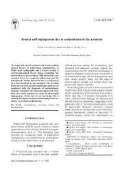
696 N. VANNET, P. KEMPSHALL, J. DAVIESFig. 1 (A+B). — MR Imaging <strong>of</strong> the knee. T 1 and T 2 weightedimages showing the area <strong>of</strong> <strong>bone</strong> bruising in the <strong>lateral</strong> <strong>femoral</strong><strong>condyle</strong>.Fig. 2. — CT scan showing <strong>collapse</strong> <strong>of</strong> the <strong>lateral</strong> <strong>femoral</strong><strong>condyle</strong>.CASE REPORTThe <strong>following</strong> case presentation is that <strong>of</strong> an18-year-old male motorcyclist who sustained aninjury to his left knee when he fell <strong>of</strong>f his bikewhilst negotiating a corner at approx 15 mph andslid into a car.Initially no fracture was identified on plain radiographs.Subsequent review in fracture clinicrevealed an effusion, marked tenderness over themedial col<strong>lateral</strong> ligament and difficulty reachingfull extension. He was advised to mobilise pain permitting,and an MRI scan was arranged to assess theintra and extra articular ligaments around the knee.Following the MRI scan (fig 1), prior to subsequentoutpatient appointment, the swelling <strong>of</strong> theknee improved, and the boy returned to normalactivity, although he was experiencing some pain.He presented again to the accident and emergencydepartment 12 days after the MR scan with apainful swollen left knee <strong>following</strong> a subsequentminor trauma when riding a pedal cycle. Plain filmsshowed a possible depression fracture <strong>of</strong> the <strong>lateral</strong><strong>femoral</strong> <strong>condyle</strong>, this was confirmed by a CT scanwhich showed a marked depression in the <strong>lateral</strong><strong>femoral</strong> <strong>condyle</strong> (fig 2).The previous MR image, when reviewed, showeda tear <strong>of</strong> the posterior horn <strong>of</strong> the <strong>lateral</strong> meniscus,a rupture <strong>of</strong> the medial col<strong>lateral</strong> ligament (MCL),a probable rupture <strong>of</strong> the anterior crruciate ligament(ACL), and a large <strong>bone</strong> <strong>bruise</strong> in the <strong>lateral</strong><strong>femoral</strong> and tibial <strong>condyle</strong>s with no cortical depression.The patient was treated surgically, with arthroscopicassisted elevation <strong>of</strong> the <strong>lateral</strong> <strong>femoral</strong><strong>condyle</strong> under image intensifier control (fig 3).A depression in the articular surface <strong>of</strong> the <strong>lateral</strong><strong>femoral</strong> <strong>condyle</strong> was visualised at arthroscopyand measured to a depth <strong>of</strong> five millimetres. Theimpaction extended from the notch increasing progressively<strong>lateral</strong>ly. The ACL was indeed ruptured ;anterior draw, Lachman’s test and pivot shift wereall positive on examination under anaesthesia. Abucket handle tear <strong>of</strong> the <strong>lateral</strong> meniscus was alsoconfirmed. Punches were used under image intensifiercontrol through a window in the <strong>lateral</strong> femur.The aim <strong>of</strong> this presentation is to emphasise thatthe <strong>bone</strong> <strong>bruise</strong> is not necessarily an innocuous incidentallesion that can be forgotten about.ClassificationThe first classification <strong>of</strong> <strong>bone</strong> <strong>bruise</strong>s was constructedby Mink and Deutsch, who used the termoccult to mean hidden. Fractures <strong>of</strong> the marrow orarticular surface that are undetectable by conventionalradiographs become visible on MRI (8).Subsequently modified by Vellet et al, <strong>bone</strong> <strong>bruise</strong>swere divided into three categories depending ontheir position in the subcortical <strong>bone</strong> and theirActa Orthopædica Belgica, Vol. 75 - 5 - 2009