
You also want an ePaper? Increase the reach of your titles
YUMPU automatically turns print PDFs into web optimized ePapers that Google loves.
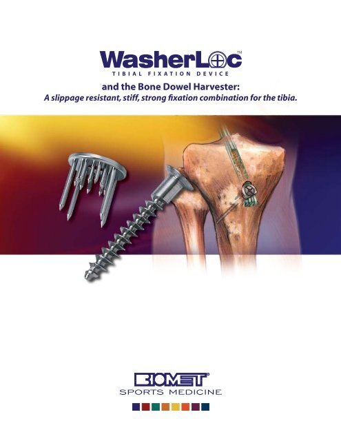
<strong>and</strong> <strong>the</strong> <strong>Bone</strong> <strong>Dowel</strong> <strong>Harvester</strong>:A slippage resistant, stiff, strong fixation combination for <strong>the</strong> tibia.
Proven Strategies for ACL Reconstruction<strong>and</strong> <strong>the</strong> <strong>Bone</strong> <strong>Dowel</strong> <strong>Harvester</strong>:A slippage resistant, stiff, strong fixation combination for <strong>the</strong> tibia.Rationale <strong>and</strong> Technique for Fixing a Soft-Tissue ACL Graft to<strong>the</strong> Tibia with <strong>the</strong> WasherLoc Device <strong>and</strong> Autogenous <strong>Bone</strong> <strong>Dowel</strong>At <strong>the</strong> time of implantation, <strong>the</strong> WasherLoc Tibial FixationDevice <strong>and</strong> autogenous bone dowel have <strong>the</strong> mechanicaladvantage of providing <strong>the</strong> most slippage resistant, stiffest, <strong>and</strong>strongest fixation combination for tibial fixation of a soft tissue4, 12, 17, 20, 23anterior cruciate ligament graft.After implantation, <strong>the</strong> WasherLoc Device <strong>and</strong> autogenousbone dowel used toge<strong>the</strong>r have <strong>the</strong> biologic advantage ofencouraging circumferential tendon-tunnel healing, 22 reducing<strong>the</strong> potential for tunnel widening, 18 maintaining stability in <strong>the</strong>face of <strong>the</strong> obligatory tension loss in <strong>the</strong> graft, 7 <strong>and</strong> lowering <strong>the</strong>tension in <strong>the</strong> graft during open-chain exercise, 13 all of which canallow for brace-free, aggressive rehabilitation.Combining <strong>the</strong> EZLoc Femoral Fixation Device with <strong>the</strong>WasherLoc Tibial Fixation Device <strong>and</strong> autogenous bone dowelprovides a comprehensive fixation combination to resist slippageof <strong>the</strong> soft-tissue graft with respect to <strong>the</strong> fixation device <strong>and</strong>slippage of <strong>the</strong> fixation device in bone during <strong>the</strong> first month ofbrace-free, aggressive rehabilitation. 23Although <strong>the</strong> WasherLoc <strong>and</strong> EZLoc devices are considered“distal fixation devices,” <strong>the</strong>y have demonstrated restoration of <strong>the</strong>same anterior laxity 16 <strong>and</strong> provide better stiffness than intratunneldevices. 16 Distal fixation devices have less slippage, 2,5,14 less tunnelwidening, 1,18 <strong>and</strong> provide greater surface area (i.e. circumferentialcontact) for tendon-tunnel healing, than intratunnel devices. 12,18,22This brochure is presented to demonstrate <strong>the</strong> surgical technique utilized by Stephen M. Howell, M.D. <strong>Biomet</strong> Sports Medcine, as <strong>the</strong> manufacturer of this device, doesnot practice medicine <strong>and</strong> does not recommend this or any o<strong>the</strong>r surgical technique for use on a specific patient. The surgeon who performs any procedure is responsiblefor determining <strong>and</strong> utilizing <strong>the</strong> appropriate techniques for such procedure for each individual patient. <strong>Biomet</strong> Sports Medicine is not responsible for selection of <strong>the</strong>appropriate surgical technique to be utilized for an individual patient.Rehabilitation activities vary depending on <strong>the</strong> individual patient <strong>and</strong> physician’s recommendations.The WasherLoc Tibial Fixation Device <strong>and</strong> <strong>the</strong> EZLoc Femoral Fixation Device were developed in conjunction with Stephen M. Howell, M.D., Sacramento, California.
Advantages of Soft Tissue ACL GraftsThe double-looped semitendinosus <strong>and</strong> gracilis (DLSTG)hamstring graft is <strong>the</strong> strongest <strong>and</strong> stiffest autogenous graftavailable, 8, 17 <strong>and</strong> <strong>the</strong> single-looped tibialis allograft is <strong>the</strong> strongest<strong>and</strong> stiffest allograft available. 9, 19 Both of <strong>the</strong>se soft-tissue graftshave less harvest morbidity compared to o<strong>the</strong>r autogenousgraft sources. The DLSTG autograft <strong>and</strong> <strong>the</strong> tibialis allograftare not normally associated with <strong>the</strong> complications of a bonepatellartendon-bone graft which include quadriceps weakness,patellar fracture, patellar tendon rupture, anterior knee pain, <strong>and</strong>degeneration of <strong>the</strong> articular surface of <strong>the</strong> patella-femoral joint.Most surgeons prefer <strong>the</strong> soft-tissue ACL graft because of itsstructural properties, minimal or non-existent harvest morbidity,<strong>and</strong> ease of rehabilitation. 10,11Limitation of Tendon-Tunnel HealingWays to improve <strong>the</strong> fixation strength of a tendon graft areneeded because <strong>the</strong> biologic bond is only half as strong as abone-patellar tendon-bone graft at three weeks. 24 By four weeks<strong>the</strong> tendon-tunnel interface provides most of <strong>the</strong> fixation strengthoffloading <strong>the</strong> mechanical strength provided by <strong>the</strong> fixationdevice. 25 However, it isn’t until six weeks that <strong>the</strong> strength of <strong>the</strong>biologic bond of <strong>the</strong> tendon-tunnel interface equals that of <strong>the</strong>bone-patellar tendon-bone graft. 24 Therefore, during <strong>the</strong> first sixweeks of implantation, <strong>the</strong> fixation method of a soft-tissue graftmust provide more slippage resistance, stiffness, <strong>and</strong> strength than<strong>the</strong> fixation used with a bone-patellar tendon-bone graft. 24Enhancing Tendon Tunnel HealingThere are several ways to improve <strong>the</strong> healing, strength, <strong>and</strong>stiffness of <strong>the</strong> tendon-tunnel biologic bond within <strong>the</strong> firstsix weeks of <strong>the</strong> reconstruction that might benefit clinicaloutcome. 18,22,24 The use of a longer tunnel provides more surfacearea for bonding. 6,22 The addition of autogenous biologic materialinside <strong>the</strong> tunnel alongside <strong>the</strong> soft-tissue graft tightens <strong>the</strong> fit<strong>and</strong> improves tendon-tunnel contact. 15 Finally, <strong>the</strong> use of distalfixation devices instead of intratunnel fixation devices, allowscircumferential ra<strong>the</strong>r than one-sided healing of <strong>the</strong> tendon to <strong>the</strong>tunnel wall. 22 Each one of <strong>the</strong>se factors are known to improve <strong>the</strong>healing, strength, <strong>and</strong> stiffness of <strong>the</strong> biologic bond. 6,15,16, 22 Thesefindings suggest that <strong>the</strong> principles for improving early healing<strong>and</strong> biologic fixation include <strong>the</strong> use of slippage resistant, stiff, <strong>and</strong>strong distal fixation devices in conjunction with compaction ofbone into <strong>the</strong> tunnel alongside <strong>the</strong> soft tissue graft. Collectively,<strong>the</strong> fixation combination of <strong>the</strong> WasherLoc Device, EZLoc Device,<strong>and</strong> <strong>Bone</strong> <strong>Dowel</strong> provide long, tight fitting tunnels that allowcircumferential tendon-tunnel healing.Reducing <strong>the</strong> Need forHardware RemovalRemoval of painful prominent hardware continues to be <strong>the</strong> mostcommon reason for additional surgery using a hamstring graft,with <strong>the</strong> incidence ranging from 12 to 26%. 3,11,21 The WasherLoc Device may minimize discomfort with kneeling or impact, 11because <strong>the</strong> average prominence of <strong>the</strong> WasherLoc Device is only2mm, which is significantly less than o<strong>the</strong>r tibial fixation methodssuch as staples (7mm) <strong>and</strong> washers (7 to 8mm). 17
Surgical TechniqueFigure 1Figure 3Figure 2Figure 4Harvest a <strong>Bone</strong> <strong>Dowel</strong> from<strong>the</strong> Tibial TunnelPlace <strong>the</strong> 2.4 mm guidepin for <strong>the</strong> tibial tunnel. Choose acannulated reamer matching <strong>the</strong> diameter of <strong>the</strong> soft tissue ACLgraft. Advance <strong>the</strong> cannulated reamer until it just breaks through<strong>the</strong> distal tibial cortex (Figure 1).Assemble <strong>the</strong> <strong>Bone</strong> <strong>Dowel</strong> <strong>Harvester</strong> bylocking <strong>the</strong> collet in <strong>the</strong> quick release h<strong>and</strong>le.Next, lock <strong>the</strong> 8mm in diameter harvestertube into <strong>the</strong> collet (Figure 2). Slide <strong>the</strong>calibrated plunger over <strong>the</strong> tibial guidepinuntil it rests inside <strong>the</strong> tibia (Figure 3). Slide<strong>the</strong> 8mm in diameter harvester tube over<strong>the</strong> plunger until it rests on cancellous bone.Impact <strong>the</strong> <strong>Bone</strong> <strong>Dowel</strong> <strong>Harvester</strong> over <strong>the</strong>guidepin to subchondral bone (Figure 4 & Figure 55). Rotate <strong>the</strong> <strong>Bone</strong> <strong>Dowel</strong> <strong>Harvester</strong> several times clockwise <strong>and</strong>counterclockwise to break off <strong>the</strong> cylindrical bone dowel <strong>and</strong> <strong>the</strong>npull to remove (Figure 6).Figure 6
Figure 7Figure 8Disengage <strong>the</strong> collet from <strong>the</strong> h<strong>and</strong>le. Read <strong>the</strong> calibrated plunger<strong>and</strong> determine <strong>the</strong> length of <strong>the</strong> bone plug (typically 25–35 mm)(Figure 7). If <strong>the</strong> guidepin was removed with <strong>the</strong> <strong>Bone</strong> <strong>Dowel</strong><strong>Harvester</strong>, <strong>the</strong>n re-insert <strong>the</strong> guidepin into <strong>the</strong> notch by insertingan 8mm femoral reamer into <strong>the</strong> tibial tunnel <strong>and</strong> <strong>the</strong>n passing aguidepin through <strong>the</strong> cannulation <strong>and</strong> through <strong>the</strong> tibial plateau(Figure 8). Finish drilling <strong>the</strong> tibial tunnel with <strong>the</strong> cannulatedreamer that matches <strong>the</strong> diameter of <strong>the</strong> graft (Figure 9).Figure 9
Figure 13Figure 15Figure 14Figure 16Select <strong>the</strong> Correct SizeWasherLoc DeviceSelect <strong>the</strong> 16mm Extended Spike WasherLoc for a 7 or 8mmdiameter graft. Select <strong>the</strong> 18mm Extended Spike WasherLoc for a9 or 10mm diameter graft. Thread <strong>the</strong> WasherLoc Device on <strong>the</strong>drill guide <strong>and</strong> thread <strong>the</strong> drill guide on <strong>the</strong> awl (Figure 13).Tension <strong>the</strong> Graft <strong>and</strong> Impact <strong>the</strong>WasherLoc DeviceAfter fixing <strong>the</strong> graft to <strong>the</strong> femur with <strong>the</strong> EZLoc FemoralFixation Device, manually tension <strong>the</strong> graft <strong>and</strong> cycle <strong>the</strong> knee fullextension to maximum flexion. Tensioning <strong>the</strong> graft <strong>and</strong> cycling<strong>the</strong> knee seats <strong>the</strong> EZLoc Device <strong>and</strong> removes slack in <strong>the</strong> graft.Place <strong>the</strong> knee in maximum hyperextension <strong>and</strong> maintain thisposition by resting <strong>the</strong> heel on Mayo st<strong>and</strong>. Place an impingementrod between <strong>the</strong> sutures attached to <strong>the</strong> tendons <strong>and</strong> have anassistant tension <strong>the</strong> graft in line with <strong>the</strong> tibial tunnel by pullingon <strong>the</strong> rod (Figure 14). Rotate <strong>the</strong> WasherLoc Device so that <strong>the</strong>flat edge faces <strong>the</strong> distal end of <strong>the</strong> tibial tunnel (Figure 15). Place<strong>the</strong> tip of <strong>the</strong> awl between <strong>the</strong> limbs of each tendon <strong>and</strong> use aright-angle clamp to maneuver <strong>the</strong> graft under <strong>the</strong> WasherLoc Device so that all four bundles are contained within <strong>the</strong> fourlonger extended spikes (Figure 16). Impact <strong>the</strong> WasherLoc Deviceusing a mallet (Figure 17).Figure 17
Figure 19Figure 18Figure 20Drill, Measure <strong>and</strong> Insert <strong>the</strong>Cancellous Compression ScrewMaintain tension on <strong>the</strong> graft. Unscrew <strong>and</strong> remove <strong>the</strong> awl from<strong>the</strong> drill guide. Insert a 3.2mm drill bit through <strong>the</strong> drill guide<strong>and</strong> push <strong>the</strong> cutting tip past <strong>the</strong> tendons (Figure 18). Protect <strong>the</strong>neurovascular structures by carefully drilling toward <strong>the</strong> fibula<strong>and</strong> through <strong>the</strong> LATERAL tibial cortex without plunging into <strong>the</strong>soft tissues. Unscrew <strong>the</strong> drill guide, insert <strong>the</strong> depth gauge, <strong>and</strong>measure <strong>the</strong> length of <strong>the</strong> tunnel (Figure 19). Momentarily leave<strong>the</strong> depth gauge in place <strong>and</strong> adjust <strong>the</strong> overhead light until <strong>the</strong>depth gauge casts a shadow on <strong>the</strong> thigh. Mark <strong>the</strong> long axis of<strong>the</strong> shadow casted by <strong>the</strong> depth gauge. Screw <strong>the</strong> appropriatesized self-tapping cancellous compression screw into <strong>the</strong> tibia.Keep <strong>the</strong> shadow of <strong>the</strong> shaft of <strong>the</strong> screwdriver in line with axis of<strong>the</strong> depth gauge while advancing <strong>the</strong> compression screw(Figures 20 <strong>and</strong> 21).Figure 21
Figure 22 Figure 23Compact <strong>Bone</strong> <strong>Dowel</strong> into<strong>the</strong> Tibial TunnelCompact any nothchplasty remnants <strong>and</strong> bone reamings into <strong>the</strong>dilated opening with <strong>the</strong> dilator or an impingement rod. Place <strong>the</strong>plastic graft protection sleeve over <strong>the</strong> cutting tip of <strong>the</strong> harvestertube (Figure 22). Position <strong>the</strong> harvester tube over <strong>the</strong> dilatedopening of <strong>the</strong> tibial tunnel. Impact <strong>the</strong> calibrated end of <strong>the</strong>plunger with a mallet until it is flush with <strong>the</strong> collet, compacting<strong>the</strong> cylindrical bone dowel into place (Figure 23). Reinsert <strong>the</strong>arthroscope into <strong>the</strong> joint <strong>and</strong> confirm that bone graft has notbeen impacted into <strong>the</strong> joint through <strong>the</strong> tibial tunnel.Completed technique with WasherLoc Device <strong>and</strong> added bone dowel.
Assess Placement of WasherLoc Device <strong>and</strong> <strong>Bone</strong><strong>Dowel</strong> on Post Operative RadiographsThe WasherLoc compression screw should course toward <strong>the</strong>fibula from anteromedial to posterolateral <strong>and</strong> exit through<strong>the</strong> LATERAL cortex of <strong>the</strong> tibia (open circle), which avoidsneurovascular structures. On <strong>the</strong> AP view <strong>the</strong> WasherLoc Deviceshould be parallel to <strong>the</strong> back wall of <strong>the</strong> tibial tunnel. The bonedowel should fill <strong>the</strong> anteromedial section of <strong>the</strong> tibial tunnel(irregular line).References1. Buelow JU, Siebold R, Ellermann A. A prospective evaluation of tunnel enlargementin anterior cruciate ligament reconstruction with hamstrings: extracortical versusanatomical fixation. Knee Surg Sports Traumatol Arthrosc. 2002;10(2):80-85.2. Caborn DN, Br<strong>and</strong> JC, Jr., Nyl<strong>and</strong> J, Kocabey Y. A biomechanical comparison of initialsoft tissue tibial fixation devices: <strong>the</strong> Intrafix versus a tapered 35-mm bioabsorbableinterference screw. Am J Sports Med. 2004;32(4):956-961.3. Clark R, Olsen RE, Larson BJ, Goble EM, Farrer RP. Cross-pin femoral fixation: a newtechnique for hamstring anterior cruciate ligament reconstruction of <strong>the</strong> knee.Arthroscopy. 1998;14(3):258-267.4. Coleridge SD, Amis AA. A comparison of five tibial-fixation systems in hamstringgraftanterior cruciate ligament reconstruction. Knee Surg Sports TraumatolArthrosc. 2004;12(5):391-397.5. Giurea M, Zorilla P, Amis AA, Aichroth P. Comparative pull-out <strong>and</strong> cyclic-loadingstrength tests of anchorage of hamstring tendon grafts in anterior cruciate ligamentreconstruction. Am J Sports Med. 1999;27(5):621-625.6. Greis PE, Burks RT, Bachus K, Luker MG. The influence of tendon length <strong>and</strong> fit on <strong>the</strong>strength of a tendon-bone tunnel complex. A biomechanical <strong>and</strong> histologic study in<strong>the</strong> dog. Am J Sports Med. 2001;29(4):493-497.7. Grover DM, Howell SM, Hull ML. Early tension loss in an anterior cruciate ligamentgraft. A cadaver study of four tibial fixation devices. J <strong>Bone</strong> Joint Surg Am.2005;87(2):381-390.8. Hamner DL, Brown CH, Jr., Steiner ME, Hecker AT, Hayes WC. Hamstring tendon graftsfor reconstruction of <strong>the</strong> anterior cruciate ligament: biomechanical evaluationof <strong>the</strong> use of multiple str<strong>and</strong>s <strong>and</strong> tensioning techniques. J <strong>Bone</strong> Joint Surg Am.1999;81(4):549-557.9. Haut Donahue TL, Howell SM, Hull ML, Gregersen C. A biomechanical evaluationof anterior <strong>and</strong> posterior tibialis tendons as suitable single-loop anterior cruciateligament grafts. Arthroscopy. 2002;18(6):589-597.10. Howell SM, Taylor MA. Brace-free rehabilitation, with early return to activity, for kneesreconstructed with a double-looped semitendinosus <strong>and</strong> gracilis graft. J <strong>Bone</strong> JointSurg Am. 1996;78(6):814-825.11. Howell SM, Deutsch ML. Comparison of endoscopic <strong>and</strong> two-incision techniquesfor reconstructing a torn anterior cruciate ligament using hamstring tendons.Arthroscopy. 1999;15(6):594-606.12. Howell SM, Roos P, Hull ML. Compaction of a bone dowel in <strong>the</strong> tibial tunnelimproves <strong>the</strong> fixation stiffness of a soft tissue anterior cruciate ligament graft: an invitro study in calf tibia. Am J Sports Med. 2005;33(5):719-725.13. Karchin A, Hull ML, Howell SM. Tension in a double loop tendon anterior cruciategraft during a simulated open chain knee extension exercise. J Orthop Res.2005;23(1):77-83.14. Khan R, Konyves A, Rama KR, Thomas R, Amis AA. RSA can measure ACL graftstretching <strong>and</strong> migration: development of a new method. Clin Orthop Relat Res.2006;448:139-145.15. Kyung HS, Kim SY, Oh CW, Kim SJ. Tendon-to-bone tunnel healing in a rabbit model:<strong>the</strong> effect of periosteum augmentation at <strong>the</strong> tendon-to-bone interface. Knee SurgSports Traumatol Arthrosc. 2003;11(1):9-15.16. Liu-Barba D, Howell SM, Hull ML. High stiffness distal fixation restores anterior laxity<strong>and</strong> knee stiffness as well as joint line fixation with an interference screw. Am JSports Med. 2007:In Press.17. Magen HE, Howell SM, Hull ML. Structural properties of six tibial fixation methods foranterior cruciate ligament soft tissue grafts. Am J Sports Med. 1999;27(1):35-43.18. Matsumoto A, Howell SM, Liu-Barba D. Time-related changes in <strong>the</strong> cross-sectionalarea of <strong>the</strong> tibial tunnel after compaction of an autograft bone dowel alongside ahamstring graft. Arthroscopy. 2006;22(8):855-860.19. Pearsall AWt, Hollis JM, Russell GV, Jr., Scheer Z. A biomechanical comparison ofthree lower extremity tendons for ligamentous reconstruction about <strong>the</strong> knee.Arthroscopy. 2003;19(10):1091-1096.20. Roos PJ, Hull ML, Howell SM. Leng<strong>the</strong>ning of double-looped tendon graft constructsin three regions after cyclic loading: a study using Roentgen stereophotogrammetricanalysis. J Orthop Res. 2004;22(4):839-846.21. Siegel MG, Barber-Westin SD. Arthroscopic-assisted outpatient anterior cruciateligament reconstruction using <strong>the</strong> semitendinosus <strong>and</strong> gracilis tendons.Arthroscopy. 1998;14(3):268-277.22. Singhatat W, Lawhorn KW, Howell SM, Hull ML. How four weeks of implantationaffect <strong>the</strong> strength <strong>and</strong> stiffness of a tendon graft in a bone tunnel: a studyof two fixation devices in an extraarticular model in ovine. Am J Sports Med.2002;30(4):506-513.23. Smith CK, Hull ML, Howell SM. Leng<strong>the</strong>ning of a single-loop tibialis tendon graftconstruct after cyclic loading: a Study using Roentgen stereophotogrammetricanalysis. J Biomech Eng. 2006;128(3):437-442.24. Tomita F, Yasuda K, Mikami S, Sakai T, Yamazaki S, Tohyama H. Comparisons ofintraosseous graft healing between <strong>the</strong> doubled flexor tendon graft <strong>and</strong> <strong>the</strong>bone-patellar tendon-bone graft in anterior cruciate ligament reconstruction.Arthroscopy. 2001;17(5):461-476.25. Zacharias I, Howell SM, Hull ML, Lawhorn KW. In vivo calibration of a femoral fixationdevice transducer for measuring anterior cruciate ligament graft tension: a study inan ovine model. J Biomech Eng. 2001;123(4):355-361.
Package Insert<strong>Biomet</strong> Sports Medicine, Inc. 212820024861 E. Airport Dr. Rev. DOntario, CA 91761, USA Date: 02/07<strong>Biomet</strong> Sports Medicine Internal Fixation DevicesATTENTION OPERATING SURGEONDESCRIPTION<strong>Biomet</strong> Sports Medicine manufactures a variety of internal fixation devices intended to aidin arthroscopic <strong>and</strong> orthopedic reconstructive procedures requiring soft tissue fixation, dueto injury or degenerative disease. Implants used for this application include: screws, washers,anchors, pins, <strong>and</strong> suture. Specialty implants are available for specialized treatments.Materials316 LVM Stainless SteelTitanium AlloyUltra-High Molecular Weight Polyethylene (UHMWPE)PolyesterPolypropyleneINDICATIONS<strong>Bone</strong> Mulch Screws are intended for use in fixation of semitendinous <strong>and</strong>/or gracile tendongrafts in ACL reconstruction only.Interference Screws <strong>and</strong> Set Screws are intended for use in fixation of patellar bone-tendon-bonegrafts in ACL reconstruction.Screw <strong>and</strong> Washers are indicated for soft tissue fixation to bone, <strong>and</strong> bone-to-bone fixation inorthopedic procedures specifically during Ligament reconstruction.Toggle anchors (i.e., ToggleLoc , ToggleLoc buttons <strong>and</strong> EZLoc ) are indicated for use forfixation of tendons <strong>and</strong> ligaments during orthopedic reconstruction procedures such as AnteriorCruciate Ligament (ACL) reconstruction.Patient selection factors to be considered include: 1) need for soft tissue to bone fixation, 2)ability <strong>and</strong> willingness of <strong>the</strong> patient to follow postoperative care instructions until healing iscomplete, <strong>and</strong> 3) a good nutritional state of <strong>the</strong> patient.CONTRAINDICATIONS1. Infection.2. Patient conditions including blood supply limitations, <strong>and</strong> insufficient quantity or quality ofbone or soft tissue.3. Patients with mental or neurologic conditions who are unwilling or incapable of followingpostoperative care instructions.4. Foreign body sensitivity. Where material sensitivity is suspected, testing is to be completedprior to implantation of <strong>the</strong> device.WARNINGS<strong>Biomet</strong> Sports Medicine internal fixation devices provide <strong>the</strong> surgeon with a means to aid in <strong>the</strong>management of soft tissue to bone reattachment procedures. While <strong>the</strong>se devices are generallysuccessful in attaining <strong>the</strong>se goals, <strong>the</strong>y cannot be expected to replace normal healthy bone orwithst<strong>and</strong> <strong>the</strong> stress placed upon <strong>the</strong> device by full or partial weight bearing or load bearing,particularly in <strong>the</strong> presence of nonunion, delayed union, or incomplete healing. Therefore, it isimportant that immobilization (use of external support, walking aids, braces, etc.) of <strong>the</strong> treatmentsite be maintained until healing has occurred. Surgical implants are subject to repeated stressesin use, which can result in fracture or damage to <strong>the</strong> implant. Factors such as <strong>the</strong> patient’s weight,activity level, <strong>and</strong> adherence to weight bearing or load bearing instructions have an effect on<strong>the</strong> service life of <strong>the</strong> implant. The surgeon must be thoroughly knowledgeable not only in <strong>the</strong>medical <strong>and</strong> surgical aspects of <strong>the</strong> implant, but also must be aware of <strong>the</strong> mechanical <strong>and</strong>metallurgical aspects of <strong>the</strong> surgical implants.1. Correct selection of <strong>the</strong> implant is extremely important. The potential for success in softtissue to bone fixation is increased by <strong>the</strong> selection of <strong>the</strong> proper type of implant. Whileproper selection can help minimize risks, nei<strong>the</strong>r <strong>the</strong> device nor grafts, when used, are designedto withst<strong>and</strong> <strong>the</strong> unsupported stress of full weight bearing, load bearing or excessiveactivity.2. The implants can loosen or be damaged <strong>and</strong> <strong>the</strong> graft can fail when subjected to increasedloading associated with nonunion or delayed union. If healing is delayed, or does not occur,<strong>the</strong> implant or <strong>the</strong> procedure may fail. Loads produced by weight bearing <strong>and</strong> activitylevels may dictate <strong>the</strong> longevity of <strong>the</strong> implant.3. Inadequate fixation at <strong>the</strong> time of surgery can increase <strong>the</strong> risk of loosening <strong>and</strong> migrationof <strong>the</strong> device or tissue supported by <strong>the</strong> device. Sufficient bone quantity <strong>and</strong> quality are importantto adequate fixation <strong>and</strong> success of <strong>the</strong> procedure. <strong>Bone</strong> quality must be assessedat <strong>the</strong> time of surgery. Adequate fixation in diseased bone may be more difficult. Patientswith poor quality bone, such as osteoporotic bone, are at greater risk of device loosening<strong>and</strong> procedure failure.4. Implant materials are subject to corrosion. Implanting metals <strong>and</strong> alloys subjects <strong>the</strong>m toconstant changing environments of salts, acids, <strong>and</strong> alkalis that can cause corrosion. Puttingdissimilar metals <strong>and</strong> alloys in contact with each o<strong>the</strong>r can accelerate <strong>the</strong> corrosion processthat may enhance fracture of implants. Every effort should be made to use compatible metals<strong>and</strong> alloys when marrying <strong>the</strong>m to a common goal, i.e., screws <strong>and</strong> plates.5. Care is to be taken to insure adequate soft tissue fixation at <strong>the</strong> time of surgery. Failure toachieve adequate fixation or improper positioning or placement of <strong>the</strong> device can contributeto a subsequent undesirable result.6. The use of appropriate immobilization <strong>and</strong> postoperative management is indicated as partof <strong>the</strong> treatment until healing has occurred.7. Correct h<strong>and</strong>ling of implants is extremely important. Do not modify implants. Do notnotch or bend implants. Notches or scratches put in <strong>the</strong> implant during <strong>the</strong> course ofsurgery may contribute to breakage. Intraoperative fracture of screws can occur if excessiveforce (torque) is applied while seating bone screws.8. Do not use excessive force when inserting suture anchors. Excessive force (e.g., long, hardhammer blows) may cause fracture or bending of <strong>the</strong> device. Prior to insertion of <strong>the</strong>implant, predrill, awl, or tap.9. DO NOT USE if <strong>the</strong>re is a loss of sterility of <strong>the</strong> device.10. Discard <strong>and</strong> DO NOT USE opened or damaged devices, <strong>and</strong> use only devices that are packagein unopened or undamaged containers.11. Ensure contact of tissue to bone when implanting. DO NOT OVERTIGHTEN <strong>the</strong> screw. Structuraldamage to <strong>the</strong> tissue <strong>and</strong> implant may occur if <strong>the</strong> screw is overtightened.12. Adequately instruct <strong>the</strong> patient. Postoperative care is important. The patient’s ability <strong>and</strong>willingness to follow instructions is one of <strong>the</strong> most important aspects of successful fracturemanagement. Patients effected with senility, mental illness, alcoholism, <strong>and</strong> drug abuse maybe at a higher risk of device or procedure failure. These patients may ignore instructions<strong>and</strong> activity restrictions. The patient is to be instructed in <strong>the</strong> use of external supports, walkingaids, <strong>and</strong> braces that are intended to immobilize <strong>the</strong> fracture site <strong>and</strong> limit weight bearingor load bearing. The patient is to be made fully aware <strong>and</strong> warned that <strong>the</strong> device doesnot replace normal healthy bone, <strong>and</strong> that <strong>the</strong> device can break, bend or be damaged asa result of stress, activity, load bearing, or weight bearing. The patient is to be made aware<strong>and</strong> warned of general surgical risks, possible adverse effects, <strong>and</strong> to follow <strong>the</strong> instructionsof <strong>the</strong> treating physician. The patient is to be advised of <strong>the</strong> need for regular postoperativefollow-up examinations as long as <strong>the</strong> device remains implanted.13. The ToggleLoc Buttons are used with a size #2 polyester suture or one of equivalent orgreater strength, unless o<strong>the</strong>rwise indicated.PRECAUTIONSDo not reuse implants. While an implant may appear undamaged, previous stress may havecreated imperfections that would reduce <strong>the</strong> service life of <strong>the</strong> implant. Do not treat withimplants that have been, even momentarily, placed in a different patient.Instruments are available to aid in <strong>the</strong> accurate implantation of internal fixation devices.Intraoperative fracture or breaking of instruments has been reported. Surgical instrumentsare subject to wear with normal usage. Instruments, which have experienced extensive use orexcessive force, are susceptible to fracture. Surgical instruments should only be used for <strong>the</strong>irintended purpose. <strong>Biomet</strong> Sports Medicine recommends that all instruments be regularlyinspected for wear <strong>and</strong> disfigurement.If device contains MaxBraid suture, refer to manufacturer package insert for fur<strong>the</strong>rinformation.POSSIBLE ADVERSE EFFECTS1. Nonunion or delayed union, which may lead to breakage of <strong>the</strong> implant.2. Bending or fracture of <strong>the</strong> implant.3. Loosening or migration of <strong>the</strong> implant.4. Metal sensitivity, or allergic reaction to a foreign body.5. Pain, discomfort, or abnormal sensation due to <strong>the</strong> presence of <strong>the</strong> device.6. Nerve damage due to surgical trauma.7. Necrosis of bone or tissue.8. Inadequate healing.9. Intraoperative or postoperative bone fracture <strong>and</strong>/or postoperative pain.STERILITY<strong>Biomet</strong> Sports Medicine internal fixation implants are supplied sterile <strong>and</strong> are sterilized byexposure to a minimum dose of 25kGy of gamma radiation or by Ethylene Oxide Gas (ETO) ifdevice contains MaxBraid PE suture. Do not resterilize.Caution: Federal law (USA) restricts this device to sale by or on <strong>the</strong> order of a physician.Authorized Representative: <strong>Biomet</strong> U.K., Ltd.Waterton Industrial Estates,Bridgend, South WalesCF31 3XA, U. K.0086The information contained in this package insert was current on <strong>the</strong> date this brochure was printed. However, <strong>the</strong> package insert may have been revised afterthat date. To obtain a current package insert, please contact <strong>Biomet</strong> Sports Medicine at <strong>the</strong> contact information provided herein.
Ordering InformationWasherLoc Tibial FixationWasherLoc Tibial Fixation Device908469 16mm Extended Spike908468 18mm Extended SpikeWasherLoc Cortical Screw908624 24mm908626 26mm908628 28mm908630 30mm908632 32mm908634 34mm908636 36mm908638 38mm908640 40mm908642 42mm908644 44mm908646 46mm908648 48mm908650 50mm908652 52mm908654 54mm908656 56mm908658 58mm908660 60mmWasherLoc Cancellous Screw908824 24mm908826 26mm908828 28mm908830 30mm908832 32mm908834 34mm908836 36mm908838 38mm908840 40mm908842 42mm908844 44mm908846 46mm908848 48mm908850 50mm908852 52mm908854 54mm908856 56mm908858 58mm908860 60mm<strong>Bone</strong> <strong>Dowel</strong> <strong>Harvester</strong>Harvesting Tubes (Disposable)900737 7mm900738 8mm900739 9mm900740 10mm900741 11mmThis material is intended for <strong>the</strong> <strong>Biomet</strong> Sports Medicine Sales Force <strong>and</strong> surgeons only.It is not intended to be redistributed without <strong>the</strong> express written consent of <strong>Biomet</strong> Sports Medicine.EZLoc, Howell, MaxBraid <strong>and</strong> WasherLoc are trademarks of <strong>Biomet</strong> Sports Medicine, Inc.P.O. Box 587, Warsaw, IN 46581-0587 • 800.348.9500 ext. 1501 • ©2007 <strong>Biomet</strong> Sports Medicine, Inc. All Rights ReservedBSM0129.0 REV123107www.biometsportsmedicine.com