Doctors' Newsletter - Winter 2004 - Douglass Hanly Moir Pathology
Doctors' Newsletter - Winter 2004 - Douglass Hanly Moir Pathology
Doctors' Newsletter - Winter 2004 - Douglass Hanly Moir Pathology
- No tags were found...

You also want an ePaper? Increase the reach of your titles
YUMPU automatically turns print PDFs into web optimized ePapers that Google loves.
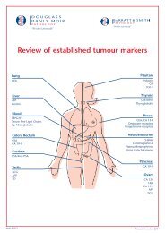
Doctors’ <strong>Newsletter</strong><strong>Winter</strong>Page 2EditorialDr Colin GoldschmidtPages 3-5Diagnostic Faecal TestingDr Miriam PaulPages 6-8UnderstandingElectrophoresis - Part 2Dr Karl BaumgartPages 9-11Anti-D in the prevention ofhaemolytic disease of thenewbornDr Jonathan BlackwellPage 12Latest News• ThinPrep Imaging System- A <strong>Douglass</strong> <strong>Hanly</strong> <strong>Moir</strong><strong>Pathology</strong> First forAustralia• Hepatitis C (HCV) PCRTesting
EditorialDr Colin GoldschmidtChief Executive OfficerCore Values in the Modern Worldof <strong>Pathology</strong>The practice of pathology has changed dramatically overthe past decade. Laboratories have consolidated underthe influences of increasing demand and diminishingavailable funds per test and it would be fair to say that itis no longer feasible to provide a fully-fledged pathologyservice without the advantages of scale that high turnoverlaboratories offer.The macro industry changes have been positive forAustralian pathology. In general, services have improvedand efficiencies have been enhanced. On the other hand,the changes have introduced new levels of complexity,which present fresh challenges in the running ofcontemporary pathology practices. Some of thesubstantive challenges in today’s setting include theenormous range of test platforms on offer, automationand its impact on laboratory design and workflow, ITmainframe systems and networks and electronictransmission of results.Perhaps the most significant modern challenge is therequirement to create cohesion from, and to maintain thestandards of, the large number of people now involved inindividual pathology operations. <strong>Douglass</strong> <strong>Hanly</strong> <strong>Moir</strong><strong>Pathology</strong> is Australia’s largest pathology practice,employing in excess of one thousand people. Out of themyriad functions carried out by our staff, we hope thatour final product presents to you as a rounded, highquality service that meets or even exceeds yourexpectations.Despite the varied roles of our staff, there has been aneed to formally unify our behaviour at work as a meansto coalesce our endeavours into a single outcome –consistent, high quality pathology service. This needculminated in the elaboration of a set of Core Values,determined by our own people as the benchmark of ourperformance at work, the essential “way of life” at DHM.These five plain English statements are now set in stoneas our guiding lights, designed to apply to every singleperson in the practice, regardless of job description orseniority. And, while for internal use only, our core valuesmay also serve to signify to you, our customer, howseriously we take our jobs and how personally we takeour responsibility to you and your patients.I wish to thank you very much for your ongoing supportand hope you will enjoy this edition of our Newletter.With warm regards,Dr Colin GoldschmidtChief Executive Officer<strong>Douglass</strong> <strong>Hanly</strong> <strong>Moir</strong> <strong>Pathology</strong>/Barratt & Smith <strong>Pathology</strong>2
Diagnostic Faecal Testingroutinely reported. Aeromonas and Plesiomonas arereported when present but it is debatable whether theseagents cause diarrhoea, as they may be isolated fromasymptomatic people.Dr Miriam PaulMicrobiologistVibrio selective agarThe Medicare Item Numbers formicrobiological examination of faeceswere changed in mid-2003. The mostsignificant changes are the restriction toone culture and two parasiteexaminations per week instead of three.There is now provision for additionalspecific testing, however, for pathogenssuch as Giardia, Cryptosporidium,Rotavirus and Adenovirus.Bacterial GastroenteritisSalmonella selective agarsTraveller’sdiarrhoea ismost frequentlycaused by enterotoxigenic E.coli.However, because these strains of E.coli are notreadily distinguishable from commensal E.coli, theycannot be identified in a routine examination of faeces.Either norfloxacin or cotrimoxazole can be used if empirictreatment is required.Microscopy for white and red cells is no longer beingperformed routinely. This is because the test is unreliable,with white cells being only intermittently present andunevenly distributed through the faecal specimen. Cellsare also broken down during specimen transport. Thepresence of blood in the specimen can be confirmed byoccult blood testing if necessary.Culture is always performedfor Salmonella, Shigella andCampylobacter. If the patienthas travelled overseas (pleasespecify in clinical notes) or has verywatery diarrhoea, then the specimen is also inoculated onto selective agar for Vibrio.Approximately 90% of cases will be positive on a singlespecimen. Salmonella and Campylobacter usually causeself-limited diarrhoea that does not require antibiotics,but sensitivity testing is available if the patient has severeor prolonged illness. Salmonella carriage may persist forseveral weeks after diarrhoea settles so the patientshould be meticulous about hand washing over this time.Antibiotic treatment is always recommended for Shigella,as it is a highly infectious organism, and sensitivities areProtozoan GastroenteritisGiardia is a common cause of diarrhoea. The laboratory isnow routinely performing an antigen test for this organismon all stool culture samples. This test is 95% sensitive ona single stool specimen, in comparison to microscopywhich is only 50-70% sensitive on a single specimen.Cryptosporidium is another common agent ofgastroenteritis. It causes self-limited diarrhoea, whichlasts 10-14 days and has a high cross-infection rate of80% within families. No treatment is required inimmunocompetent people. The laboratory is nowperforming routine antigen testing for this agent on allstool samples sent for culture.Cyclospora causes prolonged diarrhoea for many weeksin overseas travellers. If suspected, examination for thisshould be specifically requested or the history of travelmade clear. Treatment is with cotrimoxazole.3
Diagnostic Faecal TestingViral GastroenteritisViral testing must be requested specifically. Faeces canbe tested for Rotavirus, which is the commonest causeDietamoeba fragilis is also more often a commensal thana pathogen, but may be treated with doxycycline ormetronidazole. For optimal examination for this agent,faeces should be placed in transport mediumimmediately after collection and mixed well.Entamoeba histolytica, which causes amoebic dysentery,is morphologically indistinguishable from another amoebacalled Entamoeba dispar, which is nonpathogenic. Theclinical significance of finding cysts of E.histolytica/disparin the faeces depends on the travel history as well as thepresence of symptoms. Amoebic serology is only positivein about 50% of invasive amoebic dysentery.Antibiotic Associated DiarrhoeaThis can be caused by many different antibiotics.Clostridium difficile toxin testing should be requestedspecifically. Treatment is usually with oral metronidazole.Giardia and cryptosporidium antigen testingof gastroenteritis in infants, and also for Adenovirus. Inan outbreak situation, Norovirus testing may be useful.Faecal Occult Blood TestingA Medicare rebate is still available for three specimensfor this testing. Two different methods are used, asdirected by the Medicare schedule, as they have differentsensitivities. Problems that can occur include falsepositiveguaiac test results due to dietary intake such asred meat, etc. The monoclonal (immunochemical) methodis specific for human haemoglobin. It is more sensitivefor lower GIT bleeding but less sensitive for upper GITbleeding because the digestive process destroyshaemoglobin.Other Intestinal ParasitesInitial faecal specimens are all examined for ova, cystsand parasites using a concentration technique to improvethe yield. However, parasite excretion may be intermittentand repeated examination required, so if infection withhelminths, amoebae, etc. is suspected then examinationof a second specimen is recommended. Medicare willonly cover two parasite examinations in a one-weekperiod.Blastocystis is a frequently identified parasite and isregarded as a nonpathogenic commensal organism innearly all cases. It is rarely the cause of diarrhoea andshould only be treated if all other causes have beenexcluded, eg. after normal colonoscopy. Treatment iswith either metronidazole or cotrimoxazole.Helicobacter Faecal AntigenThis test has slightly lower sensitivity than the ureabreath test. However it is particularly useful for peoplewho are unable to perform a breath test, such as childrenand the intellectually disabled, as a positive result isreliable. There are no Medicare restrictions on theordering of this test. Ideally people should have a similarperiod off antibiotics and proton pump inhibitors prior tocollection, as with the breath test, otherwise falsenegative results may occur.For any enquiries, please contactDr Miriam Paul, Microbiologiston 98 555 287.4
Diagnostic Faecal TestingFaeces sample processing5
Understanding Electrophoresis - Part 2Minor plasma proteins in this zone also include fibrinogen(which also occurs in the beta region), C-reactive proteinand lysozyme. Low levels of gammaglobulins occur withreduced synthesis in hypogammaglobulinaemia andwith protein-losing states while elevated levels occurwith increased production which can be monoclonal orpolyclonal.Dr Karl BaumgartDirector of ImmunologyThis is the second of two articles explaining serumprotein electrophoresis and serum protein tests. Theprevious article was published in our Autumn <strong>2004</strong>issue and deals with abnormalities of the globulins,including paraproteins; this article describesabnormalities of non-immunoglobulin proteins.Part Two: Abnormalities ofnon-Immunoglobulin ProteinsThe five zones of the EPG: what proteinsare in these zones?There are five major zones in the EPG. The first zone onthe EPG mainly consists of albumin and someprealbumin.The next zone, alpha-1-globulin, mainly consists ofalpha-1-antitrypsin, and deficiency of alpha-1-antitrypsinis usually easily identified on the EPG. Minor plasmaproteins in this zone include alpha-lipoproteins (HDL),alpha-1-antichymotrypsin, orosomucoid andalpha-fetoprotein. Acute inflammation results inincreased levels of alpha-1-globulins.Alpha-2-globulin consists of alpha-2-macroglobulin andhaptoglobin. Minor plasma proteins in this zone includeceruloplasmin and Gc-globulin (Vitamin D bindingprotein). Elevation of alpha-2-globulin occurs withinflammation and in the nephrotic syndrome whenalpha-2-macroglobulin levels are markedly increased.Beta globulin mainly consists of beta-lipoprotein (LDL),transferrin and the C3 component of complement. Minorplasma proteins in this zone include beta-2-microglobulin, haemopexin and fibrinogen. Isolatedincreases in the beta zone can occur with chronicinflammation as well as with plasma cell dyscrasias inwhich a paraprotein (usually IgA or IgM) haselectrophoretic mobility outside the gamma region.Albumin Alpha-1 Alpha-2 Beta GammaThe gammaglobulins mostly consist 5. POLYCLONAL of the GAMMOPATHYimmunoglobulins although the variable regions of thesemolecules may result in their also migrating into otherzones in the EPG, usually in trace amounts, unless theyare produced in excess as a paraprotein.What information do I get from the EPG?The important qualitative information the EPG canprovide includes different patterns of proteins with acuteand chronic inflammation, protein-losing enteropathies,nephrotic syndrome, chronic liver disease, alpha-1-antitrypsin deficiency, hypogammaglobulinaemia and thedetection of paraproteins. These patterns are shown inFigures 1-8. In these figures, the normal pattern is shownas a black line. Relevant tests for the most likelyconditions causing these patterns are listed in the table.For a discussion on paraproteins, please refer to theprevious article in our Autumn <strong>2004</strong> issue.The quantitative information provided by the EPGincludes the concentration of paraproteins as well as theconcentration of groups of proteins within a zone orregion of the EPG tracing.How should we best use theseinvestigations in clinical practice?The qualitative information from the EPG can be useful inassessing the likelihood, severity and progress of theclinical conditions identified by these different patterns.Identification of alpha-1-antitrypsindeficiencyThe quantitative information from the EPG is very usefulfor assessment of the likelihood of alpha-1-antitrypsindeficiency since alpha-1-globulin is almost entirely alpha-1-antitrypsin. Usually all of the other zones are normal inthis instance (Figure1).Albumin Alpha-1 Alpha-2 Beta Gamma1. LOW ALPHA-1-GLOBULINIf the alpha-1-globulin level is low, then quantitation of thealpha-1-antitrypsin and genotyping studies are indicated.6
Albumin Alpha-1 Alpha-2 Beta GammaAlbumin Alpha-1 Alpha-2 Beta Gamma1. LOW ALPHA-1-GLOBULINUnderstanding ElectrophoresisAlbumin Alpha-1 Alpha-2 Beta Gamma- Part 25. POLYCLONAL GAMMOPATHY5. POLYCLONAL GAMMOPATHYAlbumin Alpha-1 Alpha-2 Beta Gamma1. LOW ALPHA-1-GLOBULINAlbumin Alpha-1 Alpha-2 Beta Gamma5. POLYCLONAL GAMMOPATHYAlbumin Alpha-1 Alpha-2 Beta Gamma1. LOW ALPHA-1-GLOBULINAcute and Chronic Inflammatory StatesAcute inflammatory states Albumin are characterised Alpha-1 Alpha-2 Beta by a Gammareduction in albumin with 1. moderate MONOCLONAL increase GAMMOPATHYinalpha-1-globulin and a markedAlbuminincreaseAlpha-1 Alpha-2inBeta Gammaalpha-2-globulin (Figure 2). 1. MONOCLONAL GAMMOPATHYAlbumin Alpha-1 Alpha-2 Beta Gamma1. MONOCLONAL GAMMOPATHYAlbumin Alpha-1 Alpha-2 Beta Gamma2. ACUTE INFLAMMATIONAlbumin Alpha-1 Alpha-2 Beta Gamma2. ACUTE INFLAMMATIONAlbumin Alpha-1 Alpha-2 Beta Gamma2. ACUTE INFLAMMATIONChronic inflammatory states are characterised byreduction in albumin, borderline reduction in betaglobulin and substantial increases in alpha-1, alpha-2Albumin Alpha-1 Alpha-2 Beta Gammaand gammaglobulin (Figure 3).8. HYPOGAMMAGLOBULINAEMIAChronic Liver Disease and PolyclonalHypergammaglobulinaemiaChronic liver disease is usually associated with a patterncalled “beta-gamma bridging” in which there ispolyclonal hypergammaglobulinaemia and the normallyAlbumin Alpha-1 Alpha-2 Beta Gammadiscernible gap between the beta and gamma regions is“filled in” (Figure 4). 6. NEPHROTIC SYNDROMEPolyclonal gammopathy, mainly IgG, is more evenlydistributed within the gamma region and occurs with avariety of chronic inflammatory disorders of whichSjogren’s syndrome would be quite common (Figure 5).Protein-Losing StatesAlbumin Alpha-1 Alpha-2 Beta Gamma8. HYPOGAMMAGLOBULINAEMIAAlbumin Alpha-1 Alpha-2 Beta Gamma8. HYPOGAMMAGLOBULINAEMIAAlbumin Alpha-1 Alpha-2 Beta Gamma6. NEPHROTIC SYNDROMEAlbumin Alpha-1 Alpha-2 Beta Gamma6. NEPHROTIC SYNDROMEAlbumin Alpha-1 Alpha-2 Beta Gamma7. PROTEIN-LOSING ENTEROPATHYAlbumin Alpha-1 Alpha-2 Beta Gamma7. PROTEIN-LOSING ENTEROPATHYAlbumin Alpha-1 Alpha-2 Beta Gamma7. PROTEIN-LOSING ENTEROPATHYPatients with nephrotic syndrome lose albumin, and to alesser extent, larger proteins from the other regions intothe urine but alpha-2-macroglobulin is such a largemolecule that the alpha-2-globulins actually risedramatically (Figure 6).Albumin Alpha-1 Alpha-2 Beta Gamma1. ADNANCED LIVER DISEASEAlbumin Alpha-1 Alpha-2 Beta GammaIn protein-losing enteropathy,1. ADNANCEDalpha-2-globulinLIVERisDISEASEalsolost so all zones are markedly Albumin reduced Alpha-1 Alpha-2 (Figure Beta 7). Gamma1. ADNANCED LIVER DISEASEAlbumin Alpha-1 Alpha-2 Beta GammaAlbumin Alpha-1 Alpha-2 Beta Gamma5. POLYCLONAL GAMMOPATHY5. POLYCLONAL GAMMOPATHYAlbumin Alpha-1 Alpha-2 Beta Gamma3. CHRONIC INFLAMMATIONAlbumin Alpha-1 Alpha-2 Beta Gamma3. CHRONIC INFLAMMATIONAlbumin Alpha-1 Alpha-2 Beta Gamma3. CHRONIC INFLAMMATIONAlbumin Alpha-1 Alpha-2 Beta GammaAlbumin Alpha-1 Alpha-2 Beta Gamma1. MONOCLONAL GAMMOPATHY1. MONOCLONAL GAMMOPATHYAlbumin Alpha-1 Alpha-2 Beta Gamma4. BETA-GAMMA BRIDGINGAlbumin Alpha-1 Alpha-2 Beta Gamma4. BETA-GAMMA BRIDGINGAlbumin Alpha-1 Alpha-2 Beta Gamma4. BETA-GAMMA BRIDGINGAlbumin Alpha-1 Alpha-2 Beta GammaAlbumin Alpha-1 Alpha-2 Beta Gamma8. HYPOGAMMAGLOBULINAEMIA8. HYPOGAMMAGLOBULINAEMIAAlbumin Alpha-1 Alpha-2 Beta Gamma5. Albumin POLYCLONAL Alpha-1 Alpha-2 Beta GAMMOPATHYGamma1. POSSIBLE MALIGNANCYAlbumin Alpha-1 Alpha-2 Beta Gamma1. POSSIBLE MALIGNANCYAlbumin Alpha-1 Alpha-2 Beta Gamma1. POSSIBLE MALIGNANCYAlbumin Alpha-1 Alpha-2 Beta Gamma6. NEPHROTIC SYNDROME6. NEPHROTIC SYNDROMEAlbumin Alpha-1 Alpha-2 Beta GammaAlbumin Alpha-1 Alpha-2 Beta Gamma1. MONOCLONAL GAMMOPATHYAlbumin Alpha-1Alpha-11. LOW A1. LOW AAlbuminAlbumin Alpha-1AlbuminAlpha-12. ACUT2. ACUTAlbumin Alpha-1Alpha-13. CHRO3. CHROAlbuminAlbumin Alpha-11. LOW AAlbumin AlphaAlbuminAlpha4. BET4. Alpha-1 BET2. ACUTAlbuminAlbumin Alpha-1 Alpha-2 Beta GammaAlbumin Alpha-1 Alpha-2 Beta Gamma7. PROTEIN-LOSING ENTEROPATHY7. PROTEIN-LOSING ENTEROPATHYAlbumin AlphAlph1. POS71. POSAlbuminAlbumin Alpha-1 Alpha-2 Beta Gamma Albumin Alpha-1
Understanding Electrophoresis - Part 2Albumin Alpha-1 Alpha-2 Beta Gamma1. MONOCLONAL GAMMOPATHYAlbumin Alpha-1 Alpha-22. ACUTE INHypogammaglobulinaemiaLone hypogammaglobulinaemia, however, almost alwaysresults from synthetic failure either secondary tounderlying haematological malignancy or primaryantibody deficiency (Figure 8).Albumin Alpha-1 Alpha-2 Beta Gamma8. HYPOGAMMAGLOBULINAEMIAAlbumin Alpha-1 Alpha3. CHRONICDIAGNOSTIC TESTS TO CONSIDER WHEN THE EPG IS ABNORMALPATTERN MAIN DISORDERS IMPORTANT TESTSACUTE INFLAMMATION Infection, Inflammatory disordersESR, CRP, MSU, Throat swab, Bloodcultures, Arthritis markers; if relevant,Viral serology, ASOT, DNAseB.Albumin Alpha-1 Alpha-2 Beta Gamma6. NEPHROTIC SYNDROMEAlbumin Alpha-1 Alp4. BETA-GCHRONIC INFLAMMATIONAutoimmune diseases (Sjogren'ssyndrome, autoimmune liver disease,connective tissue disease), chronic liverdisease, bronchiectasis, osteomyelitisand malignancy.ANA, ENA, RF, SMA, AMA, LFT,Iron studies, Haemochromatosisgene assay. Imaging for possiblemalignancy.BETA-GAMMA BRIDGINGLFT, Albumin, INR, Ammonia,Albumin Alpha-1IronAlpha-2studies,BetaCopper,GammaCaeruloplasminChronic liver disease of any aetiology7. PROTEIN-LOSINGHaemochromatosisENTEROPATHYgene assay, Hep B, Cmarkers; SMA, AMA.Albumin Alpha-1 Al1. POSSIBPOLYCLONAL GAMMOPATHYChronic inflammatory disordersANA, ENA, RF, Thyroid function andantibodies.NEPHROTIC SYNDROMEPROTEIN-LOSING ENTEROPATHYNephrotic syndromeGastrointestinal protein lossFBE, ESR, CRP, Biochemistry, ANA, ENA,RF, ANCA, GBM Ab, C3, C4, 24 hour urineProtein, Creatinine, Creatinine clearance.EPG/IFE serum, urine.Albumin Alpha-1 Alpha-2 Beta Gamma1. ADNANCED LIVER DISEASEIgG, IgA, IgM, Gliadin, TTG antibodies,FBE, ESR, CRP, Biochemistry, Vit D, B12,Folate, Iron studies.HYPOGAMMAGLOBULINAEMIAPrimary or secondary antibody deficiencydisordersIgG, IgA, IgM, IgG subclasses, FBE, ESR,CRP, Biochemistry, T, B, NK lymphocytesubsets, IFE serum, urine; Urine lightchains, Beta-2-microglobulin.LOW ALPHA-I-GLOBULIN Alpha-1-antitrypsin deficiency Alpha-1-antitrypsin level and genotype.For any enquiries, please contact Dr Karl Baumgart, Director of Immunology on 98 555 286.8
Anti-D in the prevention of haemolytic diseaseof the newbornUltimately this may end in perinatal death due to HydropsFetalis. HDN may persist up to three months after thebaby is born due to persistence of the maternalantibodies in the fetal circulation.Dr Jonathan BlackwellHaematologistAbout 20% of babies to mothers with detectable maternalanti-D will be severely affected either by Hydrops Fetalisor severe HDN. About half the susceptible pregnanciesare not affected or only mildly affected. Jaundice maydevelop early after delivery, i.e. within one or two days.The development of severe jaundice may peak at 3 to 5days postpartum. HDN may require exchange transfusionif severe, or phototherapy in milder cases.Haemolytic Disease of the Newborn andthe Role of Anti-D ProphylaxisHaemolytic disease of the newborn (HDN) is a result ofsensitisation of the mother’s immune system to foreignred cell antigens. This sensitisation results in the passageof maternal antibodies across the placenta to the fetus.These antibodies bind to fetal red cells causinghaemolysis, leading to morbidity and sometimesmortality in the infant.Rh D disease is the most common form of haemolyticdisease of the newborn. It occurs only in Rh negativewomen.HDN IncidenceHaemolytic disease of the newborn consists of asensitising episode and a subsequent pregnancy.The most common sensitising event is fetomaternalhaemorrhage (FMH). This occurs in up to 96% of allpregnancies and particularly in the third trimester and atthe time of delivery. Usually the volume of FMH is small i.e.less than 1mL, but is often sufficient for sensitisation. Theantibody develops within six months. Sensitisation occursin about 16% of at risk women for each pregnancy.Infusion of a blood product, e.g. red cells, may also bethe sensitising event. In this instance a single unit of Rhpositive blood will immunise up to 70% of susceptiblemothers.Development of Rh antibodies andClinical EffectsThe Rh D form of HDN occurs when a Rh negativemother who has been sensitised to Rh D has asubsequent pregnancy with a baby that has Rh Dantigens expressed on its red blood cells. The maternalIgG antibodies against Rh D cross the placenta. Afterapproximately 24 weeks this may lead to haemolysis ofthe fetal red blood cells predominantly in the fetal spleen.In the fetus this leads to compensatory extramedullaryhaemopoiesis in the liver, spleen, kidneys and adrenals.This in turn may lead to portal or umbilical venoushypertension and the baby may subsequently developascites and pleural effusions and hypoproteinaemia.Initial VisitJaundice in haemolytic disease of the newborn.INDICATIONRh Antibodies foundOther Antibodies foundRh D negative motherThird TrimesterAntibody screenANTENATAL TESTINGTESTABO & Rh DAntibody Screen (withidentification and titration ofantibody)Repeat at 18-20 weeks.Further testing will be requiredin consultation withobstetrician.Additional testing at 28weeks, prior to first dose ofRh D immunoglobulinSuggested if mother hasa history of antibodies,traumatic delivery or bloodtransfusionsAll women should be tested for ABO and Rh D as earlyas possible during each pregnancy, preferably at the firsttrimester visit. Weak Rh D should be considered as Rh Dpositive.9
Anti-D in the prevention of haemolytic diseaseof the newbornAntibody Screening (see table 1)All women should be tested during pregnancy forclinically significant unexpected antibodies and anyalloantibodies detected should be fully identified.Antiglobulin testing with IgG should be done to identifythose antibodies with potential to cross the placenta andcause HDN.Repeat testing at 28 weeks is currently controversial. Therisk of developing Rh D alloantibodies between the firsttrimester visit and 28 weeks is low. However repeattesting of Rh D negative women at 28 weeks prior toadministering Rh D is becoming accepted, as is theelimination of routine antibody screening at 34 weeks.Repeat testing in the third trimester is appropriate whenthere is a history of significant antibodies, bloodtransfusion or traumatic deliveries.Detection of Fetomaternal HaemorrhageFetomaternal haemorrhage is not usually manifestclinically, but does occur in nearly all pregnancies. Thepresence of fetal cells in the maternal circulation isdetermined by the Kleihauer test or by flow cytometry.The Kleihauer test is based on fetal haemoglobin beingresistant to elution by acid. On a suitably stained bloodfilm the fetal cells show up brightly whilst the adult cellsare pale “ghost” cells.Testing at DeliveryINFANT CORD BLOODINDICATIONTESTRh D negative mother Rh D (including weak D)Potentially SignificantMaternal AntibodiesABO & Rh D and DAT(Coombs)Neonate with clinicalHDN but no maternalantibodiesABO & Rh D and DAT(Coombs)Cord blood eluates, andtesting of Paternal blood afterconsultationBlood film in HDN showing numerous nucleated red blood cells.Other Forms of Haemolytic Diseaseof the NewbornINDICATIONRh D negative motherwith Rh D positive babyNo previous maternal bloodgroupMATERNAL TESTINGTESTTest for fetomaternalhaemorrhageeg. Kleihauer testABO & Rh D and antibodyscreenRh D haemolytic disease is the most common form ofHDN. With the use of Rh immunoglobulin prophylaxis,other forms of haemolytic disease will be as important asclassic Rh disease.Similar mechanisms for haemolytic disease of thenewborn can occur with other components of the Rhsystem, such as c, C, e and E, as well as the Kell, Duffy,Kidd and M and S systems.Anti-c and anti-Kell and anti-E may induce haemolyticdisease as severe as Rh disease, i.e. the baby maydevelop HND or even Hydrops Fetalis. In addition, anti-C,anti-e, and anti-k may also be associated with severeHDN. Other blood group antibodies tend to beassociated with milder HDN.Antibodies to Lewis, Lutheran, and anti-P do not lead toclinical disease.10
Anti-D in the prevention of haemolytic diseaseof the newbornPrevention of Rh D Haemolytic Disease ofthe NewbornHaemolytic disease of the newborn due to Rh D antibodymay be prevented by using Rh D immunoglobulin. Byadministering Rh immunoglobulin to non-sensitised Rhnegative women with a Rh positive infant at the time ofdelivery, the incidence of Rh immunisation has beenreduced from around 12% to 2%. Further reduction in Rhimmunisation may be achieved by giving antepartum Rhimmunoglobulin prophylaxis at 28 and 34 weeks.Similarly, if the mother has a miscarriage, antepartumhaemorrhage or any manipulations of the baby or thepregnancy, then further Rh immunoglobulin should begiven at that time, the amount depending on the stage towhich the pregnancy has progressed.The National Health and Research Council has issuedguidelines recommending that all women at risk of Rhdisease receive prophylaxis. Until recently there has beeninsufficient Rh immunoglobulin to fully implement theseguidelines. With greater availability of Rh immunoglobulinthe guidelines are now being implemented.For best practice the 1999 NHMRCGuidelines recommend:Administer Rh D Immunoglobulin as soon as possibleafter the sensitising event. Should be given within72 hours for successful immunoprophylaxis.If more than 72 hours has elapsed, a dose offered within9-10 days may provide protection.Rh D Immunoglobulin should not be given to women withpreformed anti-D antibodies (unless the preformed anti-Dis due to the antenatal administration of Rh DImmunoglobulin).Rh D Immunoglobulin 100 IU will protect against afetomaternal haemorrhage of 1.0 mL of fetal red cells(2.0 mL whole blood).The magnitude of the FMH following a sensitising event(including delivery) should be quantified to ensure anadequate dose of Rh D Immunoglobulin (more than onedose may be required).FOR EACH SENSITISING EVENTIst Trimester 2nd, 3rd Trimester PostpartumRh DImmunoglobulin250 IU*Rh DImmunoglobulin625 IUWinRho600 IU* In multiple pregnancy or where the gestational age is indoubt (? >13 weeks) 625 IU should be given.ROUTINE PROPHYLAXIS FOR THE 1ST PREGNANCY28 Weeks 34 WeeksRh DImmunoglobulin625 IURh DImmunoglobulin625 IUThe dose should be given as soon as possible and within72 hours of the event. Sensitising events include normaldelivery, miscarriage, termination of pregnancy, ectopicpregnancy, chorionic villus sampling, amniocentesis,cordocentesis, abdominal trauma considered sufficientto cause fetomaternal haemorrhage, antepartumhaemorrhage and attempted external cephalic version.It is recommended that the magnitude of fetomaternalhaemorrhage is assessed and further doses given asnecessary. As a guide, a dose of approximately 600 IUwill protect against a fetomaternal haemorrhage of upto 6 mL of Rh D positive fetal red blood cells. Forhaemorrhages greater than 6mL, the recommended doseis 100 IU per mL Rh D positive fetal red blood cells.Our ability to prevent of HND has resulted in anoutstanding reduction in numbers of affected babies; itscontinuing success with depend on the on-goingavailability of Rh D immunoglobulin and appropriate usewithin the community.Tests to assess the volume of FMH include the Kleihaueracid elution test and flow cytometry. Currently, the formertest is used in our laboratory. The majority of fetal bleedsare less than 5 mL of red blood cells. In about 50% ofcases, FMH is less than 0.05 mL. Rarely, in up to 0.6% ofcases, FMH is 30 mL or greater.For any enquiries, please contactDr Jonathan Blackwell, Haematologiston 98 555 363.11
Latest News• ThinPrep Imaging System - A <strong>Douglass</strong> <strong>Hanly</strong> <strong>Moir</strong> <strong>Pathology</strong> First for Australia• Hepatitis C (HCV) PCR TestingThinPrep Imaging System - A <strong>Douglass</strong> <strong>Hanly</strong> <strong>Moir</strong> <strong>Pathology</strong> First for AustraliaWe are pleased to announce that <strong>Douglass</strong> <strong>Hanly</strong> <strong>Moir</strong> <strong>Pathology</strong> has recently installed the ThinPrep ImagingSystem, the first installation of its kind outside the United States and Europe. Following a period of validation,we anticipate using this new and exciting technology to screen our ThinPrep samples from most of our laboratoriesby late winter <strong>2004</strong>.The ThinPrep Imaging System is an interactive computer system that helps cytotechnologists and cytopathologists toscreen and diagnose ThinPrep Pap Test slides. Its significant innovation is that it presents to the cytotechnologist onlythose portions of each slide that contain the most meaningful data for review. This permits rapid resolution of the vastmajority of slides that have no cellular abnormalities, so the cytotechnologist can focus on the slides that show potentiallysuspicious cells.With our commitment to women’s health, <strong>Douglass</strong> <strong>Hanly</strong> <strong>Moir</strong> <strong>Pathology</strong> has had a particular interest in cytologyautomation for many years with much experience gained from our use of Papnet, an earlier automated system designedto read conventionally prepared smears.In June 2003, the US Food and Drug Administration (FDA)announced the approval of the ThinPrep Imaging Systemas a primary screening tool, based on the results of amulti-site trial designed to evaluate the routine screening ofthe ThinPrep Pap Test slides using the ThinPrep ImagingSystem compared to manual review of ThinPrep Pap Testslides. Cytotechnologist screening rates were alsoevaluated during the study to determine workload limits.It appears that this, and possibly other automated systems,will offer significant benefits, eventually allowing a Medicarerebate for liquid based cytology. The data from overseas isexcellent and allows us to feel confident in its use. Inaddition, we will be performing ongoing local assessmentsand the data will be collected in a form suitable forappropriate scientific analysis.Senior cytologist reviewing ThinPrep slides.Australia has every right to be very proud of its Cervical Screening Program, which has been very successful in thereduction in incidence and mortality from cervical cancer over the last ten years. Although this success has been basedentirely on the conventional Pap smear, Australia should not ignore major developments occurring overseas. This is a timeof great flux in the area of cervical screening and with improved testing, women may need fewer smears and screeningmay be more targeted. Automation does offer great potential within the laboratory and we anticipate that the ThinPrepsystem will be one of the major systems that offer these significant advantages.For any enquiries, please contact Dr Annabelle Farnsworth, Director of Cytopathology on 98 555 180.Hepatitis C (HCV) PCR TestingQualitative Hepatitis C (HCV) PCR testing is used to detect viraemia. As such, it provides information about currentinfectivity and the likelihood of progression to chronic liver disease. From May 1st, <strong>2004</strong>, there is a Medicare rebatefor one qualitative HCV PCR test in a 12 month period for any patient who is seropositive for Hepatitis C. Therequirement for two normal liver function tests has been removed.For any enquiries, please contact Dr Ian Chambers, Director of Microbiology on 98 555 330.12DOUGLASS HANLY MOIR PATHOLOGY • ABN 80 003 332 858A subsidiary of SONIC HEALTHCARE LIMITEDBARRATT & SMITH PATHOLOGY • ABN 49 452 500 422A subsidiary of SONIC HEALTHCARE LIMITED95 EPPING ROAD • MACQUARIE PARK • NSW 2113 • AUSTRALIATEL 98 555 222 • FAX 9878 5077MAIL ADDRESS • LOCKED BAG 145 • NORTH RYDE • NSW 1670 • AUSTRALIA37 BROMLEY ROAD • EMU PLAINS • NSW 2750 • AUSTRALIATEL 4735 6166 • FAX 4735 6189MAIL ADDRESS • PO BOX 443 • PENRITH • NSW 2751 • AUSTRALIA