clevelandclinicmagazine - Best Hospitals, US News best hospitals
clevelandclinicmagazine - Best Hospitals, US News best hospitals
clevelandclinicmagazine - Best Hospitals, US News best hospitals
- No tags were found...
Create successful ePaper yourself
Turn your PDF publications into a flip-book with our unique Google optimized e-Paper software.
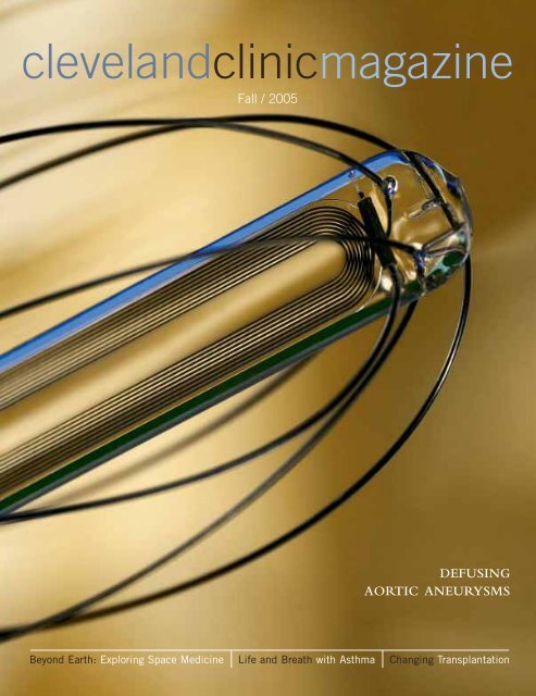
<strong>clevelandclinicmagazine</strong>Fall / 2005DEF<strong>US</strong>INGAORTIC ANEURYSMSBeyond Earth: Exploring Space Medicine Life and Breath with Asthma Changing Transplantation
Sophisticated.The finest traditions of internationalservice and elegance live at the ClevelandClinic InterContinental Hotel & MBNAConference Center.Elegant guest rooms, high-tech amenitiesand easy access to medical facilities makethis world-class hotel your choice for theperfect accommodations in Cleveland.Classics, the hotel’s AAA Five-Diamondrestaurant, offers the mostsophisticated dining experience betweenNew York and Chicago.For reservations, call1.877.707.8999
contentscontentscontentscontentscontentscontentscontentscontentscontentscontentscontentsIn This IssueThe First Word 3Metrics Make SenseThe Medicine ChestOf Masks and Men 4Artificial DiscProvides Real Relief 4Metal-Free Heart Patch 5New View forDetecting Heart Blockages 6Better Foot Healthand Diabetes 7On the WebDiagnosis Challenge 8The Patient With the Can’t-Stop CoughPhysicians see hundreds of patients a year,with every day bringing new challenges. Inthis section, we offer our readers the chanceto follow a physician through the diagnosisprocess. What diagnosis would you make?Living Healthy 9The Unhealthy TanLast year, more than 50,000 Americans werediagnosed with melanoma, most of them overthe age of 40. But what’s catching the eyesof the experts is that now more teenagersand young adults are learning they have thedisease, too.Readers’ Survey 9We want to hear from you.Readers’ Poll 9The Gift of Life: Organ DonationAsk the Experts 32Statin IntoleranceMillions of patients take statins to treat theirhigh cholesterol. While most patients have noproblems taking these drugs, a small numberexperience intolerable side-effects. Heartspecialist Steven Nissen, M.D., discusses thealternatives these statin-intolerant patientshave to lower cholesterol and the risk of heartattack and stroke.PhilanthropiaAlbert Maroone: 34Good Business in Cars and MedicineKaren Wilson: 35Building Hope Through Cancer ResearchDana Hamel: 36Helping Others When Fortune SmilesRobert Tomsich: 37Sharing the Passion to GiveFlorida Focus 38A Good Night’s SleepFor many people, getting enough sleep iseasier said than done. The Sleep DisordersProgram at Cleveland Clinic Florida Westondiagnoses and treats patients for sleep apneaand other sleep disturbances.On the HorizonLights, Camera ...Prevent Knee Injuries 41The Chemotherapy Clock 42Turning Off IBD Triggers 42Sniffing Out Cancer 43Tissue Damage Earlyin Multiple Sclerosis 43My Story 44This Soldier’s BattleBy Corey Carteras told to Cleveland Clinic Magazine“I knew something was going down. Therewere too many people standing around. Juststanding and looking. We had stoppedtraffic to let the convoy pass. Suddenly, a taxipulled up. Two men got out and startedwalking away...”To read more stories from this issue goto our Web site: www.clevelandclinic.org/<strong>clevelandclinicmagazine</strong>2 cleveland clinic magazine
themedicinechestthemedicinechestthemedicinechestthemedicinechestthemedicinechestMETAL-FREE HEART PATCHFor several decades doctors have been tryingto find ways to close holes in the heartwithout the need for open-heart surgery.Now, pediatric heart specialists at The Children’s Hospital at TheCleveland Clinic have a completely metal-free patch that uses thebody’s own healing power to seal up the hole. This option wasespecially appealing to the parents of seven-year-old Kelly Horn.Kelly was diagnosed with a special kind of hole in her heart,called an atrial-septal defect or ASD, which is located betweenthe heart’s left and right filling chambers. Without repair, oxygenrichblood can back up into the lungs, causing breathing difficulty,heart rhythm problems, heart failure or even stroke.Although surgery was once standard for the four percent ofchildren like Kelly born with ASD, Kelly and her family were offeredthree non-surgical treatment possibilities. The option theychose, the “transcatheter patch,” is made of stretchy polyurethanefoam that bonds to the hole’s edges without sutures or wire.Placed LeBron against James the heart defect, the patch is held in place for 48hours by a small balloon. Gradually the patient’s own tissue growsover the patch, making it stick to the edges of the hole. The tissuecontinues to grow over several months, eventually coveringthe patch.This treatment option also appealed to the Horn family becauseKelly has a nickel allergy, discovered when she developeda reaction to nickel earrings. Although the presence of nickel inthe metal-based devices has not been shown to cause significantproblems, Kelly’s parents felt more comfortable not having nickelor any other kind of metal in her heart. After the procedure, Kelly’smother kept her active daughter in bed for two days in thehospital, while the patch fused with the lining of Kelly’s heart,permanently sealing the hole.“We feel that this patch closure is a great improvement. It’salmost like having the body’s own tissue covering the defect,”says Lourdes Prieto, M.D., the interventional pediatric cardiologistwith the team who performed Kelly’s procedure.while preserving motion. This newsurgical procedure is designed toaddress pain and movement issues aswell as reduce healing time.”Dr. Orr, who performed this procedurefor several years in Canadabefore joining the Clinic, says thatthe ideal patient is between the agesof 35 and 55, has not already had surgeryin the lumbar (lower) spine andhas no signs of osteoporosis and noscoliosis. Although not for everyone, the surgery may benefitpatients who have a single-level degenerative disc in the lowerback and who are not experiencing nerve compression.The procedure generally takes two and a half to three hoursand patients remain in the hospital one or two days. Dr. Orrnotes that most people find that their original back pain is gonewithin a day or so; other pain resulting from the surgery usuallysubsides within four to six weeks. Although the procedure isrelatively new in the United States, about 12,000 have beenperformed worldwide.www.clevelandclinic.org 5
themedicinechestthemedicinechestthemedicinechestthemedicinechestthemedicinechestNew View for DetectingHEART BLOCKAGESThe coronary computed tomographyangiogram (CTA) is revolutionizingcardiology by revealingcoronary artery blockages withinminutes and for about one-tenth ofthe cost of invasive catheterization.Studies have shown that earlydetection of calcium deposits in thearteries can help predict whether apatient is likely to have a heartattack. Usually a patient arrivingin the emergency room with chestpains is admitted for 24-hourobservation and diagnostic testing,including X-rays and scans. Imagesproduced by the coronary CTAillustrate whether there are calciumdeposits in the arteries.“The speed, processing and qualityof images compared to othercardiac imaging, such as ultrasound,cardiac catheterization or MRI, issuperior,” explains Mario Garcia,M.D., Co-director of the Center forIntegrated Noninvasive CardiovascularImaging. The CTA scanproduces 64 images in about five to10 seconds.Just before the scan, techniciansadminister a beta-blocker to slowdown the patient’s heartbeat. Usingan electrocardiogram, the scan istimed to photograph the heart at thesame point in the pumping cycle forsix or seven beats. Once the image isreceived on the computer screen,technicians use tools similar to thosefound in graphics editing software toisolate the coronary arteries.“We can determine if soft, unstableor calcified deposits are presentin the arteries,” says Dr. Garcia,adding that those results determinehow a cardiologist can treat thepatient - with close monitoring andlifestyle changes, use of statin drugs,angioplasty, surgery or some combinationof those treatments.The coronary CTA is not withoutits downside, namely that itrequires training and expertise tomaximize its effectiveness. “It’s morecommon for physicians untrained inthis technology to see things thataren’t there,” explains Dr. Garcia.“Training is crucial.”Cardiologists also are concernedthat the coronary CTA could beoverused and lead to incorrect diagnosesand unnecessary surgical treatments.Patients need to be awarethat, as with most computed tomographyscans, it involves higher dosesof radiation exposure. In addition,some patients may be allergic to thecontrast dye used. The technology’slimitations also make it unsuitablefor extremely obese patients andthose with irregular heart rhythms.This three-dimensional CTA image showssevere narrowing of the first diagonal coronaryartery.During the cardiac catheterization procedureof the same patient, this standardtwo-dimensional angiogram image confirmedthe narrowed artery.6 cleveland clinic magazine
themedicinechestthemedicinechestthemedicinechestthemedicinechestthemedicinechestBetterFOOTHealth and DiabetesIn the United States, some 60,000 to 80,000 people withdiabetes lose part of their leg each year to amputation -usually resulting from a foot ulcer.People who have lived with diabetes for 10 to 15 years candevelop long-term complications such as peripheral neuropathy,which is a loss of nerve function and deadening of sensationin the legs and feet, and peripheral vascular disease, whichis impaired circulation in the legs and feet.“What happens is that people will injure themselves and notknow it,” says Peter Cavanagh, Ph.D., D.Sc., Academic Directorof the Clinic’s Diabetic Foot Care Program. “A commonexample is walking too far in a shoe that doesn’t fit right ordoesn’t have any cushioning.” Patients end up with first a blisteror abrasion which, if unnoticed and untreated, can becomean ulcer - a wound that penetrates through the deeper layersof skin to the underlying tissue. Then, once they have such awound, their compromised vascular status may cause them toheal more slowly.The Diabetic Foot Care Program sees about 50 to 60 peopleeach week. “When people first come to our clinic, wemap the pressure underneath their foot when they’re walking,since it’s mostly the pressure that causes ulcers. That map-(Image, below) This profile of the pressure distribution under apatient’s foot during walking - with the heel to the left and toes to theright - shows a peak of high pressure under the ball of the foot,indicating an area at high risk for ulceration.ping gives us the ability to know exactly where on the footthere may be problems in the future,” Dr. Cavanagh explains.“We know how to heal ulcers,” says Dr. Cavanagh. “Wecan do it within about six weeks, using a special fiberglasscast proven to be one of the most effective ways of healinga foot ulcer.”Patients also benefit from advanced wound-healing methodssuch as “off-loading devices,” which are inserted in theshoe to redistribute weight off the affected foot, and sophisticatedantibiotic therapy. “Bioengineered skin substitutes,which are cells grown in culture to form an artificial, livingskin, also can be grafted to heal a stubborn ulcer,” addsGeorgeanne Botek, D.P.M., Medical Director of the DiabeticFoot Care Program.The design of therapeutic footwear is a cornerstone area ofresearch for the program. “Footwear becomes absolutely key tosolving problems in the diabetic foot. We have computer modelsthat help us better understand how we can manage thesefeet to prevent them from ulcerating,” Dr. Cavanagh says.“Many people with diabetes feel that amputation is inevitablebut, in fact, it isn’t. If we can prevent the foot ulcer, there is astrong possibility that we can prevent the amputation.”www.clevelandclinic.org 7
onthewebonthewebonthewebonthewebonthewebonthewebonthewebonthewebonthewebDiagnosis ChallengePhysicians are often called upon to diagnose a wide range of symptoms andconditions. In this section, we offer our readers the chance to follow a physicianthrough the diagnosis process. What diagnosis would you make?The Patient with theCan’t-Stop CoughGary* was exhausted. He could barely keep his eyes on theroad. Not even the strongest cup of coffee could perk himup. Several times during the drive to work he caught himselfveering off the road, jerking the wheel in the other direction toget his car back on the highway.For weeks he had been suffering severe and repetitivecoughing fits. His sleep was constantly being interrupted byspasms of coughing. Midnight. 2 a.m. 4 a.m. He had hackingbouts during the day, too, often ducking out of meetings toavoid the annoyed stares of his boss and co-workers.Gary assumed it was nothing more than a cold, writing offhis symptoms as a typical winter-weather bug. But after threeweeks of excusing himself from the table at restaurants, suppressingcoughing fits in public, and waking up with chokingepisodes so violent he sometimes gagged, he decided a visitto the doctor was in order.The Office VisitCleveland Clinic physician Camille Sabella,M.D., listened to Gary explain his symptoms:several weeks of violent coughing, sometimesto the point of vomiting, and feelings of dehydration.Gary didn’t experience sinus-relatedproblems - no stuffy head or trouble breathing- other than when he couldn’t catch hisCamille Sabella, M.D.breath during a coughing fit.Gary has no history of asthma or respiratory infections,and he has no fever or abnormal temperature. Dr. Sabellainquired about Gary’s lifestyle and discovered that he isnot a smoker and maintains a fairly healthy diet, avoidinglate-night dinners or excessive alcohol consumption. Gary ismarried, without children, and his wife has not developed acough or similar symptoms. A thorough physical examinationof Gary was completely normal.Based on the described symptoms, Dr. Sabella formed afew theories about what Gary’s condition might be:Bronchitis – Gary’s cough alerted Dr. Sabella to the possibilityof this common breathing condition in which the bronchialtree inside the lungs becomes infected by a virus. Symptomsof bronchitis come on quickly and can persist for weeks.Common Cold – Winter weather and an office atmosphereharbor a slew of common cold viruses. Gary might havepicked up a virus from a co-worker. Fatigue and stress cancause colds to linger.Gastroesophageal Reflux – Characterized by a backwash ofstomach contents and acid into the airway, symptoms of thiscondition include coughing, gagging and vomiting. Also, it iscommon for these symptoms to occur at night.Pertussis (Whooping Cough) – Pertussis is a highly contagiousbacterial disease that affects the respiratory system. It producesspasms of coughing that may end in a high-pitched, deep inhalationin children, although the “whoop” is rare in adults.Asthma – With asthma, inflammation of the airways causesairflow into and out of the lungs to be restricted, producingthe characteristic wheezing sound. Mucus production also isincreased.As a next step, Dr. Sabella ordered a test for whoopingcough bacteria from the back of Gary’s nose to culture in thelaboratory. In addition, he ordered a chest X-ray.Dr. Sabella then continued with his assessment. After acareful history and physical examination, Dr. Sabella determinedthat Gary did not have bronchitis. Although bronchitissymptoms can last for a couple of weeks, Gary’s symptomswere too severe and prolonged for bronchitis.A cold was the least likely conclusion. Gary didn’t displayrelated complications, such as a stuffy or runny nose, sore throator fever. Also, colds generally run their course in two weeks.Because Gary is careful not to eat late or consume excessiveamounts of alcohol, the likelihood of gastroesophagealreflux was minimal. Reflux is common following meals, andGary experiences coughing symptoms most of the time.In addition, asthma seemed unlikely since Gary has nopast history of asthma, no history of wheezing or shortness ofbreath, and his physical examination was normal.Gary’s chest X-ray was normal and the nasal culture cameback negative for the whooping cough bacteria, meaning Dr.Sabella did not find anything in the culture that alerted himto pertussis.So, what could be the cause of Gary’s condition?For Dr. Sabella’s diagnosis, go to our Web site atwww.clevelandclinic.org/<strong>clevelandclinicmagazine</strong>You can also send us an email at<strong>clevelandclinicmagazine</strong>@ccf.org and we will emailyou the diagnosis.*The patient and history presented in “Diagnosis Challenge” are fictional.8 cleveland clinic magazine
onthewebonthewebonthewebonthewebonthewebonthewebonthewebonthewebonthewebLiving HealthyThe Unhealthy TanIn the United States, the percentage of people who developmelanoma, a form of skin cancer caused by sun exposure,has more than doubled in the past 30 years. Each year, morethan 50,000 new cases are diagnosed.While those numbers are disturbing, another alarm issounding: young people, without years of sunlight exposurelike the majority of skin cancer sufferers, are increasinglybeing diagnosed with melanoma.“Although it is still fairly rare in children, I’ve seen studieswhere cases of melanoma are found in children as young asfour years old,” says Julian Kim, M.D., a staff physician whospecializes in surgical oncology. “The concern now is thatwe are seeing people in their late teens and early 20s beingdiagnosed more and more with melanoma.”In this day and age, the mental connection between tannedskin and good health still prevails. This concept has drivenpeople to seek a sun-kissed look, whether it’s through sweatingon the beach, using artificial tanners, or “fake baking”- spending10 or 20 minutes in a tanning bed.“Although the cause of this trend of more young peoplewith melanoma is not yet known, tanning booths are probablya contributing factor,” says Dr. Kim. The major concernwith tanning beds is the extremely intense, damaging ultra-Microscopic image of skin cancer.violet (UV) rays. Tanning beds can provide hours of sunlightin minutes, and that is highly dangerous for a person’s skin,especially when the practice begins at younger ages.Some people use tanning beds to get a “base tan” so theydon’t burn on sun-filled vacations. However, a person doesn’tneed to have excessive sunlight to develop melanoma, Dr.Kim warns. “I have diagnosed patients with melanoma whohave had very brief exposure to sunlight and may have onlyvisited a tanning bed once or twice.”Unfortunately, many young people still sunbathe and visittanning beds regularly. Dr. Kim believes this is because “theconcept of cancer hasn’t sunk in to young people yet. I haveto tell college students they have a deadly disease, and theylook at me like it’s impossible.”With all the pressure to have that sun-loving look, what dodoctors tell their young patients to encourage them to avoidoverexposure to the sun?“I tell them that the damage the sun can do to their skin isirreparable, even if it doesn’t show up until years later,” saysDr. Kim. “You only have one set of skin, and it has to last youyour whole life.”For more information about melanoma risks and safeguards, go toour Web site at www.clevelandclinic.org/<strong>clevelandclinicmagazine</strong>Readers’ SurveyReaders’ PollInformation From YouMakes the Informationin Here BetterWe want to hear what you think aboutCleveland Clinic Magazine. Please take a fewminutes to go online and fill out our survey.As a special thanks, we’ll send you our exclusiveCleveland Clinic Magazine 7-day pillbox.Go to www.clevelandclinic.org/<strong>clevelandclinicmagazine</strong>The Gift of Life:Organ DonationOn our Web site, readers are invited to share theiropinions about subjects from the magazine.In the “Changing Transplantation” story (this issue, page16), we talk about innovations on several medical fronts thatare changing the way we approach organ donations andtransplantation. Tell us what you think in our next Readers’Poll “The Gift of Life: Organ Donation” atwww.clevelandclinic.org/<strong>clevelandclinicmagazine</strong>You can also visit our Web site to find out what readershad to say in our last poll, “A New Kind of Office Visit:Shared Medical Appointments.”www.clevelandclinic.org 9
At first, my chest is only lightly compressed; an unseen hand pushesinward against my breastbone. Reflexively, I draw a deeper breath andmy ribs expand with air. Then the inflow stops prematurely. To go on feelslike my lungs are being seared, as if, ahhhh, I have stepped too close to afire and breathed in hot flame. My throat begins to tighten. I exhale andtry again, the panic building. Where is my inhaler?Oh, God, where is my inhaler?Anonymous, asthma suffererLife and Breath wiha thmaAsthma is one of the most common,chronic diseases in the United States,affecting approximately one in 20 people.It claims the lives of 12 peopleevery day in the United States and thetotal number of deaths and illnessesrelated to asthma each year is increasing.Although asthma can develop atany age, it is growing fastest among children- incidence among the very younghas almost doubled since 1980 and thereare now nine million children living withthe disease. This means that not only aremore people living with asthma, but theyalso will be living longer with the disease.Twenty-year-old Nikki Nguyen hasknown the perils of asthma since shewas three years old. “It’s been withme my whole life. I live with it everyday,” she says. Her asthma triggers aremany and include grass and weeds,pollen, animal hair and viruses. Shealso can suffer an attack if she getsupset about something and starts crying.“It’s not one thing or anotherthat brings on an asthma attack. I haveto be extremely careful about where Iam and what’s going on around me,”says the College of Wooster junior andchemistry major.10 cleveland clinic magazine
Despite all her precautions andcompliance with her doctor’s orders,Nguyen still has an asthma attack aboutevery other month. She can usuallyend these attacks herself, within a halfhour, using an assortment of medications.Every now and then, though,she’ll have an attack so severe that itsends her to the emergency room.Once, she got off a roller coaster atan amusement park and felt a headachecoming on. A friend handed her apainkiller that contained aspirin and,soon after she swallowed it, all herfriends were looking at her with alarm.“They noticed that my eyes were swelling,”she recalls. “Then my chest tightenedand my throat closed up. I was inthe middle of a full-blown attack.”It was Hippocrates, the Greek physician,who first described asthma usingthe Greek word for “panting.” Severalhundred years later, a Greco-Romandoctor named Galen determinedthat asthma was caused by bronchialobstruction. Galen’s treatment ofchoice was owl’s blood in wine.By the 1800s, asthma treatmentswere a little more effective, thoughno less unusual. The most popularcures of the time frequently containedalcohol, cocaine or morphine. In thelate 19 th century, atropine - derivedfrom the deadly nightshade plant -was added to cigarettes to help treatasthma. Smoking a medicinal cigarettemade from jimson weed and choppedcamphor, drinking strong coffee oralcohol, taking a daily potion made ofgarlic, mustard seed and “vinegar ofsquills” - a dried plant also used forrat poison - were all methods used toward off “the asthma.”Fortunately, advances in asthma treatmentwere made in the early- and mid-20 th century. The big breakthroughcame in the 1960s when asthma wasdiscovered to be an inflammatory disease- an overreaction of the immunesystem to benign substances like pollen,pet dander and mold spores. This overreactioncauses immune cells throughoutthe body to increase their numbersin the lungs, setting off a chain ofevents that produces swelling in theairways, heavy mucus, and spasms andconstriction in the smooth musclessurrounding the airways (see illustration,page 15). When this inflammationbecomes severe, the asthma suffererhas an attack.The discovery that asthma is aninflammatory disease changed the waythe disease is treated. Instead of limitingtreatment to opening the airways,doctors now treat the underlyinginflammation as well.Many aspects of asthma remain amystery. Just as no one is sure why someimmune systems overreact to certaingenerally harmless triggers, no one issure why the incidence of asthma keepsincreasing in the United States andother westernized countries.One area under investigation isheredity. About one-third of all personswith asthma share this conditionwith another member of theirimmediate family. It is believed thatinherited asthma is more likely to comefrom the mother than from the father.Both allergies and asthma are stronglyassociated with hereditary factors andthey share certain genetic markers, butthey are not always inherited together.Research, then, on the genetics of theseconditions is confusing and difficult.While studies suggest that asthmaruns in families, researchers don’tbelieve there is only one gene thataccounts for it. “There are manygene regions involved, depending onthe population studied,” says SerpilErzurum, M.D., Chairman of theDepartment of Pathobiology. “Manydifferent genes are capable of leadingto the same observable characteristicsof the asthma phenotype.”Another hypothesis addressing therise of asthma in westernized nationsis called the “hygiene” theory. Thistheory notes that in countries that havewon the battle against dirt, microbesThe Annual Price of AsthmaSource: Asthma and Allergy Foundation of Americaand infections, the incidence of asthmahas risen.“The hygiene theory is based onearly studies that showed that childrenwho were around many otherchildren or animals early in life, andso were exposed to more germs, hadless incidence of asthma than childrennot exposed,” explains Dr. Erzurum.“These findings led to the hypothesisthat we may have not one, buttwo types of immune defense systemsagainst microbes. The thought, then,is that if one system lacks practicefighting bacteria and viruses early inlife - perhaps due to a very clean lifestyle- the other defense system compensates,becoming overly powerfuland leading to unusual reactions toharmless substances like pollen and catdander. Those overreactions becomeallergic reactions and asthma.”A study done in 2000 adds anotherelement to the hygiene theory - theoveruse of antibiotics in very youngchildren. This study found that sevenandeight-year-old children who weregenetically predisposed to allergic disorderswere more likely to have asthmaand allergies if they were given antibioticsin the first year of life.Today, the hygiene theory remainsjust that - a theory. “We need to considerthe flip side,” says Dr. Erzurum.“Asthma is at its worst in poor neighborhoods,where there is likely tobe more exposure to infections earlyin life, as well as greater exposureto environmental pollutants. Thisevidence runs counter to what thehygiene theory proposes. We still arenot sure what’s causing the increasingincidence in asthma.”• Treating asthma ................................................ $9.4 billion• Missed days of work ......................................... 14.6 million• Missed days of school ....................................... 10 million• Restricted days of activity ................................. 100 million• ER visits ........................................................... 1.9 million• Deaths (approximate) ....................................... 5,000www.clevelandclinic.org 11
“It’s one of the mostfrightening things a personcan go through. It’salmost as if someone, tome, is putting me in abottle and then they puton the top of that bottleto cut off the circulation…no air whatsoever.”JACKIE JOYNER-KERSEEOLYMPIC GOLD MEDALISTSerpil Erzurum, M.D.What is known is that asthma isa chronic condition with continualrelapsing. Even between outrightattacks, constant inflammation cancause progressive long-term damagein the lungs, including the permanentnarrowing of the airways. This, in turn,leads to further breathing problems.“Asthma requires chronic therapy,”says Herbert Wiedemann, M.D., Chairof Pulmonary, Allergy and CriticalCare Medicine. “Our goal when wetreat asthma is the right medicationat the right time. If we only rely ona patient’s symptoms to guide theirtreatment, then we’re lagging behind.Their asthma-induced inflammationcan get worse before their breathingcapability changes and other symptomsshow up.“However,” Dr. Wiedemann continues,“we don’t want patients takingmedication if they don’t need it.”Bronchodilators, which can beadministered as pills, liquids or inhalers,are still used as “rescue” medicationsfor quickly opening up the airway.Over-reliance on bronchodilators,however, can be dangerous.“In some studies, overuse of bronchodilatorinhalers has been linkedto mortality,” notes Dr. Erzurum.“Clinically, we understand how thishappens: sometimes patients areoverly reliant on their rescue inhalersfor daily control. They may usethem as much as ten or twelve timesa day. What that really means isthat their asthma is out of control.”If these patients have an asthmaattack, the outcome can be severe -requiring emergency room care andhospitalization.With the exception of a few extremelyinvasive tests, there hasn’t been a goodway to monitor ongoing inflammationin the lungs. “New tests for asthmainflammation would help us preventover-treatment, as well as avoid undertreatment,”says Dr. Wiedemann.“With such a tool,” adds Dr.Erzurum, “patients could make useof their medications before they getsymptoms and ward off an attack or, ifthere was no ongoing inflammation,prevent patients from unnecessarilymedicating.”Two new tests that can sound an earlyalert are currently under development.Both are tools that detect tracesof inflammation byproducts using lessinvasive methods - one through aperson’s breath and the other throughblood or urine.The first tool is a breathalyzer that- instead of sniffing out alcohol in aperson’s exhaled breath - detects minutelevels of nitric oxide, a byproductof inflammation in the lung.Nitric oxide is mostly known as anenvironmental pollutant, produced inthe high-temperature combustion offuel, especially in cars. In the late1980s, researchers determined thatnitric oxide also is produced by enzymeswithin the cells of the human body.12 cleveland clinic magazine
“Nitric oxide is the chemical mediatorfor signaling in the brain - this ishow our brain does much of its neurotransmission,”says Dr. Erzurum,who is the principal investigator of afive-year National Institutes of Healthstudy to determine the factors leadingto asthma. “Nitric oxide also isproduced by the cells lining the bloodvessels of our body, and it is the primaryvasodilator of our system. Bycausing the muscle walls of the bloodvessels to dilate, it prevents hypertension.Overall, nitric oxide is essentialfor our body to function normally.”By the mid-1990s, new, highly sensitivetools were developed to detectnitric oxide in the much more preciseparts-per-billion range. Usingthese tools, researchers found thatnitric oxide was present in the exhaledbreath of all humans and elevated inthe lungs of patients with asthma.According to Raed Dweik, M.D.,staff physician with the Department ofPulmonary, Allergy and Critical CareMedicine, there are several theoriesabout the role of this excessive amountof nitric oxide. One theory is thatthe nitric oxide found in the lungs ofasthmatics is produced by a differentkind of cellular enzyme than the onesthat produce the “good” nitric oxide,and that this nitric oxide acts as a freeradical, causing damage to the cells inthe airways. A second theory holds thatthe high level of nitric oxide in thelungs of those with asthma is merely aharmless by-product of the inflammatoryprocess and plays no significantrole, either good or bad. “The thirdtheory, suggested by some researchhere and elsewhere, is that this nitricoxide is protective,” Dr. Dweik says.“There is some evidence that it bindswith more toxic oxidants in the lungsand neutralizes them.”While the exact role of nitric oxidein asthma remains a clinical question,the Food and Drug Administrationhas approved a machine - called aNIOX system - for clinical measurementsof nitric oxide in thelungs. Patients take a deep breath,blow into the NIOX machine, andthe level of nitric oxide is registeredinstantly on a screen. Whenthe patients undergo anti-inflammatorytherapy, the machine showsthat their nitric oxide levels drop.While the machine has been usedmostly in laboratories thus far, itsmanufacturer is developing a smallhand-held model that can be usedby asthma patients at home. Withthis device, patients can monitor thelevel of inflammation in their lungsonce a day or even more frequently,just as diabetics monitor their bloodsugar at home, and modify theirtherapy accordingly. The ClevelandClinic and three other research centersbegan clinical trials to test thehand-held device earlier this year.An even newer tool for identifyingasthma inflammation involves a test fordetecting a by-product of inflammationin the airways, called bromotyrosine,in an asthmatic’s blood or urine.Bromotyrosine is formed within thelungs and airways when a group of whiteblood cells known as eosinophils areactivated and release an enzyme calledeosinophil peroxidase (EPO).“Eosinophils are the professional hitmen in the body,” says Stanley Hazen,M.D., Ph.D., Section Head, PreventiveCardiology and Director of the Centerfor Cardiovascular Diagnostics andPrevention. “Their job is to kill invadingparasites and bacteria.”“‘Why, bless my lifeand soul!’ said Mr.Omer, ‘how do youfi nd yourself? Takea seat. Smoke notdisagreeable, I hope?’‘By no means,’ said I.‘I like it - in somebodyelse’s pipe.’‘What, not in your own,eh?’ Mr. Omer returned,laughing. ‘All the better,sir. Bad habit for ayoung man. Take aseat. I smoke, myself,for the asthma.’”DAVID COPPERFIELDBY CHARLES DICKENSwww.clevelandclinic.org 13
Researchers have long known thateosinophils play a role in allergic conditions,presumably causing tissuedamage and contributing to asthmaticinflammation. However, the exactnature of eosinophil-triggered tissuedamage had been unclear.In the late 1990s, Dr. Hazen andseveral researchers at the Clinic,including Dr. Dweik, Dr. Erzurumand Mani Kavuru, M.D., ran aseries of studies in which asthmaticswere subjected to an allergic trigger.During the initial study, theresearchers discovered EPO-damagedproteins in asthmatic lungs, forminga specific product, bromotyrosine.In additional studies, Dr. Hazen andhis team confirmed that the presenceof bromotyrosine indicated thateosinophil-related inflammation washappening in asthmatic lungs. They alsowere able to prove that the amount ofbromotyrosine directly correspondedto the amount of inflammation.In 2000, Clinic researchers publishedan article in the Journal of ClinicalInvestigation on their discovery, notingthat this by-product of tissue damagecaused by eosinophils was detectableby a sophisticated form of analysis,called mass spectrometry, in the bloodand urine of asthmatics. Mass spectrometryis a device used by scientiststo define the precise chemical structureand composition of a material,and to quantify trace levels of specific“The lungs suffer andthe parts which assistrespiration sympathizewith them.”ARETAE<strong>US</strong> THE CAPPADOCIAN2ND CENTURY ADmolecules like poisons or drugs inbodily fluids.Subsequent studies confirmed thatlevels of bromotyrosine relate to theseverity of asthma and may be useful asa check on the effectiveness of asthmatherapy. A more portable means forphysicians to detect bromotyrosine,using blood or urine, is under developmentat PrognostiX, Inc., a companylaunched by the Clinic in 2004to commercialize diagnostic tests andtreatments for inflammatory diseases.Dr. Hazen is the primary inventor ofthese diagnostic tests and presently heserves as a member of PrognostiX’sBoard of Directors, Scientific AdvisoryBoard and management team.The mass spectrometry-based testingfor bromotyrosine is currentlyavailable through the specialty referencelab at PrognostiX. Scientiststhere are also intent on creating versionsof the test that can be morewidely used by general clinical laboratories.The hope is that this new,portable method will help physiciansgauge asthma severity and responsesto therapy, which in turn will help tomore accurately tailor therapies forindividual asthmatics.While inhaled corticosteroids arethe staple of current asthma therapy,new drugs are under development asresearchers painstakingly identify thecascade of events behind the inflammatoryprocess in the lungs and determinewhich chemicals can block someof these events.“For many years, asthma therapy waspretty stagnant,” says Mark A. Aronica,M.D., a staff physician in Pulmonary,Allergy and Critical Care Medicinewho spends 80 percent of his timeon asthma research. “Not any more.Potentially, over the next five to tenyears, there will be a large number ofimmune-modulating therapies.”Dr. Aronica himself is studying therole of the extracellular matrix - themixture of proteins and moleculesbetween the cells that keeps our bodiesglued together - in the lungs duringchronic inflammation. Some studieshave shown that long-term inflam-mation causes a rearrangement of thelung’s matrix components that, inturn, leads to the remodeling andnarrowing of the airways, as well as todecreased elasticity. This remodelinghas been found even in childrenwith asthma, and some researchersbelieve these changes to the extracellularmatrix may be a factor in thepersistence of asthma into adulthood.Dr. Aronica hopes to determine howthese particular molecules contributeto inflammation and the developmentof severe, unremitting asthma.Also at the Clinic, Fred Hsieh,M.D., associate staff in Pulmonary,Allergy and Critical Care Medicine, isstudying the role of the mast cell - yetanother immune cell - in asthma.In an allergic individual, mast cellsmove into the airways, proliferate onthe inner, mucous-laden surface andhelp set off asthma attacks in thepresence of allergens like pollen ormold spores. “Our goal is to identifyspecific proteins or receptors on themast cells that direct their function inasthma,” says Dr. Hsieh. “Then, thenext step is to develop a therapeutictool to specifically block this mast cellactivation in the airways.” He smiles inanticipation, “That would be a noveltreatment for asthma.”Until some of this research leadsto new treatments, asthma suffererNikki Nguyen will stick to the regimenthat’s working for her: trackingthe outdoor pollen counts, changingher routine when they’re high, keepingher inhalers handy, and goingto regularly scheduled appointmentswith her asthma and allergy specialistwith questions and prescriptionsthat need refilling. “When I was littleand had an asthma attack it was reallyfrightening. I would gasp and gasp andnever get enough air - like somethingwas blocking my throat.” She frownsthoughtfully, then smiles. “It still canbe scary, but I work hard to stay healthyand keep my symptoms under control.With my inhalers and my meds, I’m incharge, not my asthma.”To read more about asthma, go to www.clevelandclinic.org/<strong>clevelandclinicmagazine</strong>14 cleveland clinic magazine
ALLERGIC ASTHMA ATTACKAllergic triggers are encountered.Inside the bronchioles of the lungs,immune cells are activated to fightthe trigger, releasing chemicals thatamplify inflammation.mast cellsnitric oxide moleculesNitric oxide also triggers secretionand helps to activate mast cellsthat signal the smooth musclebands around the airways toconstrict, narrowing the airway itself.inflammationInflammation increases the nitricoxide production, leading to toxicreactions that further inflame theairway.mucusAs the attack escalates,wheezing and coughing occur.www.clevelandclinic.org 15
ChangingTransplantationPhoto by Lilias Hahn
A teenage girl shouldn’t have yellow eyes. But Christine Tabar was bornwith biliary atresia - a liver disease where the bile ducts designed todrain bile into the small intestine aren’t functioning properly. When shewas eight weeks old, Tabar had surgery, called a Kasai procedure, toreconstruct her liver and create a new bile duct. This was a stopgapmeasure, a “bridge” that would allow her to strengthen and grow untilshe was old enough to tolerate a liver transplant.Throughout Tabar’s life, health problemspersisted. From as young as five monthsold, she had attacks of bacterial infections,called cholangitis, caused by herscarred bile ducts. When she was in sixthgrade, her malfunctioning liver causedsevere breathing problems as the bloodvessels in her esophagus dilated. Despitethese and other setbacks, Tabar’s childhoodwas a happy and adventurous time.She excelled at sports and never fearedtrying new things.“I never let it stop me from doing thethings I wanted,” says Tabar. At age 14,however, her liver problems were onceagain threatening her life. Blood wasbacking up outside her lungs. She couldn’tbreathe. The time had come: Tabar neededa liver transplant. She was placed onthe waiting list for an available organ.Image, left: Kidney surgeryImage, above: Christine TabarThe Long Wait for OrgansDespite continuing advances in medicineand technology, the demand for organsdrastically outstrips the number of organdonors. According to the United Networkfor Organ Sharing (UNOS), a privatenonprofit charitable organization, thechronic shortage of organ donors is themost critical issue facing the field of organtransplantation today. It’s easy to see why:The number of deceased organ donors hasincreased by 26 percent over the past 10years (from 5,099 in 1994 to 6,455 in2004), while the number of patients onthe waiting list has increased by 160 percentover the same period of time (from35,751 to 82,882 candidates).In addition, donor characteristics havechanged dramatically, with fewer deathsfrom trauma, such as motor vehicle accidents,and more deaths due to age-relateddiseases, such as stroke. Lastly, consent rateshave remained stagnant, with only 57 percentof families of suitable donors givingconsent for donation.A look at the official solid organ transplantwaiting list for the United States tellsa sobering story. More than 88,000 patientsare currently awaiting a transplant.More than 61,000 need a kidney, andmore than 17,000 need a new liver. Theaverage wait nationally for a new kidney isnow approaching three years.Fifty-nine-year-old Helen Schtscherbakis one of those 17,000 hopefuls eagerlyawaiting a new liver. Five years ago, sheaccidentally walked into a table in herParma, Ohio apartment. She thought littleof it, until awakening the following morningto the sight of one leg that was blackand blue from ankle to hip. “It looked likea truck had hit me,” she recalls.Still, she ignored it, putting in a full day ofwork at an American Greetings factory. Butshe soon went to the emergency room ofher local hospital, where she learned she hadliver problems, and was quickly put on medication.It didn’t help. Instead, she says, shesuffered through two comas, from which herdoctors didn’t expect she would recover.“My whole lifestyle changed,” she says.“I had to take early retirement, which wasdifficult. I worked there 35 years - it wasmy second home.”In the fall of 2001, she went on thewaiting list for a new liver. “It was thesickest time of my life,” she says. “It wasright after the September 11 terrorist attacksand I turned on the TV and saw allthose people die, and I’m thinking I’mgoing to die, too.” Instead, this singlewoman was surrounded by a warm andsupportive network that helped her getthrough her personal turmoil.Today, Schtscherbak lives on about onethirdof her normal liver function. “Youhave to be really sick to get a transplant,”she says. Every three months she goes infor blood work and every six months shesubmits to a larger battery of tests. Theseyield a seriousness scale from six to 40.“I’ve gotten as high as 13. When you getup in the 20s, they tell you to be ready fora transplant.”“The imbalance between patients waitingfor an organ and the available organsisn’t as bad as it could be, but it’s still insufficientto meet the demands of the waitinglist,” says John Fung, M.D., Ph.D., Directorof the Transplant Center and Chairman ofthe Department of General Surgery. Thelingering problem has prompted the Clinicto expand its on-site Transplant HospitalityUnit, a 38-room guesthouse wheretransplant patients and their families whocome from out of town can live near thehospital campus. “It’s a way to providelow-cost housing for patients who sometimesstay months while waiting for anorgan,” says Dr. Fung.www.clevelandclinic.org 17
Dr. Fung,who joinedthe Clinic in2004, is aheavy hitterin the worldof transplantsurgery.Dr. Fung, who joined the Clinic in 2004,is a heavy hitter in the world of transplantsurgery. A member of the team that performedthe world’s first successful baboonto-humanliver transplant, he arrived at theClinic from the University of Pittsburghwhere he served as chief executive officer ofthe medical center’s renowned TransplantationInstitute. His impressive resume includesalmost 20 years of practice with one of themost prominent transplantation specialists inthe world, Thomas Starzl, M.D., who pioneeredorgan transplantation techniques inthe 1950s and performed the world’s firstsuccessful liver transplant in 1967. The Starzl-Fung team’s breakthrough work is standardreading in many a medical textbook.John Fung, M.D., Ph.D.A Short HistoryThe concept of transplanting a healthy organto replace a failing one is quite ancient.Reports going back as far as 800B.C. mention skin grafting for new nosesand the replacement of tissue damagedfrom burns, injury and disease. Yet it wasn’tuntil the early 20th century that the transplantof vital, solid organs became scientificallydocumented.In 1954, Joseph E. Murray, M.D., performedthe first successful human-to-humankidney transplant. Earlier in his careeras a surgeon at an army hospital, Dr. Murrayhad noticed that the skin grafts fromunrelated patients died quickly, while thegrafts between identical twins survivedlong enough for the patient’s own skin toheal. Dr. Murray and other surgeons discussedthis observation, hypothesizing thatthe closer the patients were genetically, the18 cleveland clinic magazine
slower the graft dissolution would be.After proving his hypothesis in animals,Dr. Murray performed the world’s firstsuccessful kidney transplant between theidentical Herrick twins, Richard andRonald, at the Peter Bent Brigham Hospitalin Boston.Despite new transplant successes, rejectioncontinued to be a source of frustrationand exploration for Dr. Murray andother transplant physicians, includingChristiaan Barnard, M.D., who is creditedwith the world’s first successful humanto-humanheart transplant. In 1967, Dr.Barnard transplanted the heart of a youngfemale automobile accident victim into a59-year-old man, Louis Washkansky.Washkansky, however, did not survivevery long. The drugs used to suppress hisimmune system had so weakened him thatpneumonia set in and he died 18 days afterthe operation. Dr. Barnard continued advancingthe science of cardiac transplantation,though the lack of effective immunesuppression hampered his progress.Tricking the BodyEven today, one of the most vexing butcritical issues in organ transplantation involvesbeating the body’s natural tendencyto reject the new organ or tissue. Thebody’s immune response is one of the keyways it fends off invaders. Unfortunately, itcannot distinguish between a harmful infectionand the “beneficial” invasion of atransplanted organ.It was this hurdle, in fact, that stymiedtransplantation advances for years after thefirst rush of excitement in the field withthe successes of doctors Murray, Barnardand Starzl. Transplant specialists noticed atthe time that, while they could successfullyperform the transplant surgery, patientsurvival rates were dismal.That picture changed for the better inthe wake of a new and more powerfulgeneration of immunosuppressive drugs -therapies that prevent the body’s immunesystem from rejecting foreign tissue. In1983, the Food and Drug Administrationapproved cyclosporine, which had an instanteffect on the field. “It was the drugthat most people would say revolutionizedsolid organ transplantation,” says Dr. Fung.“Everything took off from there.”Cyclosporine works by reducing thebody’s natural immunity, thereby preventingwhite blood cells from rejecting thenewly implanted organ.Even with the success of cyclosporine,chronic organ rejection remains a problem.The majority of transplant patientsrequire long-term treatment of large dosesof immunosuppressants, depressing theirimmune system and increasing the chancefor infection and malignancies.“The idea oftransplanttolerance is totrick your bodyinto thinkingthe new organisn’t fromanother body.”Peter Heeger, M.D.Peter Heeger, M.D., and colleagues, whoare part of an emerging transplant researchunit at the Clinic, are working on this problem.The Cleveland Clinic is one of fiveacademic medical institutions in NorthAmerica collaborating on a major five-yeartransplant study funded by the National Institutesof Health (NIH). This study intendsto follow the immune responses of heartand kidney transplant patients, and thensuggest ways to better calibrate and tailortherapies so that future transplant patientswill have positive responses.“Your body has a low-grade inflammatoryresponse to a new organ, thus creatingthe need for immunosuppressive drugs.We try to understand why an organ is rejected,what the cellular and other complicationsare,” explains Dr. Heeger. “Theidea of transplant tolerance is to trick yourbody into thinking the new organ isn’tfrom another body.”The goal driving much of the clinicalresearch on transplantation, he adds, is tounderstand how to improve the long-termsurvival rates of patients who undergothese procedures. As recently as 1985, theone-year survival rate for a transplantedkidney was between 50-75 percent. Today,the national average is well over 90 percent.“We’ve made great strides in shorttermsurvival for transplant patients,” Dr.Heeger notes. “But not for long-term.”The ultimate goal is to go beyond theuse of anti-rejection drugs. “What we needare ways to minimize the drug treatmentsand still have good results,” says Dr. Heeger.“It would be helpful to be able to predictthe immune response of a particular recipientto a particular donor organ beforea transplant occurs. This way, you couldpredetermine the strength of the immuneresponse, then tailor the [post-transplant]therapy to that response. By being able tomeasure the immune response, we havesomething to hang our hat on - before,during and after the transplant.”Building BridgesOrgan shortage and immune suppressionare the core motivators for innovation intransplantation. One of the areas showingpromise is in the development of variousassist apparatus. These are biomedicallyengineereddevices that can serve as a“bridge” strategy, helping patients survivelonger while waiting for an eventualorgan transplant.This strategy has worked, perhaps <strong>best</strong> ofall, in cardiac transplants. Nicholas Smedira,M.D., Director of the Clinic’s Heart TransplantProgram, says that with all the attentionbeing paid to innovative forms ofother therapies, the number of heart transplantshas stabilized in recent years. “Weused to do about 70 heart transplants a year,and in recent years it’s been closer to 60 ayear. My sense is that the number of hearttransplants is down all across the country.”What’s on the rise, however, is the implantationof innovative cardiac assist devices.Dr. Smedira has been a key innovatorin one such device, the left ventricularassist device, or LVAD. This battery-operatedsynthetic pump is surgically implantedand regulates the heart’s left ventricle,the source of most heart attacks and otherchronic cardiac problems.Heart pump devices have been used sincethe 1950s, but a new generation of these assistdevices, some of which purr along at anastounding 10,000 revolutions per minute,offer a real alternative to the need for transplantinga completely artificial heart. “Obviously,if you could put a device that’s thesize of a wallet onto the left ventricle, you’drather do that than transplant a totally artificialheart,” says Dr. Smedira.While ventricular assist device (VAD)technology for adults has constantly improved,currently available devices aremuch too large for children - in fact, someadult VADs are larger than the entire bodyof the smallest newborn.Last year a team of Clinic pediatric specialistsand biomedical engineers wereawarded a $4.2 million, five-year governmentcontract from the National Heart,Lung and Blood Institute, a part of theNIH, to continue the development of thePediPump pediatric VAD designed especiallyfor infants and small children.www.clevelandclinic.org 19
“Heart disease is very different in childrencompared to adults – extreme abnormalitiesof heart structure are common,”says Brian Duncan, M.D., lead investigatorfor the PediPump project and staff surgeonwith the Department of Pediatricand Congenital Heart Surgery. “For example,some of our children are born witha single pumping chamber of the heart,while in others the heart may actually becompletely backward.”The PediPump, which is still in thedevelopment phase, is about the size of agolf tee. It is designed to support the widerange of patient sizes encountered in pediatrics,and to provide circulatory supporteven in the case of extreme physical abnormalitythat frequently occurs in infantsand small children with heart disease.The next four years will see the earlydevelopment and testing of the pump, followedby clinical studies. Although eligibilityfor FDA approval is still a long wayoff, the PediPump research team hopes toeventually have a fully implantable systemthat could support newborns with heartsno larger than a walnut.Living DonorsWhile the shortage of available organsfrom deceased donors shows little sign ofabating, one of the more beneficial movementsin the field is the number of transplantscoming from living donors. Livingdonors increased from 3,102 in 1994 to6,820 in 2004.Keith Libby and his brother Craig arejust one example of this trend. Keith wasborn with a rare malady that mostly affectsboys, called prune belly syndrome. His abdominalmuscles failed to develop whilehe was still in the womb, leading to anumber of childhood kidney infections,among other problems. Many who sufferfrom this syndrome die in their first twoyears of life, or suffer chronic renal failureor clubfoot. “I spent a lot of time in thehospital as a kid,” he says today.In June 2004, after a long wait Keithreceived a kidney transplant at the Clinic.The donor was older brother Craig, a formerMarine officer who served in the firstGulf War. When questioned how Craig feltabout offering up a kidney to save hisbrother’s life, Keith says with a shrug,“Craig’s attitude is, ‘see the hill, take thehill.’ You just do it. Even if it’s a really bighill. He’s tough.” But even this tough Marinecame to respect the grueling regimenthat his civilian brother endured to preparefor an organ transplant. As a living organdonor, he went through several medicalscreenings, blood work and countless otherThe PediPump, an artificial heart pump designed specifically to treat end-stage heart failure inchildren, would be placed inside the blood vessels to assist in pumping blood. Brian Duncan,M.D., holds a PediPump prototype.20 cleveland clinic magazine
kinds of preparation, prompting him to askhis younger brother: “Is this what you’vebeen going through your whole life?”Since the first living donor transplant byDr. Murray on the Herrick twins in 1954,thousands of these transplants have beenperformed. In 2001, history was madewhen more transplanted organs came fromliving than from deceased donors, with themost common donation being a kidney.“In the last five years, there’s been a shiftin where kidneys are coming from,” saysDavid Goldfarb, M.D., a transplant surgeonwith the Glickman Urological Institute.“More are coming from living donors andthe outcomes are better. All of these thingsare pushing us toward living donors.”Today, 50 percent of all kidney transplantsare performed using living donors.“The long-term safety of living with onekidney has been established by the data,”says Dr. Goldfarb. “And the emergence ofminimally invasive surgery for donors hashelped, because there’s less pain and disruptioninvolved in being a donor now.”The ability to perform transplant surgeriesusing minimally invasive techniquesalso has helped increase the use of livingdonors for other organs, including theliver, lung and pancreas. “With the liver,we remove only a section from the livingdonor to use as a transplant,” explains Dr.Fung. “And because the liver has the abilityto regenerate, it eventually becomeswhole again.”Though the lung cannot regenerate itself,a single lobe can be donated and theremaining lung tissue expands to fill thedonated area.With living organ donations accountingfor only 10 percent of all liver transplantsand less than 2 percent of lung transplants,various efforts are being made to increaseorgan donations, particularly from livingdonors. Recently, the American MedicalAssociation (AMA) adopted a new ethicspolicy to guide physicians involved intransplanting organs from living donors.According to the AMA, these are the firstnational guidelines to be developed. Theguidelines include assigning living donorsan advocacy team that is primarily concernedwith the well-being of the donor,and physician support for the developmentand maintenance of a national databaseof living donor outcomes.Other groups also continue to innovatein an effort to increase living organ donations.In Ohio, a new computer organmatchingprogram - the only one of itskind in the nation - matches would-bekidney donors and recipients by lookingat the usual transplant list variables, such asage and blood type, but with one distinctdifference. With the new program, run bythe Ohio Solid Organ Transplant Consortium(OSOTC), someone in need of akidney cannot get onto this registry alone.He or she must have another person signup as a living organ donor. This paired exchangeprogram allows for better matchesas many times a person willing to donatean organ to a loved one or a friend can’tbecause they are not a match. This programallows them to donate to someonewho is a match, as long as that person hassomeone who can donate back to theoriginal person in need.Dr. Goldfarb, who performed the secondsuch paired kidney exchange transplantmade possible through the OSOTC program,says the program is a “win-win to allparties involved.” He hopes the program attractsmany more people to join and thatother states develop similar programs oftheir own.Splitting LiversIn November 2004, the Clinic’s livertransplant team learned there was a donororgan available in Michigan that was amatch for Christine Tabar, the 14-year-oldborn with biliary atresia whose liver wasrapidly failing. The organ, however, wasdesignated for a local child. Not wantingto forfeit this rare opportunity, Liver TransplantProgram Director Charles Miller,M.D., and his Clinic colleagues proposedperforming a split-liver transplant. “Thisway,” says Dr. Miller, “both lives could besaved using this one organ.”In a split-liver transplant, the liver is dividedalong the lines of its sections or lobes,so that instead of one person receiving adonated liver, two people can benefit.Most split-liver transplants involve cuttingthe liver into a three-quarter sectionand a separate one-quarter piece, with thelarger piece going to an adult and thesmaller to a child. Dr. Miller, however, hasperfected a method of bisecting the organinto two equal halves. “The advantage ofthis method is that two people still canbenefit, but one doesn’t have to be a verysmall child.”It was this innovative technique thathelped to save Christine Tabar’s life. Withprecise timing, Dr. Miller flew to Michiganand removed the necessary tissue and bloodvessels, while Dr. Fung began the surgery inCleveland to remove Tabar’s malfunctioningliver. When Dr. Miller returned withthe split liver, Dr. Fung completed thenearly day-long operation that transplantedthe new organ. Today, Tabar is back at homeplaying the sports she enjoys most.“Dr. Miller contributed significantly tothe split-liver technique used with livingdonors and he continues to improve uponthis same technique,” says Dr. Fung. “Whensplit-liver transplants were first performedin the early 1990s, the results were not optimal.With more experience and the determinationof the surgical profession, theseprocedures now produce results that arecomparable to whole liver transplants.”Expanding the Donor PoolAs transplant specialists develop a betterunderstanding of what constitutes ahealthy donor organ, many are sheddingthe more rigid parameters that might haveruled out accepting organs from donorsover a certain age. “We now use liversfrom people who are up to 86 years old,”says Dr. Miller. “That change is a result ofour better understanding of what makes agood liver good. We now have better informationregarding the risks over time ofany one part of the donor pool.”Some of that increasing sophisticationin assessing the potential donor organ poolcomes as a result of better data from researchers,and some comes from simplyhaving more medical expertise in-house.“The liver transplant team here, composedof six surgeons, has a collective experienceof more than 100 years. Withmore experience and better data, we canmake better judgments,” says Dr. Miller.Benefits of the improvements intransplantation are being felt by donorseverywhere.A year after his surgery, Keith Libbycalls himself “a blessed man. They say theaverage transplanted kidney lasts just fiveto ten years. But, personally, I think I’mgoing to go 30 years, based on how wellit’s going so far.”Helen Schtscherbak, meanwhile, hopesto join Libby some day soon. She remainsas upbeat as she can, as she patientlyawaits her new liver. “You have to trustyour doctor and the whole team that’slooking out for you, because you’re intheir hands now.”www.clevelandclinic.org 21
Center forSpaceMedicineExploresSolutionsfor Long-Term SpaceTravelStrange things happen to astronauts after long periods in space:THEIR FACES APPEAR SWOLLEN, THEIR LEGS GET THINNER, THEIR BLOOD PRESSURE DROPS.22 cleveland clinic magazine
The lack of gravity leaches away strength and muscle tone,while radiation from solar winds, flares and galactic cosmicrays increases the possibility of cancer.Risks aside, humans have longdreamed of getting off the “rock” andexploring the worlds around the Earth- not to mention traveling throughoutthe rest of the Milky Way. As the spacerace of the 1960s propelled us forwardto the current day space shuttleprogram and the International SpaceStation, scientists and researchershave attacked the problems that arepart and parcel of living and workingin space. Building and testing solutionson the ground and in orbit, theywork toward our most ambitious goalyet: the odyssey to Mars.At The Cleveland Clinic, the newlycreated Center for Space Medicine islending the National Aeronautics andSpace Administration (NASA) a handin solving some of the many medicalproblems experienced by humans duringlong-term space flight. Headed byPeter Cavanagh, Ph.D., D.Sc., Chairmanof the Cleveland Clinic’s Departmentof Biomedical Engineering; andJames Thomas, M.D., Section Head ofCardiovascular Imaging, the center isworking closely with engineers and scientistsat NASA Glenn Research Centerin Cleveland.“NASA brings the rich heritage of ourunderstanding of microgravity [zerogravity] to this collaboration. Workingwith the Clinic allows us access to alarge, expert staff of biomedical researchersand clinicians,” says MarshaNall, Bioscience and Engineering Pro-gram Manager for NASA Glenn. “It’s awonderful opportunity for NASA. TheCenter for Space Medicine provides uswith a direct link to the premier researchat The Cleveland Clinic as weidentify biomedical issues that mustbe resolved in support of NASA’s ‘Visionfor Exploration’ plan, which wasoutlined by President Bush last year.”The biomedical issues involvedin long-term space flight presentmajor challenges.“Two show-stoppers for interplanetaryflight are radiation and bone loss,”says Dr. Cavanagh. “Until those twoproblems are solved, there is very littlehope we can really spend long periodsof time in space.”Radiation exposure becomes a criticalconcern for travel outside of lowEarth orbit. Intense solar flares releasevery-high-energy particles that can beas hazardous to human health as thelow-energy radiation from nuclearblasts. The Van Allen radiation belt, abroad band of magnetism that surroundsthe Earth and deflects particles,protects the planet from the harmfuleffects of solar flare radiation.“Once you travel outside the belt,you’re subject to very-high-energyparticles from the Sun and deep cosmicspaces, and these particles causedamage to the body,” says Dr. Cavanagh.“They literally destroy cells andmake a tunnel through the brain,leaving a track behind them.” Thesehigh-energy particles pass throughhuman tissue with ease, causingdamage to DNA and greatly increasingcancer risk.When they pass through the visualcortex of the brain, the perception isone of bright flashes of light. Dr. Cavanaghsays these particles are probablythe cause of the bright flashes theApollo astronauts commented on whenthey were on the moon.To protect current and future spacetravelers, two very different approachesto the radiation issue are being studied.One team at the Clinic is workingon a way to shield, screen and deflectthe particles in the same way the VanAllen belt does. This method requireshigh field magnets that have not yetbeen flown in space. When used aboardthe spacecraft, these magnets wouldprovide a safe haven for astronautswhere at least a portion of the vehiclewould be sheltered from radiation.Another team is working on interventionat the cellular level to protectsensitive tissues from radiation. SaysAndrei Gudkov, Ph.D., Chairman ofMolecular Genetics, “Specifically,we’re developing drugs that repressthe natural mechanism in each cellthat normally triggers cell death.We’ve already identified several compoundsthat, acting through thismechanism, are effective ‘radioprotectants,’allowing the cells, and ourmodels, to survive otherwise lethaldoses of radiation.”On May 19th, 2005, NASA’s Mars Exploration Rover Spirit captured this view as the sun sank below the rim of Gusev crater on Mars.www.clevelandclinic.org 23
Equally problematic for long-termspace travel is bone loss. As soon asastronauts are launched into space,they start developing a negative calciumbalance by excreting more calciumthan they can absorb. “This negativebalance, which begins immediately asfar as we know, continues unabated foras long as a human is in space,” saysDr. Cavanagh, who leads the Bone LossTeam for the National Space BiomedicalResearch Institute.Astronauts can lose as much bone ina single month as a post-menopausalwoman can lose in a year. “Think of theastronaut’s bones as a sponge whose innerwalls are getting thinner by the second,”explains Dr. Cavanagh. “If only alittle of the bone minerals are lost, say1.5 percent each month, it doesn’tseem like a lot. But after a 24-monthtime period, that sponge won’t be ableto hold up much of anything since abouta third of its support is lost.”Dr. Cavanagh and his team are currentlyconducting an experiment on theInternational Space Station that examinesthe interconnection between exerciseand bone loss in space. On boardthe station, astronauts wear a speciallydesigned suit (see top left image, page25) that measures muscle activity,joint movement, and forces on the feet.By comparing the data from the sameastronaut during a day on Earth and aday in space, researchers hope to gainmore insight into the role of exercise inpreventing bone loss.Brian Davis, Ph.D., a member of theCenter for Space Medicine, says thatthe amount of bone loss astronauts experienceis vastly underestimated bythe general public. “On Earth, boneloss occurs because people tend to becomeless active as they grow older.When astronauts are floating around onlong-duration space missions, theirlegs are subjected to no forces whatsoever.”The legs figure out that theydon’t need heavy minerals weighingthem down. The calcium that comesout of the bones goes into the bloodstreamand is filtered into the kidneys.This increases the risk that astronautswill develop kidney stones.24 cleveland clinic magazineProviding astronauts with calciumrichfoods to replace lost calcium isn’tthe solution. “That would actually compoundthe problem,” says Dr. Davis.“The blood then would be absorbingcalcium from both the stomach andthe bones. The kidneys then wouldhave to filter out even more calcium,placing the astronaut at increased riskfor kidney stones.”For now, the <strong>best</strong> solution is to reintroducephysical forces to the legswhile in space. “You can do that byperforming resistance exercise, runningon a treadmill or jumping up anddown,” Dr. Davis says. “We’re lookingat both running and jumping.” In eithercase, astronauts would have to <strong>best</strong>rapped down to do the exercise. “Youwant their legs to experience high forces,but you don’t want the surroundingspacecraft to shudder as they do it,”notes Dr. Davis.Currently, there is a treadmill on theInternational Space Station, but it is acomplex machine that requires constantmaintenance. The astronautsalso have a bicycle and a resistanceexercise device that, while effective,haven’t been successful in completelywarding off bone loss.“NASA doesn’t send people intospace to spend hours and hours exercising,”comments Dr. Davis. Existingguidelines provide about two hoursper day for each astronaut to exercise,including set-up and tear-downtime. “For four astronauts, that’seight hours of time when they couldbe doing experiments,” says Dr. Davis.“We’re trying to learn how to keeptheir skeletons healthy, but reduce thetime required to do it.”To accomplish this goal, Dr. Davisemploys a concept called “daily loadstimulus,” which measures the amountof work required by the body to keep ahealthy skeleton. “It depends on thenumber of times you put your legsdown - as well as the magnitude of theforces. If you double the magnitude,you may need only 1/10th the amountof exercise. For example, if you movefrom walking to jumping, you maydouble your forces, and therefore reducethe amount of exercise you needby tenfold.”One possible solution to developing amore efficient exercise system for astronautsis a machine Dr. Davis inventedcalled the Dynamic Exercise CounterMeasure Device (see lower image,page 25). “I call it the ‘Jolly Jumper,’”laughs Dr. Davis. “It’s not unlike theinfant jumper parents attach to a doorframeto allow their infants to hop upand down in. While it’s fun for the infant,it’s also strengthening the infant’slegs.” The Jolly Jumper would allow astronautsto exercise through jumping,knee bends and calf-raises.“Not only will it cut down on theamount of time needed to exercise,”adds Dr. Davis, “but because it is avery simple device, it needs no electricityand it’s easy to fix when thingsgo wrong. That’s also key when you’remillions of miles away from the nearesthardware store.”As part of their space life research,Dr. Cavanagh and his team also areconducting a bed-rest study that simulatesconditions in space. Previously,researchers noticed that patients whowere bed-ridden for prolonged periodsof time experienced bone loss andchanges in heart function similar tothose experienced by astronauts. “Thisstudy will help us to better understandwhy these changes occur and what wemight be able to do to correct them,”says Dr. Cavanagh.The study involves placing twentyfourpeople in bed for three monthswith their bodies tilted in a slightlyhead-down position. “The lack of gravityin space causes fluid to flow aroundand pool in the head, so we use thehead-down position to simulate thespace condition,” says Dr. Cavanagh.During the study, half of the test subjectsare placed each day in a ZeroGravity Locomotion Simulator (see image,page 26). Using this device, createda decade ago by Dr. Cavanagh,Dr. Davis and a team at The PennsylvaniaState University, subjects aresuspended horizontally off the floor,simulating weightlessness. With a(continued on page 26)
ASTRONAUT MIKE FOALE WEARS SPECIAL PANTS THAT RECORD HIS PHYSICAL DATA WHILEHE IS PERFORMING AN EXPERIMENT WITH THE SURROUNDING EQUIPMENT.ASTRONAUT KENNETH BOWERSOX ON THE INTERNATIONALSPACE STATION TREADMILL.DYNAMIC EXERCISE COUNTER MEASURE DEVICEwww.clevelandclinic.org 25
ZERO GRAVITY LOCOMOTION SIMULATORcombination of elastic cords andsprings attached by cuffs to theirarms, legs, torso, chest, and head, thesubjects will run on a treadmill mountedvertically to the wall, pushing off alittle with each step.In space, this push would beenough to send the subject shootingacross the room. But the springs andpulleys of the device bring the subjectsback to the “floor,” simulatinggravity. “This way we’re replacinggravity. We’ll find out if, by replacingthe load, we can prevent the boneloss,” explains Dr. Cavanagh.Dr. James Thomas also is looking towork with these bed-confined subjects,but with a slightly different focus.“Maneuvers to help preservebone mass may also work for cardiachealth. We’re hoping to get two solutionsfor the price of one.”Astronauts who have been in spacefor long periods of time experience cardiovascularde-conditioning. “They returnto Earth weakened,” Dr. Thomassays. “They have low blood pressures,less blood volume and loss of tone tothe blood vessels that haul bloodaround the body.”While researchers have not studiedthe long-term effects of this deconditioningon the heart, they do knowthat the absence of gravity makespumping blood much easier in space.“The heart begins to think it’s on vacation,and the blood vessels get lessfirm,” says Dr. Thomas. Without gravity,more blood circulates to the head26 cleveland clinic magazinecausing it to swell slightly, while lessblood travels to the legs, causingthem to shrink, a condition dubbed“Puffy-Head, Bird-Legs” syndromeby the astronauts.“By the time astronauts get back toEarth, some of them can barely sit upwithout fainting. We need to find outjust what happens to the heart in spaceand how to keep it working properlyonce back on Earth,” says Dr. Thomas.“It’s still a mystery exactly why thesethings happen.”Because it is so difficult to work inspace, Dr. Thomas and his team lookfor ground-based analogs - where theload on the heart is suddenly changed- to see if they can learn to predict whatwill happen in space.For example, because the heart hasless workload in space, blood pressuregoes down. Dr. Thomas says this is similarto what happens in the body withaortic valve replacement. “Take the caseof someone on the ground who has aorticstenosis [blockage of the aortic arteryvalve]. You remove that tiny valveand put in a prosthetic one: the patient’sblood pressure drops - similar to the waythe astronaut’s blood pressure drops inspace.” By studying the loss of musclemass experienced by people with aorticvalve replacement, Dr. Thomas and histeam can make predictions about theheart in space.“We’re also trying to find out how todeliver plain old medical care inspace,” adds Dr. Thomas. “We don’thave the option of a twelve-hour returnto Earth to get to a hospital. Ifthey’re halfway to Mars, they’re aboutthree years away.” In March 2001,the space shuttle Discovery broughtalong an echocardiograph machine -which takes pictures of the heart usingsound waves - to be placed on theInternational Space Station. This ispart of the plan to find new ways todiagnose and treat heart problemsfrom long distance.“NASA knew they needed medicalimaging. But most of the usual choiceson earth, such as x-ray and CT [computedtomography] scan machines, arenot options in space because of weight,power and safety reasons,” says Dr.Thomas. “The echocardiograph was theobvious choice.”The long-term goal is for physiciansto be able to make a space “housecall.” Prior to their space flight, astronautswould be given a three-dimensionalCT or total body scan. Once inspace, imaging tools such as the echocardiographsent up in 2001 will sendimages of the astronauts’ hearts backdown to Earth. “We’ll be able to downloadtheir heart data from space andcompare the before, during and after,”says Dr. Thomas.Working with NASA, the Center forSpace Medicine team also has beenable to develop the largest echocardiogramlaboratory in the world. The referenceinformation from this repositorywill allow physicians to take data abouthow an astronaut’s heart is functioningin space, make comparisons with informationfrom the lab and prescribe acourse of action.All this new imaging technologywill help planet-bound physiciansbetter study the function of the heartand detect unseen leakages throughheart valves and subtle abnormalitiesthat can lead to congestive heart failure.“There is great value in understandinghow the heart responds toan increased load, as on Earth, or adecreased load, as in space. It mayallow doctors to intervene in diseaseboth in space and on Earth much earlier,”Dr. Thomas says.To read more about the health challenges oflong-term de-conditioning, go to www.clevelandclinic.org/<strong>clevelandclinicmagazine</strong>
“ The Cleveland Clinicmade traveling for mycare so much easier.”LORI RUMBERG Tampa, FloridaTHE CLEVELAND CLINIC MEDICAL CONCIERGEA special complimentary service for our out-of-state patients and their familiesFrom coordinating multiple medical appointments to arranging airline reservations,ground transportation and hotel accommodations, our Medical Concierge serviceis here to assist you before, during and after your Cleveland visit.FOR INFORMATION ABOUT OUR MEDICAL CONCIERGE SERVICE,please call 1.800.223.2273 EXT. 55580or email us at medicalconcierge@ccf.org
It affectstwo millionpeoplenationallyand cankill in aminute.Ninetypercent ofpeople whohave onedon’t evenknow it.Silent BombDEF<strong>US</strong>INGTHE AORTICANEURYSM“It’s a hidden time bomb,” says Lars Svensson, M.D., Directorof the Center for Aortic Surgery and Marfan and ConnectiveTissue Disorder. “When a patient is told they have an aorticaneurysm, their fi rst reaction is usually one of shock.”
An aortic aneurysm is a bubble or bulgein the aorta, the body’s main artery,which extends from the heart throughthe chest and stomach and splits intothe iliac arteries that feed the pelvis andlegs. The aorta is roughly the thicknessof a garden hose and resembles a largehorseradish or ginger root. Aneurysmsform where the three-layer artery wallhas weakened from a breakdown inelastin or collagen (see images, lower right).This weakening may be caused by smoking,arteriosclerosis, hypertension (highblood pressure) or various genetic diseasessuch as Marfan’s Syndrome.If detected, aneurysms typically aretreated when they expand to twicethe thickness of the aorta, about 2inches. Smaller aneurysms are simplymonitored. Over a period of years, bloodpressure gradually inflates the aneurysm -like blowing air slowly into a balloon -until it bursts or dissects, separating thelayers of the aorta, and usually causingfatal internal bleeding.“In patients who rupture the abdominalaorta, between 50 and 75 percentdie immediately,” says Dr. Svensson. “Ifpatients rupture the aorta in the chest, 95percent die immediately. That’s why it’svery important that these aneurysms arepicked up and treated electively before itbecomes an emergency situation.”Because aneurysms rarely exhibitsymptoms before erupting, most peoplefeel they can’t protect themselves againstthe disease. But experts believe thatmany lives can be saved with a simpleultrasound screening.In February 2005, the U.S. PreventiveServices Task Force advised all males overage 65 who have ever smoked to havean ultrasound. Kenneth Ouriel, M.D.,Chair of the Division of Surgery, says hewould expand the recommendation. “Iwouldn’t say just males over 65 who’vesmoked. I would include other people- women, younger people.”Roy Greenberg, M.D., Director ofEndovascular Research, also believes theindication should be expanded. “I thinkanyone over age 60 with a history ofsmoking, a history of peripheral vasculardisease or a family history of aneurysmsshould be screened.” He adds that, “therisk of rupture in people who smoke ismuch higher than the risk of rupture inpeople who don’t smoke.”Dr. Greenberg cautions that an ultrasoundonly detects aneurysms belowthe renal or kidney arteries, whichconstitute about 50 percent of the cases.It takes a computed tomography (CT)scan, magnetic resonance image (MRI)or echocardiogram to detect chest orthoracic aneurysms.The number of aortic aneurysmcases in the United States has tripledover the past 20 years. Part of theincrease is due to improved early detection.“We see many more patients withchest aneurysms now because they getCT scans, MRIs or echocardiograms forother reasons, and so these aneurysmsare being picked up incidentally,” notesDr. Svensson.According to Dr. Svensson, if a patienthas an operation before an aneurysmbursts, the risk of death with surgeryis only 1 to 3 percent. “However, if apatient develops aneurysm dissection,which is different from rupture, 40percent of those patients will die immediately,and between 1 to 2 percent willdie every hour that surgery is delayed.”Long-term survival also is much betterfor patients who have surgery before ananeurysm ruptures, rather than after.Thanks to recent research, much ofit done at The Cleveland Clinic, manyaneurysm patients now can be treatedwith less risk, less invasive surgery, andbetter long-term results. One of theadvances, a sensor that measures bloodpressure in the vicinity of an aneurysm,is currently in trials at the Clinic.Like a modern-day wizard, Dr. Ourielwaves a tennis-racket shaped wand overthe abdomen of Gene Zeppernick ofSalem, Ohio, the first person in theUnited States to have a wireless sensorimplanted inside his aneurysm.Dr. Ouriel treated the 70-year-oldZeppernick’s aortic abdominal aneurysmwith a stent - also known as anendograft - in July 2004. The coiled,tube-like Dacron stent was threadedthrough Zeppernick’s groin and insertedinto his aorta where it expandedinto a section of tightly woven clothpipe only slightly smaller than the aorta,channeling blood past the aneurysm.Aortic Aneurysms in theUnited States• Approximately onein 1,500 people hasan aortic aneurysm.• 5 to 7 percent ofpeople over age 60have abdominal aorticaneurysms.• Aortic aneurysmdisease is the 13 thdeadliest disease,killing 25,000 peryear – more thanAIDS or brain tumors.CT IMAGE OF A NORMAL AORTA,THE KIDNEYS AND SPLEEN.CT IMAGE OF AN ABDOMINALAORTIC ANEURYSM.www.clevelandclinic.org 29
WIRELESS PRESSURESENSOR IMPLANTSENSOR INSIDE ANEURYSM SACWIRELESS SENSOR PROBEDuring the same surgery, a separatecatheter placed the wireless pressuresensor inside the aneurysm sac, next tothe endograft (see image, above center). Amonth later, Zeppernick returned tothe Clinic to have his sensor read.“When he passes the wand over mystomach it looks like he’s detecting formetal, for lost treasure,” says Zeppernick.Actually, Dr. Ouriel is looking for leaksin Zeppernick’s stent.“Some of these aneurysms, eventhough you think they’re fixed, reallyaren’t. The stent can leak,” says Dr.Ouriel. “Sometimes you can’t even seethe leak on a CT scan.” The new sensordetects leaks by measuring increasesin blood pressure in the aneurysmsack. A high-pressure reading indicatesthe stent has a leak and blood is seepinginto the aneurysm. If this bloodflow isn’t arrested, the aneurysm couldeventually rupture.The sensor implant, designed inconjunction with Clinic cardiologistJay Yadav, M.D., and manufactured byCardioMEMS, a private company, workslike a pressure gauge in a tire.“It’s a wireless sensor you placeoutside the endograft but inside theaneurysm,” explains Dr. Ouriel. Wavesemanating from the wand, which also isa power source, activate the sensor like asolar cell charged by sunrays. “It’s reallyslick,” says Dr. Ouriel. “The powered-upsensor measures blood pressure in theneighborhood of the aneurysm, thensends back signals to the probe, whichalso functions as an antenna, picking upand reading the waves, which are thenrelayed to a computer.”The procedure takes five to ten minutesand is painless. “I didn’t feel a thing,”says Zeppernick. “The wand passing overme was like a breeze going by.” His firstpost-operative check-up revealed thatthe stent had not leaked and that theaneurysm had actually deflated.The sensor implant has been intrials for about one year. “The FDAwill hopefully approve this within theyear,” says Dr. Ouriel, who believesthe sensor will save lives by detectingleaks that CT scans miss.Until recently, some aneurysms havebeen untreatable. “There’s a very significantnumber of people with heart conditionswho can’t survive standard opensurgery for aneurysms. It’s too invasivefor them, too risky,” says Dr. Greenberg.These same patients also may not becandidates for endovascular surgery. Ifthe patient’s aortic aneurysm is too closeto any of the major arteries branchingfrom the aorta, such as the renal or iliacarteries, a stent-graft inserted in thatsection of the aorta could obstruct thosesecondary arteries - like running a pieceof hose through a branched duct andblocking off the secondary ducts.Within the past year, Dr. Greenberghas developed a new breed of stent thatlooks like a tree trunk with croppedlimbs forking off of it (see image right,page 31). A physician threads this “ZenithFenestrated Stent-Graft” - which is compressedto the size of a pencil - using acatheter through the groin.Then, usingX-rays to guide it, the physician insertsthe stent’s trunk into the aorta and plugsthe branches into the appropriate arteries.Finally, the stent’s snug covering ispeeled off, allowing the endograft toexpand and fill the artery cavities.“We’ll go into the two branches in theintestines, the two branches to the kidneys,and the two branches to the pelvisall in one case,” explains Dr. Greenberg.“Before, we always had this question:Who is a candidate for an endograft?Now everyone is a candidate. It’s notwho can we put a stent in, it’s who shouldwe put a stent in. Ultimately, the abilityto place conventional stent-grafts, fenestratedstent-grafts, as well as performopen surgery, allows us to choose the<strong>best</strong> procedure for each patient.”Even with this revolutionary endograft,some aneurysms in the secondaryarteries remain beyond the reachWaves emanating fromthe wand, which also isa power source, activatethe sensor like a solarcell charged by sunrays.“It’s really slick,” saysDr. Ouriel.of the short arms of the fenestratedstent. In these cases, another new typeof endograft is used - a “helical” stent.This stent, which is shaped like a cylindricalspring (see image left, page 31),was designed by Dr. Greenberg and hiscolleagues in collaboration with fluiddynamics engineers at the NASA GlennResearch Center in Cleveland.“Our intention with the helicaldevice is to mimic what the bloodflow patterns would be normally, butsubstituting the implanted graft for theactual arteries,” says Dr. Greenberg. “We30 cleveland clinic magazine
HELICAL STENT WITHTWISTING BRANCH TOALLOW MORE NATURALBLOOD FLOW THROUGHTHE GRAFT ONCE IT ISIN PLACE.CT OF THE ZENITH FENESTRATEDSTENT-GRAFT, WITH THE HOLES INTHE GRAFT ALIGNING WITH THEKIDNEY ARTERIES. THE ANEURYSMSAC IS STILL VISIBLE, HOWEVER,THE BLOOD IS NOW CHANNELEDTHROUGH THE GRAFT.Endograft branchesto both kidneysdeveloped the helical design with thegoal of preserving blood flow throughthe twists and turns of the artery in the<strong>best</strong> way possible, mimicking humanphysiology.”To determine which variety of stent- straight, fenestrated or helical - <strong>best</strong>meets a patient’s needs, Dr. Greenberg,who also is a radiologist, analyzes x-rayimages of patients’ aortas.Sitting at his desk, he taps the keyboardof his computer until a colorizedX-ray of a human chest flasheson the monitor. Leaning forward, Dr.Greenberg points to the screen, “Yousubtract away everything that’s not necessary,like bones and other organs,” hesays. With a mouse click, the rib cage,heart and kidneys fade into the gray fogbackground of the X-ray, leaving theroot-like, red-brown aorta isolated andmuch easier to study.Dr. Greenberg examines the aneurysmby reviewing cross-section imagessliced through the width of the aorta.“To evaluate the patient properly, Ineed to view the cross sections likethis, sideways to the twists and turns ofthe aorta. Because it’s very contorted,I have various complicated techniquesto straighten the aorta out so that it’sperpendicular to the aneurysm. If we’regoing to put a stent in, this allows meto figure out the <strong>best</strong> positioning of thestent; it has to fit just so, or else it mayblock something else.”Studying computer-processed imagesof the aorta helps Dr. Greenbergdecide how <strong>best</strong> to treat the patientand to develop a plan for the operatingroom, whether it’s a stent or an opensurgery. “I personally will do maybe sixstents a week and maybe two open surgeriesa week,” Dr. Greenberg says.Dr. Greenberg notes that the Clinictreats more than 1,000 aneurysms a year,three times more than any other institution.“The advantage of coming to theClinic is that you’ve got two or threetop experts in their fields here and wework as a team. The result is that thepatient gets the <strong>best</strong> operation based onphysical shape and the type or stage ofaneurysm he or she has.”Sixty-six-year-old Donald Servatkawas one of the first patients to have ahelical stent. His aneurysm was discoveredby accident, although he has a familyhistory of the disease. “My father diedof the same thing in 1998,” he says.After attending a wedding in June2004, Servatka felt sick and thoughthe had contracted Legionnaires’ disease.“A couple of days later I went to thehospital. By that time I believed I hada bad case of bronchitis. The doctor ranme through an MRI, then I was sent tothe Clinic. That’s when they found aneurysms.”Dr. Greenberg inserted Servatka’shelical stent in November 2004.“I was awake during the entire surgery,”says Servatka. “They’d say, ‘breathein deep, and hold your breath.’ I wasin there for three and a half hours, butit seemed like twenty minutes. I washome three days later by two o’clock inthe afternoon. I couldn’t believe it. It’sutterly remarkable what they did.”Servatka was so impressed with histreatment that he saved the box inwhich the stent was shipped. “It camefrom Australia,” he says. “The box is fivefeet long. It looks like a model airplanecame in it.”TREATING THESMALL ONESThe Clinic is participating in astudy to test the benefits of treatingsmall aneurysms - those thatare less than five centimeters indiameter.“We don’t treat all aneurysms,”Dr. Ouriel points out. “We treat themwhen they reach a certain size. Onlylarge aneurysms rupture; you don’twant to do open surgery on a smallaneurysm that you know isn’t goingto rupture, so we use a cut-off ofabout 5.5 centimeters – a little overtwo inches. Aneurysms bigger thanthat we recommend fixing; smallerones we simply watch with ultrasoundand CT scans.”The goal of the new study,launched in collaboration withmedical device maker Medtronicand seventy other institutions, is todetermine if proactive treatment ofpatients with smaller aneurysms - 4to 4.5 centimeters - is in the <strong>best</strong>interest of the patient. The aneurysmpatients participating in thestudy are randomly distributed intotwo groups. One group is treatedwith stents; the other group is simplymonitored with ultrasound. SaysDr. Ouriel, “Three years from nowwe’re going to look and see how thepatients in the two groups did todetermine if we should be treatinganeurysms at a smaller size withendovascular grafts.”www.clevelandclinic.org 31
expertsasktheexpertsasktheexpertsasktheexpertsasktheexpertsasktheexpertsasktheQMillions of people take statins. These drugs are the only first-line treatment for highSteven E. Nissen, M.D.Medical Director, Cardiovascular Coordinating Center,Cleveland Clinic Heart CenterPresident, American College of CardiologyANSWERS QUESTIONS ABOUTStatin Intolerancecholesterol. Statins significantly lower levels of bad cholesterol (LDL) in the bloodand reduce the risk of heart attack and stroke. They also can lower inflammation,which is increasingly recognized as a risk factor for coronary heart disease. Mostpatients have no problems taking statins. But for a small number of patients, intolerableside effects may occur. For these statin-intolerant patients, what are theiralternatives to lower cholesterol and the risk of heart attack and stroke?32 cleveland clinic magazine
expertsasktheexpertsasktheexpertsasktheexpertsasktheexpertsasktheexpertsasktheAHow are side effects diagnosed and treated?They can be very hard to pin down. Onehelpful test measures the level of CPKenzymes in patients’ blood. CPK is anenzyme that is released by the muscleswhen muscle cells are injured. If the CPKenzymes are elevated, we reduce thestatin dosage. If it is very high, we stopthe statin. Thoughtful physicians don’tgive up at this point, however. They willtry a different statin. For reasons we don’tunderstand, different statins affect certainindividuals differently than others.By way of background, what are statins?Statins are a class of drugs that reduce theproduction of LDL cholesterol by the liver.How is the liver involved?The body takes in saturated fat from sourcessuch as meat fat, tropical oils (coconut,palm), butter and other sources, and theliver makes cholesterol out of them. About85 percent of the cholesterol in the body ismade, not eaten. Statins cause the liver tomake less LDL cholesterol.How effective are statins?Very. Since they were introduced in 1987,we’ve accumulated extraordinary dataon various populations that show statinsreduce morbidity and mortality from coronaryheart disease and stroke.Are all statins alike?There are a variety of statins available,but each lowers cholesterol to a differentextent. Some are, milligram for milligram,more potent than others. Some causemore side effects in some patients thanin others. Statins also differ in their abilityto turn off inflammation. Earlier this year,I published an article in the New EnglandJournal of Medicine suggesting that theability of a statin to lower CRP [C-reactiveprotein, a marker for inflammation] wasan important predictor of its ability to slowthe progression of coronary artery disease.One of the drugs I tested gave patients amuch greater reduction in CRP and alsoseemed to play a major role in slowingatherosclerosis.What are the side effects of statins?The most common side effect is musclepain or weakness. As many as 3 to 5percent of patients who take statins experiencesome muscle symptoms. The mostconcerning side effect is rhabdomyolysis,which is the breakdown of muscle tissueinto the bloodstream. Fortunately, this isextremely rare.When do you classify a patient as “statinintolerant?”Sometimes when we’ve tried every brandof these drugs at even the lowest dosages,the patient still experiences muscle pain,weakness and/or high muscle enzymes.That’s when we classify them as statinintolerant. Up until that point, we workvery hard with patients to find a drug anddosage that might work.How many people are statin intolerant?Approximately 1 to 2 percent of the peoplewho try statins cannot tolerate them. Thatmay not sound like many, but when youconsider that tens of millions of people takestatins, this becomes a significant number.What alternatives do statin-intolerantpatients have?There is a class of drugs called bile acidsequestrants – also known as a cholesterolbindingresin. These are resins that actuallybind up the cholesterol and pass it out ofthe system. They are taken several times aday as a powder mixed in fluid or pills.How effective are bile acid sequestrants?These sequestrants can remove 12 to 15percent of the cholesterol from the body.Statins, by comparison, can lower cholesterollevels by 50 percent or more. So thesequestrants are much less effective. Theyalso can cause bloating and other unpleasantgastrointestinal effects.Are there other drug alternatives?A drug called ezetimibe was introducedrecently. A small dose of this drug effectivelyblocks the system that transportsdietary cholesterol from the bowel into thebloodstream. Imagine someone eating abunch of eggs, meat or milk and havingthe cholesterol just pass through yourbody. This is how it works. However, thedrug can only lower LDL about 16 to 18percent, because most cholesterol comesfrom the liver, not the diet. Ezetimibe hasnot been shown to reduce the risk of heartattack or stroke.There’s also a treatment called niacin,which is a vitamin that is known to raiseHDL, the good cholesterol, and lower LDL.If you push the dose high enough, it canreduce cholesterol by 20 to 25 percent.But niacin has its own tolerance problems.It can produce muscle injury, intenseflushing of the skin and sometimes itching.There is some evidence for reduced morbidityand mortality with niacin.Do any other over-the-counter productslower cholesterol?Not really. There are advertising claimsfor everything from garlic products to foodsupplements like red yeast rice. Most ofwhat’s sold out there is not useful, possiblyfraudulent, and will likely not do anythingto lower levels of bad cholesterol.How can a patient find the right alternativeto statins?Work with a specialist. We have a preventionclinic here that’s very sophisticated.They will hang in there with patients andwork with them to find the right therapy.When you are a statin-intolerant patient,you’ve got to seek out an expert. You’regetting into an area where you want somebodywith a lot of experience and who’sgoing to keep trying alternatives and findsomething that works for you rather thangiving up.www.clevelandclinic.org 33
philanthropiaphilanthropiaphilanthropiaphilanthropiaphilanthropiaphilanthropiaphilanthropiaGOOD B<strong>US</strong>INESS IN CARS AND MEDICINEDealership leader Maroone givesto continue legacy of care and research.Al and Kit Marooneard work and goodservice are two of theoutstanding characteristicsAl and KitMaroone embody.These qualities have been key to theirsuccess in the last 50 years, helping themcreate a dealership network that continuestoday. The Maroones also perceivedthese same traits in Cleveland ClinicFlorida, inspiring them to pledge $2 millionin support of continuing world-classcare for generations to come.To say that in his early life Mr.Maroone was driven to succeed wouldbe an understatement. In the early 1950s,at age 26, he achieved success as one ofthe youngest general foremen at the FordMotor Company in Buffalo, N.Y.However, he and his wife, Kit, agreed heshould learn the other end of the business- selling cars.“So I quit my job where I was makinggood money and went into selling carsfor about a third of that,” Mr. Maroonelaughs. Eventually, with a little directionfrom Ford, he purchased a dealership intiny Middleport, N.Y., using his motherin-law’shouse as collateral. After that,the work really began.“The key to my success? It was allabout hard work. I was a 24/7 type ofguy - I’d leave early in the a.m., drive 32miles away to run the dealership all day,and get back home around 10:00 atnight,” says Mr. Maroone. “I’m lucky tobe married to Kit. She’s amazing. She’dhave potential customers lined up in theliving room waiting for me when I gothome. I’d sell cars at the dealership duringthe day and then sell more at homeuntil maybe midnight.”The hard work paid off and theMaroones were able to purchase a seconddealership, this time in a Buffalosuburb. Innovative advertising throughfull-page newspaper ads, personally starringin his own television commercialsand back-of-the-bus ads, and sponsorshipof numerous athletic teams broughtpeople into the dealership. But the goodservice, such as a car wash and follow-upphone calls, kept customers comingback for all of their car needs.Mr. Maroone explains his core philosophy,“Once you get a customer, youmake sure you give them the right deal,the right service and take care of themeven after the service. You want thatcustomer for life.”In 1977, the Maroones, along withson Michael, took their strategy toMiami, purchasing a bankrupt dealershipand, again applying hard work andgood service, made a profit in their firstyear. The business continued to growand in the late ‘90s became a part of theAuto Nation group.In 2003, Mr. Maroone needed spinesurgery and he became a patient ofRobert Biscup, D.O., Chairman andDirector of the Cleveland Clinic FloridaSpine Institute. Although the Maroones’admiration for the Clinic had begun inearlier years, after his surgery Mr.Maroone became a fan of Dr. Biscupand the Clinic’s exceptional work in thespine and neuromuscular areas. In supportof these endeavors, the Marooneshave dedicated $2 million to expandingthe Cleveland Clinic Florida SpineInstitute in Weston.“They just don’t come any betterthan Dr. Biscup,” Mr. Maroone saysenthusiastically. “Thanks to him, I’m inpretty good shape now. I play golf, exerciseevery day, walk, ride a bike, liftweights - I’m able to do just about anythingI want, even after two surgeries.”Just as word-of-mouth advertisingworked for their dealership business, theMaroones are hard at work for theClinic. Mr. Maroone, who recentlyretired from being Chairman of theCleveland Clinic Florida LeadershipBoard, explains, “We spread the wordabout the Clinic wherever we can. Afteryou get people to go to the Clinic andthey experience the great patient carethere, they’re a customer for life.”34 cleveland clinic magazine
philanthropiaphilanthropiaphilanthropiaphilanthropiaphilanthropiaphilanthropiaphilanthropiaBUILDING HOPE THROUGH CANCER RESEARCHWilson endowsBrain Tumor Institute chair.Karen Wilson (back) with her grandchildren (front,left to right): Morgan Lyons, Rachel Partain, HannahLyons, Stacey Partain and Samantha Partain.fter losing both her father andmother to brain tumors, KarenWilson is taking an active rolein advancing research and treatmentsat the Cleveland ClinicBrain Tumor Institute. Sherecently committed $2 millionto establish an endowed chair inpediatric brain tumor research and fundother laboratory research at the BrainTumor Institute.“This gift is dedicatedto my parents, to supportresearch, develop newtreatments and find acure,” says Ms. Wilson.“Back when my dadhad his tumor, we didn’thave the diagnostics, so wedidn’t even know whatkind of tumor he had.We’ve come such a longway in diagnosing andtreating cancer,” she notes.Ms. Wilson’s father died ofa malignant brain tumorwhen he was 49 years old.Ms. Wilson’s familyhas played an instrumentalrole in the evolution of the Brain TumorInstitute. Before her passing, her mother,Rose Ella Burkhardt – who was a patientof Gene Barnett, M.D., Chairman, BrainTumor Institute – made a gift to helpestablish the institute and create the RoseElla Burkhardt Chair, currently held byDr. Barnett. Her mother’s husband,Melvin H. Burkhardt, also continuesactive support of the Brain TumorInstitute, including the establishment ofan additional endowed chair.“My mother’s gift, given 11 yearsago, helped to create the Brain TumorInstitute. These gifts build on eachother. Through them, patient care andtreatment improve and provide for theresearch that will ultimately lead to acure. They make a difference,” saysMs. Wilson.The grandmother and former teacherfelt especially compelled to help childrenwith her endowment. In additionto the pediatric chair, a portion of hergift will support the investigative workof Robert J. Weil, M.D., the newlyrecruited Associate Director of BasicResearch at the Brain Tumor Institute.“It is my hope that this gift will helpto make a positive difference in thetreatment and survival rate of futurecancer patients,” Ms. Wilson says.Beyond her own philanthropy toexpand what her mother helped start,Ms. Wilson serves on the Brain TumorInstitute Leadership Board. As chairmanof the board and chief executiveofficer of her family-run business,Central Distributors of Beer, Inc.,Romulus, Mich., she organizes anannual golf outing, which has raised$70,000 in four years to benefit theBrain Tumor Institute.www.clevelandclinic.org 35
philanthropiaphilanthropiaphilanthropiaphilanthropiaphilanthropiaphilanthropiaphilanthropiaHELPING OTHERS WHEN FORTUNE SMILESEntrepreneur Dana Hamelpledges gift to create endowed Cosgrove Chair.t his summerhome on beautiful Lake Winnipesaukeein New Hampshire, Dana Hamel enjoyshis retirement activities: the managementof investments, along with a littlefishing, a little tennis, and lots of visitingwith his children and grandchildren.But Mr. Hamel has yet another, avid focusfor his retirement - he spends a generousamount of time and effort on philanthropy.“It’s important to rememberthat you didn’t get where you are byyourself,” he remarks. “You should helpothers if you’re fortunate enough to beable to.” An entrepreneur throughouthis life, Mr. Hamel achieved success asthe co-founder of a Princeton, N.J.,company that specialized in consumerproducts.“I try to put my money where it willhave the biggest bang for the buck,” saysMr. Hamel. One of his more recentcharitable ventures was the creation of a$1.5 million endowed chair at TheCleveland Clinic, in the name of DelosCosgrove, M.D., now the Chief ExecutiveOfficer and President of the Clinic.“I have tremendous respect for Dr.Cosgrove and I believe in supportingmedical research and education at TheCleveland Clinic. I knew the moneywould be spent wisely by the Clinic.”Mr. Hamel worked with Clinic developmentofficers to set up the endowmentin the area of cardiothoracic surgery. Thefunds have been used to bring in visitingphysicians to share their research and expertisewith Clinic physicians. “The peoplein development were friendly and gotto know me and my family very well.That’s important when you’re figuringout how to set up your philanthropic support,”Mr. Hamel says. “The service theyprovided was superb and we worked outall the details together.”Mr. Hamel’s relationship with theClinic began in 1997 when he was diagnosedat another institution with heartdisease. “When I tried to evaluate whereto go for treatment, every place I talkedto compared themselves to The ClevelandClinic,” he recalls. “A close friendin Florida recommended the Clinic andToby [Delos] Cosgrove.” At the time,Dr. Cosgrove was Chairman of Thoracicand Cardiovascular Surgery.“I knew how bad the blockages werein my arteries,” Mr. Hamel says, “and Iwas trying to figure out how long I couldwait before having surgery. It was at theend of November that I said to Dr. Cosgrove,‘How about after Christmas?’ Hecountered, ‘How about this week?’” Mr.Hamel laughs. “So our kids came toFlorida for Thanksgiving. When it wasover, they hopped on a plane to go homeand I was off to Cleveland.”Mr. Hamel was one of the first of Dr.Cosgrove’s patients to have minimally invasivedouble bypass surgery, which onlyrequired two small incisions, rather than asingle large one through the chest. “I hadsurgery on Friday and less than a weeklater I was back in Florida riding my bikethree miles,” recalls Mr. Hamel.After his heart surgery, Mr. Hameland his late wife, Kathryn, becamefriends with Dr. Cosgrove. “He [Dr.Cosgrove] has been extremely helpful tome over the years, and is always willingto make time when I need him,” Mr.Hamel says. “I enjoy visiting with him,but don’t like to bother him,” he adds.“He’s got a great hospital to run.”36 cleveland clinic magazine
philanthropiaphilanthropiaphilanthropiaphilanthropiaphilanthropiaphilanthropiaphilanthropiaSHARING THE PASSION TO GIVETomsichs’ enthusiasm and dedicationexpand national, global support for the Clinic.obert Tomsich regularlyfields calls from friendsand acquaintances acrossthe nation who are seekinga physician referral atThe Cleveland Clinic.This may seem like astrange role for thechairman of Nesco, Inc., an internationalindustrial and service business, but as Mr.Tomsich explains, it’s a natural fit whenyou consider community loyalty.Mr. Tomsich views himself and othersnot by their contributions in business, butthrough personal efforts to better theircommunities. He not only generouslysupports the arts, education and healthcare in the Cleveland region, but also giveshis time and business sense to many notfor-profitorganizations’ governing boards,including the Clinic’s Board of Trustees.As one of the Clinic’s most passionateand devoted volunteers - holdinga position on the Board of TrusteesExecutive Committee and chairing theHeart Center Campaign - Mr. Tomsichis pleased to serve as a medical messenger,connecting people in need with theClinic, whether they require medical careor philanthropic information.Mr. Tomsich alsohas taken a hands-onapproach at the Clinic asthe lead volunteer fundraiser.His involvementbegan with spearheadingthe effort to raise $15million for the ClevelandClinic Digestive Disease Center. In just 10months the campaign reached its philanthropicgoal. Soon after, Mr. Tomsich wasasked to serve as volunteer chair of the$300 million campaign for a new HeartCenter. In this capacity, he initiated theMedallion Society, a special recognitionfor those who contribute $1 million ormore to the Heart Center campaign. ToMr. Tomsich’s surprise and delight, manypeople have joined at this level.“I have found that people are so willingto donate time and money - they get thesame passion,” he marvels. “People whohave never been asked to support beforeare giving their millions to this newHeart Center.”Mr. Tomsich’s personal allegiance tothe Clinic stems back to his youth, whenCharles Brown, M.D., saved his mother’slife. “The physician dedication today isthe same as back in my mother’s time,”he explains. “The Cleveland Clinic is apowerful, moving institution of fine doctorsand nurses. They have such intensityfor their work.”Mr. Tomsich andhis wife Suzanne’senthusiasm for theClinic inspired theirpersonal pledgeat the MedallionSociety level to thecampaign for a newHeart Center. However, they feel thatthis is only the beginning of their workfor the Clinic and have offered their hospitalityto many prospective supporters,spreading the excitement for the Clinic’swork across the country.In the past few years, Mr. Tomsichalso has been instrumental in establishingorganized groups of Clinic supporters,referred to as “chapters,” across theUnited States and worldwide from SouthAfrica to the United Kingdom, generatingexcitement and international supportfor the Clinic. The Heart Center campaignhas achieved three record-breakingyears of donations and is nearing its goalof $300 million.In addition to the Clinic, Mr. Tomsichhas supported many worthy endeavors inthe community. Volunteering his time andresources to support medicine, though,has proven especially fulfilling. “It’s theinstitution itself that inspires you to help.When supporting health care, I feel there’san almost immediate, concrete benefit toeveryone. Once you’re involved as a trusteeor donor, you become very passionateabout advancing the work of the Clinic.”www.clevelandclinic.org 37
floridafocusfloridafocusfloridafocusfloridafocusfloridafocusfloridafocusfloridafocusA good night’s sleeSLEEP CENTER SETS SLEEPLESS ON PATH TO SWEET SLUMBERIn May 2004, Floridian Robert Bartolotta packed up his motorcycle and cruised to the nation’scapital where he met his father, a veteran, and other relatives for the dedication of the NationalWorld War II Memorial. He could have fl own to this historic event with the rest of his family, butinstead jumped at the chance to tour the scenic Blue Ridge Parkway.I f the dedication had been five monthsearlier, Bartolotta would have passed on thisonce-in-a-lifetime experience. His then undiagnosedsleep apnea, which involves interruptionsin breathing throughout the night,deprived him of deep sleep and turned himinto a “walking zombie” by early afternoonevery day. He simply would not have felt safeon the nearly 3,000-mile roundtrip motorcycleexcursion. He didn’t even feel entirelysafe on his daily drive to work.“At least two times on my 24-mile commuteI can remember doing a quick nod behindthe wheel,” says Bartolotta. In February,this ongoing fatigue brought him to the SleepDisorders Program at Cleveland Clinic FloridaWeston, where he was diagnosed with severeobstructive sleep apnea. Through overnightobservation, it was determined that Bartolottaactually stopped breathing several hundredtimes a night, anywhere from a few secondsto a minute at a time.“I was shocked. I knew I had apnea, butI didn’t realize I was on the ‘top ten’ list,”he says.Surprise and denial are common reactionsto an obstructive sleep apnea diagnosis,says Laurence Smolley, M.D., SleepCenter Medical Director and Chairman ofthe Department of Pulmonology at Weston.These episodes of halted breathing, referredto individually as an “apnea,” cause a dropin blood oxygen levels and trigger the brainto try to wake the body, which is when anapnea sufferer gasps for air and rouses fromsleep. But because the individual awakensfor such a short period, he or she does notremember the apnea.The effects, however, can be serious.Each apnea occurrence disruptsthe body’s natural progressionto deep sleep. In aperpetually lighter sleep, theheart does not enter its normalresting stateand thebody’s blood pressure does not temporarilydrop as it would during deeper sleep. Whenbreathing stops during an apnea, additionalstress is placed on the heart. All this effortprevents both the mind and body from rejuvenatingand can lead to cardiovascularproblems such as a round-the-clock increasein blood pressure.“It’s like exercising all night,” explainsSleep Center chief polysomnographic technologistPatrick McMahon. “That’s why excessivenight sweating is a common sideeffect of sleep apnea.”The most drastic treatments for obstructivesleep apnea include surgery to removethe tonsils, uvula (the little piece of fl eshthat hangs down in the back of the throat)or other tissue, while mild to moderate casescan be addressed with dental appliances.Losing excess weight and avoiding alcoholand smoking can eliminate or improvethe severity of obstructive sleep apnea aswell. For his severe case, Bartolotta didn’twant to undergo surgery, which is notRobert Bartolotta38 cleveland clinic magazine
floridafocusfloridafocusfloridafocusfloridafocusfloridafocusfloridafocusfloridafocusImage, left: Electrode placement fora sleep study.Image, right: A sleep technologistmonitors a patient during a study.guaranteed to fi x the problem, and insteadopted for a treatment called ContinuousPositive Airway Pressure (CPAP).Sleep Center technologists fi tted Bartolottawith a mask and machine to keep hisairway open at night. This CPAP device is ashoebox-sized air pump connected to afacemask by a long tube. It pressurizes theair he breathes just enough to prevent hisairway from collapsing as his throat musclesrelax during sleep, but not so much that hecan’t easily exhale. Bartolotta compares it towearing a scuba regulator, from the feel ofthe mask to the oddly comforting sound ofair streaming in and out.From the first night Bartolotta wore theCPAP device, the number of apneas heexperienced was significantly reduced.He felt more energetic during the dayand his daytime blood pressure wentdown as well. His commute was no longera cause for worry. After nearly a yearof using the device, he returned to theSleep Center to undergo evaluation for amore Acompact, travel-friendly version.t the Sleep Center, a technologistcarefully measures Bartolotta’s head andattaches small metal electrodes to his scalpand face with a toothpaste-like adhesive.These will measure brain waves and facialmuscle movements as he sleeps. The sleeptechnologist also adheres electrodes trailedby a rainbow of wires to Bartolotta’s legsand chest to track muscle motion andheart rate. A blood oxygen monitor clipsonto a fi nger, and a tube to measure airfl owis inserted in his nostrils. Belts around thechest and waist will measure the physicalmotion of breathing, and a video camerastands by to capture tossing and turning.Two technologists will be on hand all nightto ensure the electrodes stay attached andindicate activity in patient data logs.Though all this wiring and equipmentmight seem a bit intimidating, the room inwhich Bartolotta sleeps is anything but.With soft lighting, rich wood paneling andjewel-toned bed linens framed by an ornateheadboard, the accommodations are morereminiscent of a hotel than a medical laboratory.“Except, of course, for the whiteboard with the ‘Nursing Assistant’ note onit,” laughs Bartolotta.The unusually inviting hospital setting isone of the quality assurance requirementsthat recently earned the four-bed Sleep Centeraccreditation by the American Academyof Sleep Medicine (AASM). To gain this status,the center passed a full inspection by anaccrediting physician who scrutinized everythingfrom lab techniques to bathroom facilities.The academy standards are designed toensure the highest quality levels in patientcare and comfort.Staff credentials also are an importantfactor in AASM accreditation. After a patientsuch as Bartolotta visits the lab, one of threeregistered polysomnographic technologistspours over the sleep study, called a polysomnogram,viewing six hours’ worth ofcaptured data in 30-second increments.(see graph, page 40). As lines plotting thepatient’s life functions zigzag across thecomputer screen, the technologist notestransitions into various sleep states andsleep disturbances, such as apneas. He assignsa score based on the number of disturbances.As the American Board of SleepMedicine-certifi ed director, Dr. Smolley reviewsany conclusions made.Almost all of the more than 1,000 patientsreferred to the Sleep Center each year aresuspected sleep apnea sufferers. The SleepCenter also evaluates individuals with othersleep disorders, such as restless leg syndromeand narcolepsy, which entails extremesleepiness and a tendency to fallasleep at inappropriate times.In cases of suspected sleep apnea, thesleep study is recommended to confi rm diagnosis,which is based mainly on medicalhistory. A follow-up study, usually performedon a second night but sometimes during thesecond half of a single-night study, allowstechnologists to customize the pressure settingEon a CPAP machine.yes light up behind metal-rimmedglasses when Dr. Smolley, an exuberant pulmonologist,describes his team’s work. Thecenter’s contributions have been added to arecent wave of change in the fi eld of sleepmedicine, a development driven by the serioushealth risks posed to at least 40 millionAmericans who suffer from chronic sleepdisorders, according to the National Institutesof Health.While physicians have been aware ofcardiovascular implications of sleep apneasince the 1970s, it was only in the late1990s that physicians and researchersstarted assembling solid clinical and laboratorydata, says Dr. Smolley, who built theSleep Disorders Program from the groundup when he came to Cleveland ClinicFlorida in 1995.“There’s strong evidence today that CPAPtreatment for obstructive sleep apnea is invaluable.It not only helps a patient get agood night’s sleep and feel better the nextday, but it can abort the cardiovascular consequencesof sleep apnea - the high bloodpressure, the heart failure and cardiac arrhythmia,”says Dr. Smolley. “And there’s avery strong association between atrial fibrillationand sleep apnea.”Beyond cardiovascular consequences, inApril 2005 the Archives of Internal Medicinereported study results associatingsleeping for less than six hours or for morethan nine hours a night with increased riskwww.clevelandclinic.org 39
floridafocusfloridafocusfloridafocusfloridafocusfloridafocusfloridafocusfloridafocusTracking ApneaIn the sleep study graph below, each line represents the signal from a different electrode on thebody. By tracking brain waves, breathing, heart rate and muscle movements, technicians canidentify sleep patterns and disturbances, such as the apnea evidenced here by the halt in airflowthrough the nose and mouth (green box).BRAIN ACTIVITYFACIAL MOVEMENTSHEARTBEATLEG MOVEMENTCPAP AIRFLOWNOSE AND MOUTH AIRFLOWCHEST AND ABDOMINAL MOVEMENTBLOOD OXYGEN LEVELHEART RATEof diabetes and impaired blood sugar (glucose)tolerance.With the dangerous effects of sleep disordersbetter defi ned, the latest industry researchand development efforts have focusedon successful treatments. Recently anew sleeping pill, eszopiclone, was determinedsafe for use for up to 12 months.“The conventional wisdom was thatsleeping pills shouldn’t be taken for morethan two to three weeks,” says Dr. Smolley.“The whole sleep community is in a bit of anupheaval regarding any possible change inthe AASM’s recommendations on how longa sleeping pill can be taken.”Dr. Smolley believes the controversy isgoing to be between behavioral and cognitivetherapists and pharmacologists whostudy drug therapies. He says that he typicallyfi nds success treating disorders, suchas insomnia, with behavioral therapies includingrelaxation techniques and havingpatients get out of bed when not drowsy insteadof “trying too hard” to fall asleep.Dr. Smolley also applies his behavioral expertiseto introducing the CPAP machinegradually. The Sleep Center team regularlyholds talks on sleep apnea and CPAP for atriskgroups, such as seniors and gastric bypasssurgery candidates. Education aboutsleep apnea and the benefits of CPAP treatmentand gradual acclimation to the deviceare crucial to an individual sticking with thetreatment, says Dr. Smolley. “A CPAP machineis like a pair of dress shoes: It takessome getting used to.”To further enhance the comfort and effectivenessof CPAP treatment, Dr. Smolley expectsan explosion in engineering for newCPAP masks and interfaces in coming years,offering more consistent pressure and customizedcomfort. He also anticipates thefine-tuning of “smart” CPAP machine designsthat automatically adjust air pressureon the fly to improve the effectiveness for patientssuch as Bartolotta.“I would like to not have to use CPAPsomeday,” says Bartolotta. “But until thatday comes, I’m really glad it’s there.”In addition to the Sleep Center in Weston, Fla., TheCleveland Clinic operates a Sleep Disorders Centerwith a sleep lab in Cleveland, Ohio.RATE YOUR SLEEP ONLINEDo you wake up tired? Wonder if you should undergo a sleep study? The Epsworth Sleepiness Scale isa screening tool that helps determine whether individuals are candidates for a sleep study. Take the quiz online atwww.clevelandclinic.org/<strong>clevelandclinicmagazine</strong>.40 cleveland clinic magazine
onthehorizononthehorizononthehorizononthehorizononthehorizononthehorizononthehorizonLights, Camera...Prevent Knee InjuriesAnd the Oscar goes to...a Cleveland Clinic research scientist?That’s right. Antonie J. van den Bogert,Ph.D., with the Lerner Research Institute’sBiomedical Engineering Department, wonan Academy Award earlier this year forhis technical work in developing motioncapture software, which is being used bytop Hollywood producers to create threedimensionalanimation based on humanmovements. His software helped to buildsome of the animated characters in blockbustermovies such as “The Lord of theRings” trilogy and “I-Robot.”So what does all of this have to do withhealth care? Plenty.Knee damage is the most commoninjury for high school, college and professionalathletes. While advances in orthopedicsurgery can repair knee injuries, mostathletes who have had such injuries sufferosteoarthritis when they are only in their30s or 40s (most people don’t get arthritisuntil their 70s). What’s more, womenathletes are five times more likely to injuretheir knees, but no one knows why. Dr.van den Bogert’s research aims to find theanswer to that question and to determinewhether new athletic techniques can bedeveloped to help athletes reduce or preventknee injuries.Dr. van den Bogert recently brought in10 men and 10 women college basketballathletes to record their common movementswith special, high-speed cameras.The athletes wore tiny balls that acted asmarkers on their bodies, tracking theirmotions. The balls reflect light back to thecamera, creating a three-dimensional, highcontrast image. All of this information isthen fed into the motion capture software.“We created a computer model of eachperson and that computer model performsthe same movements as the athlete,”says Dr. van den Bogert. “Because thecomputer model moves the same waythat the athlete does, we can do hundredsof experiments on the computer modelrather than on the person.” For example,by manipulating the computer model,researchers can find out what kind ofknee stress is produced when the feet areplaced in different positions.“We think that these knee injuries occurin athletes not because they run and jumpalong, but because their movement ispoorly controlled,” he says. “We’re hopingthat our computer simulations will give usinsights about improving the control of anathlete’s movements to see whether wecan reduce the propensity for injury.”Dr. van den Bogert’s software also isbeing used to study foot and ankle injuriesincurred on uneven surfaces, and in gaitand posture rehabilitation.Data from the athlete’s motions are fedthrough motion capture software and a model(image, far left) is created that can be usedagain and again to simulate human movementsand develop injury prevention strategies.www.clevelandclinic.org 41
onthehorizononthehorizononthehorizononthehorizononthehorizononthehorizononthehorizonthe chemotherapyclockEveryone has an internal clock, a genetictimepiece that controls a broad range ofmetabolic, cellular, physiological and behavioralactivities. Understanding which genescontrol the variations in these daily activitiescould lead not only to new cancer therapies,but also to the <strong>best</strong> time of day to applythose therapies.The concept is simple: If you know whena person is more sensitive to chemotherapy- that is, when less of a toxic chemical isneeded to achieve the same cancer-fightingeffect with fewer physical side effects - drugtherapies can be directed accordingly. Theresult would be to more carefully administertherapies to the time of greater tolerance.Marina Antoch, Ph.D., Cancer Biology, isamong a group of researchers at theCleveland Clinic Lerner Research Instituteand the Howard Hughes Medical Institutewho reported on how the internal clock controlsthe response to one of the widely usedchemotherapeutic drugs - cyclophosphamide.The results were published earlier thisyear in the Proceedings of the NationalAcademy of Sciences.“Within the last eight years, the genesthat govern the circadian system have beenidentified and explored and we’ve madetremendous gains in the area,” saysDr. Antoch. “This is the first link establishedbetween the molecular mechanics of theinternal body clock and the effect of therapeuticdrugs.”Dr. Antoch and her colleagues also willbe investigating biological and medicalpossibilities for “re-setting” a person’s internalclock so that the body will think it’sreceiving therapy during the most effectivepart of its daily rhythm.Turning Off IBD TriggersInflammatory bowel disease (IBD) affects morethan one million Americans. During inflammatoryflare-ups, a hallmark symptom of IBD,erosion of the inner lining of the intestineoccurs, causing bleeding, diarrhea, fever andabdominal pain.Cleveland Clinic researchers Scott Strong,M.D., and Carol de la Motte, Ph.D., with theDepartment of Colorectal Surgery and theDepartment of Pathobiology, want to find a wayto stop the inflammation and erosion processassociated with IBD. To do this, they are focusingon how cell mechanisms within the bowelwork and how they trigger IBD symptoms.Researchers believe that environmentaltriggers, including microbiological agents andviruses, begin the process of inflammation andkeep it going in the intestine of patients whoare genetically susceptible to IBD.Dr. Strong’s team has found that when avirus infects muscle cells in the intestine,the cells will make big, long cables of acompound called hyaluronan. This compoundis essential for the body’s ability to repair itselfand is found in the joints, liver, skin, eyesand intestine.“When these long, complex cables of hyaluronanare produced, white blood cells willcome and sit on them,” says Dr. Strong. “Thewhite blood cells then become activated,releasing their own chemicals, attacking anderoding the interior lining of the intestine.”Although much more research is needed tofully understand this cell mechanism, Clinicresearchers are looking at several options thatwould essentially prevent IBD flare-ups fromoccurring. These options include halting theexcessive production of hyaluronan or promotingthe breakdown of hyaluronan. Anotherpossible option is to interfere with the whiteblood cells that attach to the hyaluronan,preventing the release of chemicals thatperpetuate inflammation.The long green hyaluronan cables attract whiteblood cells, shown here as the round blue balls,which then attach themselves to the cables.42 cleveland clinic magazine
onthehorizononthehorizononthehorizononthehorizononthehorizononthehorizononthehorizonSniffing OutCANCERThe electronic nose, a device long used forsafety and quality control in the food, wineand perfume industries, may also be usedin the future to detect early evidence oflung cancer.Known as the Cyranose, the electronicnose is a hand-sized device that usesbiosensor technology to produce a "smellprint" of the volatile organic compoundsthat comprise human breath and otherairborne compounds.Led by Serpil Erzurum, M.D., Chairmanof the Department of Pathobiology at theCleveland Clinic Lerner Research Institute,researchers speculated that the electronicnose could be used to detect and distinguishbetween lung diseases, particularlylung cancer.Testing their theory, they found theexhaled breath of lung cancer patients hadspecific characteristics that, in fact, couldbe detected with the device. In their study,Cleveland Clinic researchers examined theexhaled breath of 14 lung cancer patientsand 45 healthy patients. The electronicnose was programmed to detect certaincharacteristics in breath and used algorithmsto create patterns viewable on acomputer screen. Researchers found thepattern characterizing the breath of lungcancer patients was distinctly differentfrom that of healthy patients and of peoplewith other lung diseases. Their findingswere published in the American Journal ofRespiratory Medicine earlier this year.“Our work indicates that the electronicnose can be used as a non-invasive tool forthe early diagnosis of lung cancer and tomonitor the effectiveness of treatment onlung cancer patients,” says Dr. Erzurum.The electronic nose could enable physiciansto determine the appropriate coursefor a lung cancer patient’s treatment at anearlier stage, rather than after the cancerhas spread to other parts of the body andis more difficult to treat. The small,portable nature of the electronic nosealso will make it easy to use in physicianoffices and outpatient settings.Tissue Damage Early in Multiple SclerosisMultiple sclerosis (MS) is a debilitating disease that attacksthe brain and spinal cord, resulting in the loss of thinkingability, muscle control, balance, vision and sensation. In along-term research study that focuses on how the diseaseprogresses and how it contributes to theloss of brain tissue over time, ElizabethFisher, Ph.D., a biomedical engineer,and her colleagues have determinedthat permanent damage in the braincan be detected at an earlier stage ofMS than previously thought by usingmagnetic resonance imaging (MRI).Dr. Fisher says these findings couldhelp validate new treatments forMS patients, therapies that could beused to prevent inflammation at a muchearlier stage of the disease and therefore preventearly brain tissue loss.In 1997, Dr. Fisher developed landmark software that providesaccurate measurements of minute changes in the braineven in the early stages of MS. In collaboration with RichardRudick, M.D., and other neurologists at the Cleveland ClinicMellen Center, Dr. Fisher has been following about 60 MSpatients over a period of five to 14 years with MRI exams.“Although we’ve known that the progression of MS is associatedwith inflammation,” says Dr. Fisher, “what we didn’t knowis how and when the inflammation contributes to brain tissueloss.” These inflammatory events attack the myelin, the tissuethat covers and protects the nerve fibers inthe brain and spinal cord. Oftentimes,the patients will recover from theseinflammatory events, especially in theearly stages of the disease.“But because patients usuallyrecover from inflammatory eventsduring the early stages of the disease,it was difficult to determinewhether or not there was residualdamage to the brain,” Dr. Fisher says.“However, when we look at the images,we’re seeing severe damage that results in permanent braintissue loss. We previously thought that the severe residualdamage was a late-stage complication of MS. Now it appearsthe damage is actually happening early on in the disease.”From the same patient, the image left is from 1990 and the image rightis from 1998. With MS, the brain tissue atrophies over time and the fluidfilledspaces, the black areas in these images, expand. This is an extremecase, with a lot of atrophy. A more typical amount of atrophy could not bedetected by the eye, but is now detectable by the measuring softwarecreated by Dr. Fisher.www.clevelandclinic.org 43
mystorymystorymystorymystorymystorymy storymystorymystorymystorymystorymystoryThis Soldier’s battleBy Corey Carter as told to Cleveland Clinic MagazineI knew something was going down. There were too many people standing around. Juststanding and looking. We had stopped traffi c to let the convoy pass. Suddenly, a taxi pulledup. Two men got out and started walking away...Iraq is a hot place. Temperatures get up to120 degrees in the shade during the day.Traveling the highways is even hotter -like an infernal hair dryer blasting nonstopin your face. At night, you peel offyour gritty, sweat-ringed uniform andgrab some sleep, sharing your bunk witha bunch of sand fleas.But the worst part is that you wake upevery morning knowing you’ve got toleave the relative safety of your base andtravel out into the “Red Zone” where thefarther you go, the more dangerous it gets.The adrenaline starts pumping. Everythingis your business. You notice small details. It’snot only your life at stake, but the livesyou’re protecting.It affects you. But you have to do it. I’vebeen in the Air National Guard now for10 years, with the 121st Refueling Wing.In December 2003, I was living in Columbus,Ohio, working as a mechanic atRyder Truck Rental and taking care ofmy family. In January 2004, I was called upfor duty in Iraq.My unit shipped out to Speicher AirBase, in Tikrit, where I worked as a vehicleoperator craftsman. In civilian terms, adriver. My unit supplied traffic securityfor convoys. You see, a convoy can’t stop -not in the Red Zone. A stopped convoy isa target for insurgents. So we have to keepthem moving.We started the day at 5 a.m., escorting asmall convoy down to Balad Air Base, 68kilometers north of Baghdad. At 7 a.m., wecame to an intersection. My truck was thefirst in to block off the intersection, stoppingtraffic to let the convoy through.I was in the back of the truck scanningthe area. I noticed a bunch of people standingaround on my right. I panned back tosee what they were looking at, and that’swhen the explosion came.Shrapnel flew everywhere. I was knockedoff my feet. I tried to push myself up, butsomething was wrong: I couldn’t move myarm. I couldn’t even feel it. I’d been hit. Myarm was paralyzed and bleeding.Within an hour, I was back at the base, insurgery. Shrapnel had shredded my shoulder,slashing through the nerve and artery.I’d lost a lot of blood. But I wasn’t scared - Iknew they’d take the <strong>best</strong> care of me.The docs at the base sealed me up andsent me home. I ended up at Wright-PattersonAir Force Base in Dayton. I was gladto be back in Ohio, but I still couldn’t moveor feel my arm. I was getting concerned.The Wright-Patt docs couldn’t repair thenerve damage in my arm, so they sent meto see Dr. Nicholas Boulis at The ClevelandClinic. Dr. Boulis explained that theshrapnel had damaged a group of nervesunder my collarbone. These particularnerves, called the brachial plexus, controlmy arm, my bicep. He thought that aggressivesurgery and grafting might improve myodds for a good recovery. Dr. Boulis wasskeptical, however, that I’d get back completefunction. I was determined to provehim wrong - I wanted full feeling and fullmotion in my arm, my hand and my fingers.I was determined.We did the surgery in December. Duringthirteen long hours, they took a nerveout of my left leg and sewed it into myshoulder. A month later, I was able tomove my arm.Dr. Boulis was amazed. I was way aheadof schedule. He didn’t expect that muchmovement for another month. That was sixmonths ago. Now, the feeling is comingback into my hand. My palm is almost backto normal. I can move my fingers and wrist.I can make a fist. My tricep works. NowDr. Boulis and I are talking about a secondsurgery for my bicep. I go to physical therapytwice a week. I’m working hard, andhaven’t forgotten my goal of regaining fullfeeling and motion.Things are getting better. I’m not uptightabout what happened. I served my country.I did my job. I’d do it again – gladly.44 cleveland clinic magazine
We save lives 24/7.Now you can too.Simple to use. Tax deductible. Always available.www.clevelandclinic.org/giving
“29-year-old pediatriccardiology fellows aren’tsupposed to need hearttransplant surgery.”Over 100 doctors select The Cleveland Clinic for their heart surgery every year.Dr. Jennifer Shih of Cincinnati, Ohio was one of them.For more information on the heart center rated #1 in the nationby U.S.<strong>News</strong> & World Report, call us toll-free at 866/289-6911.Or visit www.clevelandclinic.org/heartcenter.Office of Development/UA209500 Euclid Avenue, Cleveland, OH 44195www.clevelandclinic.org/givingNon-Profit Org.U.S. PostagePAIDCleveland, OhioPermit No. 4184


