clevelandclinicmagazine - Best Hospitals, US News best hospitals
clevelandclinicmagazine - Best Hospitals, US News best hospitals
clevelandclinicmagazine - Best Hospitals, US News best hospitals
- No tags were found...

Create successful ePaper yourself
Turn your PDF publications into a flip-book with our unique Google optimized e-Paper software.
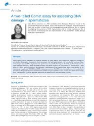
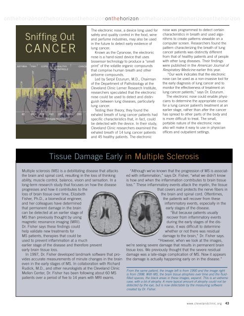
onthehorizononthehorizononthehorizononthehorizononthehorizononthehorizononthehorizonSniffing OutCANCERThe electronic nose, a device long used forsafety and quality control in the food, wineand perfume industries, may also be usedin the future to detect early evidence oflung cancer.Known as the Cyranose, the electronicnose is a hand-sized device that usesbiosensor technology to produce a "smellprint" of the volatile organic compoundsthat comprise human breath and otherairborne compounds.Led by Serpil Erzurum, M.D., Chairmanof the Department of Pathobiology at theCleveland Clinic Lerner Research Institute,researchers speculated that the electronicnose could be used to detect and distinguishbetween lung diseases, particularlylung cancer.Testing their theory, they found theexhaled breath of lung cancer patients hadspecific characteristics that, in fact, couldbe detected with the device. In their study,Cleveland Clinic researchers examined theexhaled breath of 14 lung cancer patientsand 45 healthy patients. The electronicnose was programmed to detect certaincharacteristics in breath and used algorithmsto create patterns viewable on acomputer screen. Researchers found thepattern characterizing the breath of lungcancer patients was distinctly differentfrom that of healthy patients and of peoplewith other lung diseases. Their findingswere published in the American Journal ofRespiratory Medicine earlier this year.“Our work indicates that the electronicnose can be used as a non-invasive tool forthe early diagnosis of lung cancer and tomonitor the effectiveness of treatment onlung cancer patients,” says Dr. Erzurum.The electronic nose could enable physiciansto determine the appropriate coursefor a lung cancer patient’s treatment at anearlier stage, rather than after the cancerhas spread to other parts of the body andis more difficult to treat. The small,portable nature of the electronic nosealso will make it easy to use in physicianoffices and outpatient settings.Tissue Damage Early in Multiple SclerosisMultiple sclerosis (MS) is a debilitating disease that attacksthe brain and spinal cord, resulting in the loss of thinkingability, muscle control, balance, vision and sensation. In along-term research study that focuses on how the diseaseprogresses and how it contributes to theloss of brain tissue over time, ElizabethFisher, Ph.D., a biomedical engineer,and her colleagues have determinedthat permanent damage in the braincan be detected at an earlier stage ofMS than previously thought by usingmagnetic resonance imaging (MRI).Dr. Fisher says these findings couldhelp validate new treatments forMS patients, therapies that could beused to prevent inflammation at a muchearlier stage of the disease and therefore preventearly brain tissue loss.In 1997, Dr. Fisher developed landmark software that providesaccurate measurements of minute changes in the braineven in the early stages of MS. In collaboration with RichardRudick, M.D., and other neurologists at the Cleveland ClinicMellen Center, Dr. Fisher has been following about 60 MSpatients over a period of five to 14 years with MRI exams.“Although we’ve known that the progression of MS is associatedwith inflammation,” says Dr. Fisher, “what we didn’t knowis how and when the inflammation contributes to brain tissueloss.” These inflammatory events attack the myelin, the tissuethat covers and protects the nerve fibers inthe brain and spinal cord. Oftentimes,the patients will recover from theseinflammatory events, especially in theearly stages of the disease.“But because patients usuallyrecover from inflammatory eventsduring the early stages of the disease,it was difficult to determinewhether or not there was residualdamage to the brain,” Dr. Fisher says.“However, when we look at the images,we’re seeing severe damage that results in permanent braintissue loss. We previously thought that the severe residualdamage was a late-stage complication of MS. Now it appearsthe damage is actually happening early on in the disease.”From the same patient, the image left is from 1990 and the image rightis from 1998. With MS, the brain tissue atrophies over time and the fluidfilledspaces, the black areas in these images, expand. This is an extremecase, with a lot of atrophy. A more typical amount of atrophy could not bedetected by the eye, but is now detectable by the measuring softwarecreated by Dr. Fisher.www.clevelandclinic.org 43