Ingrown Toenails - Cutis
Ingrown Toenails - Cutis
Ingrown Toenails - Cutis
- No tags were found...

Create successful ePaper yourself
Turn your PDF publications into a flip-book with our unique Google optimized e-Paper software.
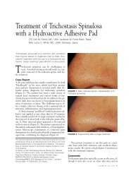
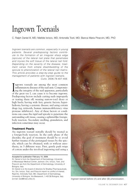
<strong>Ingrown</strong> <strong>Toenails</strong>C. Ralph Daniel III, MD; Matilde Iorizzo, MD; Antonella Tosti, MD; Bianca Maria Piraccini, MD, PhD<strong>Ingrown</strong> toenails are common, especially in youngpatients. Several predisposing factors contributeto the formation of an irregular sharp edge(spicule) of the lateral nail plate that penetratesand injures the soft tissue of the lateral nail fold.Depending on the severity of the disease, treatmentvaries from simple disembedding of thespicule to phenolization of the lateral nail matrix.This article provides a step-by-step guide to themanagement of patients with ingrown toenails.<strong>Cutis</strong>. 2006;78:407-408.<strong>Ingrown</strong> toenails are among the most commoninflammatory diseases of the nail unit. Compromisingthe integrity of the nail apparatus, particularlyof the great toe I, can cause it to become ingrown.Predisposing factors include cutting nails improperlyor tearing them off; wearing narrow-toed shoes orhigh heels; having wide feet; genetic factors; hyperhidrosis;having a systemic disease; and using certaindrugs (eg, retinoids, human immunodeficiency virusprotease inhibitors). Any of these factors or conditionscan cause the rigid nail spicule to penetrate thesurrounding soft tissue, causing a splinterlike foreignbodyreaction. Secondary swelling, granulation, andinfection sometimes may occur.ATreatment PearlsAn ingrown toenail initially should be treated asa foreign-body reaction. In the early phase of thedisorder, the goal of treatment should be to avoidfurther trauma of the periungual tissues by the spicule,which can be obtained, with or without anesthesia,in 3 different ways. First, gently push wispsof cotton under the involved ingrowing nail using aAccepted for publication February 10, 2006.Dr. Daniel is from the Department of Dermatology, Universityof Mississippi Medical Center, Jackson. Drs. Iorizzo, Tosti, andPiraccini are from the Department of Dermatology, University ofBologna, Italy.Dr. Daniel is on the board of directors for Bradley Pharmaceuticals,Inc. Drs. Iorizzo, Tosti, and Piraccini report no conflict of interest.Reprints: Antonella Tosti, MD, Department of Dermatology,University of Bologna, Via Massarenti, 1–40138 Bologna, Italy(e-mail: tosti@med.unibo.it).B<strong>Ingrown</strong> toenail before (A) and after (B) phenolization.VOLUME 78, DECEMBER 2006 407
<strong>Ingrown</strong> <strong>Toenails</strong>2-mm nail elevator or a number 1 or 2 curette, repeatingthis process if the cotton falls out. Second, gentlyease dental floss proximally to lift out the ingrownspicule and then leave it in place. 1 Patients should beinstructed to soak the toe for 10 minutes 3 times dailyfor 1 to 2 weeks in 1 L of cold water mixed with 1 to2 teaspoons of salt or Epsom salts. After each soak,patients should apply a mid- to high-potency topicalsteroid. Third, apply an anchor tape to the tip of thetoe and around the nail vertically and longitudinally.The tape will pull the bulging tissue away from the nailplate and allow nail growth to proceed unimpeded.Additional surgical tape should be applied to affix theprevious one to the nail plate and skin. 2In advanced cases, when granulation tissue, pain,and infection are present, treatment can be performedonly after cure of the inflammatory reaction, which canbe affected by using the soaking solution and regimenand by applying a mid- to high-potency topical steroidafter each soak. After this treatment, 1 of 2 techniquescan be performed using digital block anesthesia: guttersplint with formable acrylic resin 3 and phenolization. 4The former technique requires elevating the ingrownnail spicule by inserting a longitudinally incised tubealong the lateral margin of the nail plate. A formableacrylic resin should then be applied inside the tube andthen upon the plate to hold the tube in place. The lattertechnique requires cutting out the ingrown spiculewith an English anvil nail splitter and removing it.Then, ensuring a dry bloodless field, use Calgiswabs(urethral swabs with a small cotton tip and a metalhandle that can be easily bent to match the shape ofthe groove housing the proximal matrix) to apply 88%phenol to the affected nail matrix 3 times (30 secondseach time); then thoroughly cleanse out the phenolwith alcohol (Figure).An electrode also can be used to perform electrodesiccationand curettage. One side is active,and the other side is Teflon ® coated and is inactive.After surgery, a dressing consisting of bacitracin/polymyxin ointment, Telfa dressing, 434s, tubegauze, and then paper tape should be applied.Assure that the bandage is not too tight. Leave iton for 48 hours, elevating the digit as high as possible.The patient should then soak the digit with1 teaspoon of salt or Epsom salts in 1 L of warmwater, dry it off, and apply bacitracin/polymyxinointment and a bandage. This should be done3 to 4 times daily for 1 to 2 weeks. If infectionis suspected, an appropriate oral antibiotic shouldbe prescribed.All patients should be cautioned to avoid wearingnarrow-toed shoes, high heels, improperly fittingshoes, or stockings that are tight at the toe;avoid rounding toenails at the edges (they shouldbe cut straight across); and see a physician as soonas possible when an ingrown nail first starts.References1. Woo SH, Kim IH. Surgical pearl: nail edge separation withdental floss for ingrown toenails. J Am Acad Dermatol.2004;50:939-940.2. Arai I, Arai T, Nakajima H, et al. Formable acrylic treatmentfor ingrowing nail with gutter splint and sculpturednail. Int J Dermatol. 2004;3:759-765.3. Arai I, Arai T, Nakajima H, et al. Elastic gauze anchortaping method for ingrowing nail, nail loss and onychomycosis[abstract]. J Eur Acad Dermatol Venereol. 2004;18(suppl 2):579-580.4. Siegle RJ, Harkness J, Swanson NA. Phenol alcoholtechnique for permanent matricectomy. Arch Dermatol.1984;120:348-350.408 CUTIS ®