Pterygium of the Nail - Cutis
Pterygium of the Nail - Cutis
Pterygium of the Nail - Cutis
- No tags were found...

Create successful ePaper yourself
Turn your PDF publications into a flip-book with our unique Google optimized e-Paper software.
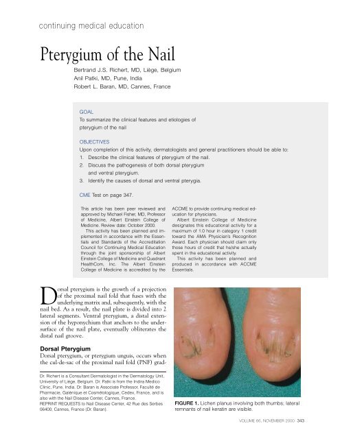
continuing medical education<strong>Pterygium</strong> <strong>of</strong> <strong>the</strong> <strong>Nail</strong>Bertrand J.S. Richert, MD, Liège, BelgiumAnil Patki, MD, Pune, IndiaRobert L. Baran, MD, Cannes, FranceGOALTo summarize <strong>the</strong> clinical features and etiologies <strong>of</strong>pterygium <strong>of</strong> <strong>the</strong> nailOBJECTIVESUpon completion <strong>of</strong> this activity, dermatologists and general practitioners should be able to:1. Describe <strong>the</strong> clinical features <strong>of</strong> pterygium <strong>of</strong> <strong>the</strong> nail.2. Discuss <strong>the</strong> pathogenesis <strong>of</strong> both dorsal pterygiumand ventral pterygium.3. Identify <strong>the</strong> causes <strong>of</strong> dorsal and ventral pterygia.CME Test on page 347.This article has been peer reviewed andapproved by Michael Fisher, MD, Pr<strong>of</strong>essor<strong>of</strong> Medicine, Albert Einstein College <strong>of</strong>Medicine. Review date: October 2000.This activity has been planned and implementedin accordance with <strong>the</strong> Essentialsand Standards <strong>of</strong> <strong>the</strong> AccreditationCouncil for Continuing Medical Educationthrough <strong>the</strong> joint sponsorship <strong>of</strong> AlbertEinstein College <strong>of</strong> Medicine and QuadrantHealthCom, Inc. The Albert EinsteinCollege <strong>of</strong> Medicine is accredited by <strong>the</strong>ACCME to provide continuing medical educationfor physicians.Albert Einstein College <strong>of</strong> Medicinedesignates this educational activity for amaximum <strong>of</strong> 1.0 hour in category 1 credittoward <strong>the</strong> AMA Physician’s RecognitionAward. Each physician should claim onlythose hours <strong>of</strong> credit that he/she actuallyspent in <strong>the</strong> educational activity.This activity has been planned andproduced in accordance with ACCMEEssentials.Dorsal pterygium is <strong>the</strong> growth <strong>of</strong> a projection<strong>of</strong> <strong>the</strong> proximal nail fold that fuses with <strong>the</strong>underlying matrix and, subsequently, with <strong>the</strong>nail bed. As a result, <strong>the</strong> nail plate is divided into 2lateral segments. Ventral pterygium, a distal extension<strong>of</strong> <strong>the</strong> hyponychium that anchors to <strong>the</strong> undersurface<strong>of</strong> <strong>the</strong> nail plate, eventually obliterates <strong>the</strong>distal nail groove.Dorsal <strong>Pterygium</strong>Dorsal pterygium, or pterygium unguis, occurs when<strong>the</strong> cul-de-sac <strong>of</strong> <strong>the</strong> proximal nail fold (PNF) grad-Dr. Richert is a Consultant Dermatologist in <strong>the</strong> Dermatology Unit,University <strong>of</strong> Liège, Belgium. Dr. Patki is from <strong>the</strong> Indira MedicoClinic, Pune, India. Dr. Baran is Associate Pr<strong>of</strong>essor, Faculté dePharmacie, Galénique et Cosmétologique, Cedex, France, and isalso with <strong>the</strong> <strong>Nail</strong> Disease Center, Cannes, France.REPRINT REQUESTS to <strong>Nail</strong> Disease Center, 42 Rue des Serbes06400, Cannes, France (Dr. Baran).FIGURE 1. Lichen planus involving both thumbs; lateralremnants <strong>of</strong> nail keratin are visible.VOLUME 66, NOVEMBER 2000 343
PTERYGIUM OF THE NAILFIGURE 2. Dorsal pterygium involvingone finger after a major trauma.FIGURE 3. Ventral pterygium affectingonly one finger.ually shortens and <strong>the</strong> nail plate thins until, eventually,<strong>the</strong> PNF fuses to <strong>the</strong> matrix and, subsequently,to <strong>the</strong> nail bed. The resulting nail plate segments progressivelydecrease in size as <strong>the</strong> pterygium widens,resulting in 2 small remnants involving <strong>the</strong> PNF.Complete involvement <strong>of</strong> <strong>the</strong> matrix and nail bedproduces a total loss <strong>of</strong> <strong>the</strong> plate and permanent atrophy<strong>of</strong> <strong>the</strong> nail apparatus. Dorsal pterygium is usuallyacquired, but exceptional congenital forms havebeen reported. In a case <strong>of</strong> nail lichen planus in 1921,Friedman first described pterygium unguis using <strong>the</strong>term navellierungsprozess. 1 Dorsal pterygium rarely affects<strong>the</strong> toes; involvement <strong>of</strong> all 20 toenails has beenreported in only one case.Although pterygium unguis is most commonlycaused by lichen planus (Figure 1), 2 it also may originatefrom burns, radiodermatitis, trauma (Figure 2),and diseases prone to developing adherence bands,including cicatricial pemphigoid, graft versus host disease,toxic epidermal necrolysis, and pemphigus foliaceus(Table I). Peripheral ischemia, intermittentlydetected in Raynaud’s phenomenon 3 or permanentlyfound in a<strong>the</strong>rosclerosis, diabetic vasculopathy, andtype 2 lepra reaction, has been identified as a vascularcause <strong>of</strong> dorsal pterygium. An idiopathic form <strong>of</strong>pterygium unguis, or idiopathic atrophy <strong>of</strong> <strong>the</strong> nails,exists, but may be a variation <strong>of</strong> lichen planus andremains a controversial cause. 4 Congenital forms maybe associated with dyskeratosis congenita, andpterygium formation has been reported in systemiclupus ery<strong>the</strong>matosus and in one case <strong>of</strong> sarcoidosisinvolving <strong>the</strong> PNF.Healing <strong>of</strong> a disease involving <strong>the</strong> PNF may lead toscarring pterygium formation; however, several factorsthat interfere with <strong>the</strong> healing <strong>of</strong> <strong>the</strong> PNF must appearsimultaneously for this to occur. In patients with dorsalpterygium (excluding <strong>the</strong> traumatic and congenitaltypes), <strong>the</strong> main factor is dilatation <strong>of</strong> <strong>the</strong> nail capillaryloops and <strong>the</strong> formation <strong>of</strong> a slender, microvascularshunt system in <strong>the</strong>m. Although pterygium that344 CUTIS ®
PTERYGIUM OF THE NAILTable I.Causes <strong>of</strong> Dorsal <strong>Pterygium</strong>(<strong>Pterygium</strong> Unguis)A<strong>the</strong>rosclerosisBurnsCicatricial pemphigoidCongenital etiologyDiabetic vasculopathyDyskeratosis congenitaGraft versus host diseaseIdiopathic atrophy <strong>of</strong> <strong>the</strong> nails 3Inadequate corticosteroid matrix infiltration forCandida paronychiaLichen planus 1OnychotillomaniaPemphigus foliaceusRadiodermatitisRaynaud’s phenomenon 3Sarcoidosis involving <strong>the</strong> PNFSystemic lupus ery<strong>the</strong>matosusToxic epidermal necrolysisTraumaType 2 lepra reactionresults from trauma is not linked to <strong>the</strong> intensity <strong>of</strong><strong>the</strong> trauma, it may be observed in severe distal injury.Also, pterygium remains exceptional in repeatedchronic trauma inflicted to <strong>the</strong> PNF in onychotillomania,and we observed one pterygium unguis arisingafter inadequate corticosteroid matrix infiltration forCandida paronychia (Richert, unpublished data, 1997).Ventral <strong>Pterygium</strong>Ventral pterygium (Figure 3) is a newly described conditionin which a distal extension <strong>of</strong> <strong>the</strong> hyponychiumanchors to <strong>the</strong> undersurface <strong>of</strong> <strong>the</strong> nail plateand, subsequently, obliterates <strong>the</strong> distal nail groove.The condition was first described in 1973 by Caputoand Prandi, 5 who coined <strong>the</strong> term pterygium inversumunguis (PIU) to describe a forward extension <strong>of</strong>Table II.Causes <strong>of</strong> Ventral <strong>Pterygium</strong>(<strong>Pterygium</strong> Inversum Unguis)Causalgia <strong>of</strong> <strong>the</strong> median nerveCongenital etiologyFamily historyFormaldehyde-containing hardeners 8Lenticular atrophy <strong>of</strong> <strong>the</strong> palmar creasesLeprosyNeur<strong>of</strong>ibromatosisParesisScarring in <strong>the</strong> vicinity <strong>of</strong> <strong>the</strong> distal nail grooveSubungual exostosisSystemic connective tissue diseasesSystemic lupus ery<strong>the</strong>matosusSystemic sclerosis<strong>the</strong> hyponychium anchoring to <strong>the</strong> undersurface <strong>of</strong><strong>the</strong> nail plate and, thus, obliterating <strong>the</strong> distal nailgroove.PIU may be ei<strong>the</strong>r congenital or acquired. Odomet al 6 first described <strong>the</strong> congenital form as a “congenital,painful and aberrant hyponychium” in 1974.In some instances, PIU has been reported as familialand, in most reported cases, patients sought medicaladvice for <strong>the</strong> pain or bleeding <strong>the</strong>y experiencedwhen trimming <strong>the</strong>ir nails. Causes <strong>of</strong> PIU are listedin Table II.Because <strong>the</strong> frequency <strong>of</strong> ventral pterygium maybe underestimated, especially in asymptomatic cases,Mello Filho 7 performed a systematic digital examinationon 2000 patients and found that 0.4% <strong>of</strong> <strong>the</strong>adult population was affected. In <strong>the</strong>se cases <strong>the</strong>re wasno family history, and <strong>the</strong> condition was observedmore frequently in women than in men. Idiopathicforms were preponderant, and involvement <strong>of</strong> <strong>the</strong>toes remains exceptional.Acquired PIU, which occurs in 16% <strong>of</strong> patients,is by far <strong>the</strong> most common form and, though idiopathic,is usually secondary to systemic connectivetissue diseases, progressive systemic sclerosis, and systemiclupus ery<strong>the</strong>matosus. One patient with congenitalPIU developed systemic lupus ery<strong>the</strong>matosusat <strong>the</strong> age <strong>of</strong> 19, but whe<strong>the</strong>r this was coincidentalVOLUME 66, NOVEMBER 2000 345
PTERYGIUM OF THE NAILComments<strong>Pterygium</strong> <strong>of</strong> <strong>the</strong> nail has been described on both dorsaland ventral aspects <strong>of</strong> <strong>the</strong> nail plate. The dorsalpterygium consists <strong>of</strong> a forward growth <strong>of</strong> a projection<strong>of</strong> <strong>the</strong> proximal nail fold that fuses with <strong>the</strong> underlyingmatrix and, subsequently, with <strong>the</strong> nail bedand divides <strong>the</strong> nail plate in 2 lateral segments. Ventralpterygium is a distal extension <strong>of</strong> <strong>the</strong> hyponychiumthat anchors to <strong>the</strong> undersurface <strong>of</strong> <strong>the</strong> nailplate and eventually obliterates <strong>the</strong> distal nail groove.Both conditions are nonspecific abnormalities <strong>of</strong> <strong>the</strong>nail apparatus.FIGURE 4. Ventral pterygium <strong>of</strong> several digits afterapplications <strong>of</strong> nail polish containing formaldehyde.or related has not been determined.PIU may be secondary to scarring in <strong>the</strong> vicinity<strong>of</strong> <strong>the</strong> distal nail groove, or it may arise from a reactionto formaldehyde-containing nail hardeners 8(Figure 4) or from subungual exostosis. PIU has beenreported once in neur<strong>of</strong>ibromatosis, which was associatedwith lenticular atrophy <strong>of</strong> <strong>the</strong> palmar creasesand causalgia <strong>of</strong> <strong>the</strong> median nerve. Unilateral PIU <strong>of</strong><strong>the</strong> fingers and toes has been reported 1 year after astroke and resulted in paresis <strong>of</strong> <strong>the</strong> same side.REFERENCES1. Friedman M. Nagelveranderungen bei lichen ruber. ArchDerm Syph. 1921;135:174-179.2. Zaias N. The nail in lichen planus. Arch Dermatol.1970;101:264-271.3. Edwards EA. <strong>Nail</strong> changes in functional and organic arterialdisease. New Engl J Med. 1948;239:362-365.4. Tosti A, Piraccini BM, Fanti PA, et al. Idiopathic atrophy<strong>of</strong> <strong>the</strong> nails: clinical and pathological study <strong>of</strong> two cases. Dermatology.1995;190:116-118.5. Caputo R, Prandi G. <strong>Pterygium</strong> inversum unguis. Arch Dermatol.1973;108:817-818.6. Odom RB, Stein KM, Maibach HI. Congenital, painful,aberrant hyponychium. Arch Dermatol. 1974;110:89-90.7. Mello Filho A. Ocorrencia do “pterygium inversum unguis”em populaçao adulta. Med Cut Ib Lat Am. 1985;13:401-405.8. Daly BM, Johnson M. <strong>Pterygium</strong> inversum unguis due to nailfortifier. Contact Dermatitis. 1986;15:256-257.DISCLAIMERThe opinions expressed herein are those <strong>of</strong> <strong>the</strong> authors and do not necessarily represent <strong>the</strong> views <strong>of</strong> <strong>the</strong> sponsor or its publisher. Please review complete prescribinginformation <strong>of</strong> specific drugs or combination <strong>of</strong> drugs, including indications, contraindications, warnings, and adverse effects before administering pharmacologic<strong>the</strong>rapy to patients.FACULTY DISCLOSUREThe Faculty Disclosure Policy <strong>of</strong> <strong>the</strong> College <strong>of</strong> Medicine requires that faculty participating in a CME activity disclose to <strong>the</strong> audience any relationship with a pharmaceuticalor equipment company that might pose a potential, apparent, or real conflict <strong>of</strong> interest with regard to <strong>the</strong>ir contribution to <strong>the</strong> program.Drs. Richert, Patki, and Baran report no conflict <strong>of</strong> interest. Dr. Fisher reports no conflict <strong>of</strong> interest.346 CUTIS ®