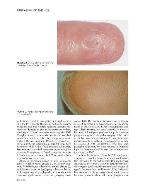
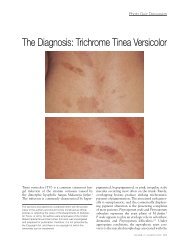
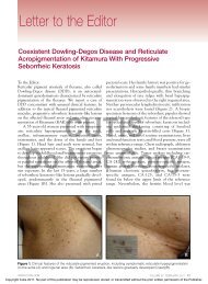
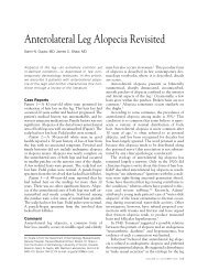
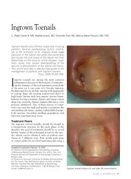
PTERYGIUM OF THE NAILFIGURE 2. Dorsal pterygium involvingone finger after a major trauma.FIGURE 3. Ventral pterygium affectingonly one finger.ually shortens and <strong>the</strong> nail plate thins until, eventually,<strong>the</strong> PNF fuses to <strong>the</strong> matrix and, subsequently,to <strong>the</strong> nail bed. The resulting nail plate segments progressivelydecrease in size as <strong>the</strong> pterygium widens,resulting in 2 small remnants involving <strong>the</strong> PNF.Complete involvement <strong>of</strong> <strong>the</strong> matrix and nail bedproduces a total loss <strong>of</strong> <strong>the</strong> plate and permanent atrophy<strong>of</strong> <strong>the</strong> nail apparatus. Dorsal pterygium is usuallyacquired, but exceptional congenital forms havebeen reported. In a case <strong>of</strong> nail lichen planus in 1921,Friedman first described pterygium unguis using <strong>the</strong>term navellierungsprozess. 1 Dorsal pterygium rarely affects<strong>the</strong> toes; involvement <strong>of</strong> all 20 toenails has beenreported in only one case.Although pterygium unguis is most commonlycaused by lichen planus (Figure 1), 2 it also may originatefrom burns, radiodermatitis, trauma (Figure 2),and diseases prone to developing adherence bands,including cicatricial pemphigoid, graft versus host disease,toxic epidermal necrolysis, and pemphigus foliaceus(Table I). Peripheral ischemia, intermittentlydetected in Raynaud’s phenomenon 3 or permanentlyfound in a<strong>the</strong>rosclerosis, diabetic vasculopathy, andtype 2 lepra reaction, has been identified as a vascularcause <strong>of</strong> dorsal pterygium. An idiopathic form <strong>of</strong>pterygium unguis, or idiopathic atrophy <strong>of</strong> <strong>the</strong> nails,exists, but may be a variation <strong>of</strong> lichen planus andremains a controversial cause. 4 Congenital forms maybe associated with dyskeratosis congenita, andpterygium formation has been reported in systemiclupus ery<strong>the</strong>matosus and in one case <strong>of</strong> sarcoidosisinvolving <strong>the</strong> PNF.Healing <strong>of</strong> a disease involving <strong>the</strong> PNF may lead toscarring pterygium formation; however, several factorsthat interfere with <strong>the</strong> healing <strong>of</strong> <strong>the</strong> PNF must appearsimultaneously for this to occur. In patients with dorsalpterygium (excluding <strong>the</strong> traumatic and congenitaltypes), <strong>the</strong> main factor is dilatation <strong>of</strong> <strong>the</strong> nail capillaryloops and <strong>the</strong> formation <strong>of</strong> a slender, microvascularshunt system in <strong>the</strong>m. Although pterygium that344 CUTIS ®
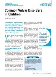
PTERYGIUM OF THE NAILTable I.Causes <strong>of</strong> Dorsal <strong>Pterygium</strong>(<strong>Pterygium</strong> Unguis)A<strong>the</strong>rosclerosisBurnsCicatricial pemphigoidCongenital etiologyDiabetic vasculopathyDyskeratosis congenitaGraft versus host diseaseIdiopathic atrophy <strong>of</strong> <strong>the</strong> nails 3Inadequate corticosteroid matrix infiltration forCandida paronychiaLichen planus 1OnychotillomaniaPemphigus foliaceusRadiodermatitisRaynaud’s phenomenon 3Sarcoidosis involving <strong>the</strong> PNFSystemic lupus ery<strong>the</strong>matosusToxic epidermal necrolysisTraumaType 2 lepra reactionresults from trauma is not linked to <strong>the</strong> intensity <strong>of</strong><strong>the</strong> trauma, it may be observed in severe distal injury.Also, pterygium remains exceptional in repeatedchronic trauma inflicted to <strong>the</strong> PNF in onychotillomania,and we observed one pterygium unguis arisingafter inadequate corticosteroid matrix infiltration forCandida paronychia (Richert, unpublished data, 1997).Ventral <strong>Pterygium</strong>Ventral pterygium (Figure 3) is a newly described conditionin which a distal extension <strong>of</strong> <strong>the</strong> hyponychiumanchors to <strong>the</strong> undersurface <strong>of</strong> <strong>the</strong> nail plateand, subsequently, obliterates <strong>the</strong> distal nail groove.The condition was first described in 1973 by Caputoand Prandi, 5 who coined <strong>the</strong> term pterygium inversumunguis (PIU) to describe a forward extension <strong>of</strong>Table II.Causes <strong>of</strong> Ventral <strong>Pterygium</strong>(<strong>Pterygium</strong> Inversum Unguis)Causalgia <strong>of</strong> <strong>the</strong> median nerveCongenital etiologyFamily historyFormaldehyde-containing hardeners 8Lenticular atrophy <strong>of</strong> <strong>the</strong> palmar creasesLeprosyNeur<strong>of</strong>ibromatosisParesisScarring in <strong>the</strong> vicinity <strong>of</strong> <strong>the</strong> distal nail grooveSubungual exostosisSystemic connective tissue diseasesSystemic lupus ery<strong>the</strong>matosusSystemic sclerosis<strong>the</strong> hyponychium anchoring to <strong>the</strong> undersurface <strong>of</strong><strong>the</strong> nail plate and, thus, obliterating <strong>the</strong> distal nailgroove.PIU may be ei<strong>the</strong>r congenital or acquired. Odomet al 6 first described <strong>the</strong> congenital form as a “congenital,painful and aberrant hyponychium” in 1974.In some instances, PIU has been reported as familialand, in most reported cases, patients sought medicaladvice for <strong>the</strong> pain or bleeding <strong>the</strong>y experiencedwhen trimming <strong>the</strong>ir nails. Causes <strong>of</strong> PIU are listedin Table II.Because <strong>the</strong> frequency <strong>of</strong> ventral pterygium maybe underestimated, especially in asymptomatic cases,Mello Filho 7 performed a systematic digital examinationon 2000 patients and found that 0.4% <strong>of</strong> <strong>the</strong>adult population was affected. In <strong>the</strong>se cases <strong>the</strong>re wasno family history, and <strong>the</strong> condition was observedmore frequently in women than in men. Idiopathicforms were preponderant, and involvement <strong>of</strong> <strong>the</strong>toes remains exceptional.Acquired PIU, which occurs in 16% <strong>of</strong> patients,is by far <strong>the</strong> most common form and, though idiopathic,is usually secondary to systemic connectivetissue diseases, progressive systemic sclerosis, and systemiclupus ery<strong>the</strong>matosus. One patient with congenitalPIU developed systemic lupus ery<strong>the</strong>matosusat <strong>the</strong> age <strong>of</strong> 19, but whe<strong>the</strong>r this was coincidentalVOLUME 66, NOVEMBER 2000 345