Plasma membrane calcium ATPases are important components of ...
Plasma membrane calcium ATPases are important components of ...
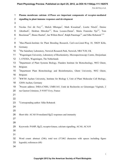
Plasma membrane calcium ATPases are important components of ...

Create successful ePaper yourself
Turn your PDF publications into a flip-book with our unique Google optimized e-Paper software.
Frei dit Frey et al.12345678910111213141516171819202122232425262728293031323334Ranf et al., 2008). The Ca 2+ burst occurs upstream <strong>of</strong> many MAMP-elicited responsesincluding the rapid production <strong>of</strong> reactive oxygen species (ROS), activation <strong>of</strong> signallingkinases, as well as changes in gene expression (Blume et al., 2000; Boller and Felix, 2009;Ranf et al., 2011; Segonzac et al., 2011). However, genetic studies and identification <strong>of</strong> theunderlying molecular <strong>components</strong> <strong>of</strong> the MAMP-induced Ca 2+ burst <strong>are</strong> largely missing (Ranfet al., 2008; Boller and Felix, 2009). In general, cytosolic Ca 2+ levels <strong>are</strong> regulated by plasma<strong>membrane</strong>- and endo<strong>membrane</strong>-bound Ca 2+ channels that mediate influx <strong>of</strong> Ca 2+ and effluxtransporters that re-establish Ca 2+ homeostasis. A number <strong>of</strong> ion channels have beenidentified, some <strong>of</strong> which have roles in plant immunity such as DEFENSE NO DEATH 1(DND1) (Clough et al., 2000; Lamotte et al., 2004; Kudla et al., 2010). Recently, ionotropicglutamate receptor-like proteins (iGLRs) were shown to regulate Ca 2+ influx at the plasma<strong>membrane</strong> and were also implicated in MAMP-induced responses (Cho et al., 2009;Kwaaitaal et al., 2011; Michard et al., 2011; Vatsa et al., 2011), and an ER-localized P2AtypeCa 2+ ATPase was described to contribute to pathogen-induced cell death and to alter theMAMP-triggered Ca 2+ burst (Zhu et al., 2010). The relevance <strong>of</strong> Ca 2+ influx in MAMPelicitedresponses is underlined by polysaccharides secreted from bacterial pathogens tochelate Ca 2+ in the apoplastic space (Aslam et al., 2008).Here, we report that the plasma <strong>membrane</strong> resident P2B-type Ca 2+ ATPase ACA8interacts with FLS2 in planta. Loss-<strong>of</strong>-function aca8 plants, the mutant <strong>of</strong> its closesthomologue aca10, and the aca8 aca10 double mutant were more susceptible to bacterialinfection. Analysing individual MAMP responses, aca8 aca10 mutant plants displayeddecreased flg22-triggered Ca 2+ influx and ROS accumulation. Importantly, flg22-triggeredgene expression downstream <strong>of</strong> MAPK signalling was increased, but reduced for geneexpression downstream <strong>of</strong> CDPK signalling. This suggests that the MAMP-induced Ca 2+burst is required for proper transcriptional reprogramming upon elicitation. According to theirfunction as Ca 2+ pumps, ACA8 and ACA10 <strong>are</strong> hypothesized to regulate Ca 2+ efflux duringthe flg22-elicited Ca 2+ burst, which suggests a molecular link between the FLS2 receptor,Ca 2+ signalling and flg22-triggered downstream responses. In addition, aca8 and aca8 aca10mutant plants showed developmental phenotypes affecting inflorescence height as well asroot length. Together with the finding that ACA8 also interacts with other RKs such as BRI1and CLV1, these results suggest that plasma <strong>membrane</strong> Ca 2+ <strong>ATPases</strong> function in multipleRK-mediated signalling pathways.4
Frei dit Frey et al.12345678910111213141516171819202122232425262728293031323334ResultsACA8 interacts with FLS2 and other RKsIn a proteomics approach, we previously isolated proteins co-localizing to FLS2 inplasma <strong>membrane</strong> microdomains (Keinath et al., 2010). To address whether some <strong>of</strong> theseproteins can associate with FLS2, we focussed on Ca 2+ <strong>ATPases</strong>, which have also beenidentified as differentially phosphorylated and transcriptionally induced in response to flg22(Zipfel et al., 2004; Benschop et al., 2007). ACA8 and ACA10 belong to the family <strong>of</strong> type2B auto-inhibited Ca 2+ <strong>ATPases</strong> consisting <strong>of</strong> ten members in Arabidopsis (Figure S1A;(Boursiac and Harper, 2007). These Ca 2+ <strong>ATPases</strong> comprise ten trans<strong>membrane</strong> spanningdomains, harbour a calmodulin binding domain for auto-inhibition <strong>of</strong> the ATPase active site,and can localize to different <strong>membrane</strong> compartments (Boursiac and Harper, 2007). ACA8,ACA9 and ACA10 group into a distinct subfamily and accumulate at the plasma <strong>membrane</strong>(Bonza et al., 2000; Hwang et al., 2000; Lee et al., 2007). While ACA9 expression isrestricted to pollen and thereby is critical for pollen tube development, ACA8 and ACA10 <strong>are</strong>expressed throughout the plant and have not yet assigned any specific function besidesinflorescence growth (Schiott et al., 2004).We transiently expressed FLS2 and ACA8 fused to the N- and C-terminal halves <strong>of</strong>YFP, respectively, in Nicotiana benthamiana and examined possible protein-proteininteractions by confocal microscopy in a so-called bimolecular fluorescence complementation(BiFC) assay (Bracha-Drori et al., 2004). In this assay, we observed reconstitution <strong>of</strong> the YFPmolecule by detection <strong>of</strong> fluorescence when expressing FLS2 fused to both <strong>of</strong> the YFP halvesindicative <strong>of</strong> FLS2 homodimerization (Figure 1A). We also observed BiFC when FLS2 wasco-expressed with ACA8. As BiFC assays <strong>are</strong> based on transient expression in a heterologoussystem, the tagged proteins can accumulate to high levels facilitating the reconstitution <strong>of</strong> aBiFC signal, and thus ACA12, BRI1 and CLV1 were included as controls. No YFPreconstitution could be detected upon co-expressing <strong>of</strong> FLS2 and ACA12, another plasma<strong>membrane</strong> resident Ca 2+ ATPase. Notably, ACA8 showed a broader interaction pattern,because BiFC was also observed with other RKs such as BRI1 and CLV1, <strong>of</strong> which the latteris functioning in stem cell identity maintenance and is normally not expressed in leaf tissue(Waites and Simon, 2000). Similar to FLS2, both BRI1 and CLV1 formed homodimers in thisassay, but unlike FLS2 they also interacted with ACA12 (Figure 1A). In all cases <strong>of</strong> BiFC, theYFP signal was recorded at the cell periphery suggesting complex formation at the plasma<strong>membrane</strong>.5
Frei dit Frey et al.12345678910111213141516171819202122232425262728293031323334To further overcome limitations <strong>of</strong> BiFC assays we performed Förster ResonanceEnergy Transfer (FRET) measurements on the basis <strong>of</strong> Fluorescence Lifetime ImagingMicroscopy (FLIM) using respective FLS2 and ACA8 fusions to CFP or YFP. FRET can bedetected using FLIM where reduction <strong>of</strong> the fluorescence lifetime <strong>of</strong> a donor-containingmolecule occurs due to proximity <strong>of</strong> an acceptor-containing molecule, which is an indication<strong>of</strong> physical interaction. We examined FLS2-ACA8 association in protoplasts from soil-grownArabidopsis plants. Under this condition we observed a significant reduction in fluorescencelifetime when FLS2-CFP and ACA8-YFP were co-expressed as comp<strong>are</strong>d to the fluorescencelifetime <strong>of</strong> ACA8-CFP alone (Figure 1B; Table S1). Similar results were obtained when weused FLS2-CFP and ACA8-YFP. This suggests that both proteins <strong>are</strong> in close proximity toeach other, indicative <strong>of</strong> protein-protein interaction. Interaction <strong>of</strong> fluorophore-tagged FLS2and ACA8 was detected at the plasma <strong>membrane</strong>, which is in line with the subcellularlocalization <strong>of</strong> the two proteins and substantiates our findings <strong>of</strong> BiFC in N. benthamiana.However, interaction <strong>of</strong> fluorophore-tagged FLS2 and ACA8 was not distributed uniformlyacross the plasma <strong>membrane</strong>, but seen as patchy <strong>are</strong>as with strongly reduced fluorescencelifetimes (Figure 1B), which indicates the presence <strong>of</strong> FLS2-ACA8 complexes were restrictedto subdomains within the plasma <strong>membrane</strong>. This observation is in agreement with the notionthat FLS2 and ACA8 can localize to flg22-induced plasma <strong>membrane</strong> microdomains (Keinathet al., 2010). Despite numerous attempts we failed to clone a full-length ACA10 cDNA, whichprecluded analysis <strong>of</strong> ACA10 by fluorophore-based interaction assays. Despite poor resultsby co-immunoprecipitation analysis, pull-down experiments <strong>of</strong> FLS2-GFP followed by massspectrometricanalysis repeatedly revealed presence <strong>of</strong> ACA8 and ACA10 peptides, furthercorroborating the existence <strong>of</strong> FLS2-ACA8 and FLS2-ACA10 complexes in planta (FigureS2). Taken together, these results indicate that FLS2 forms a complex with ACA8 at theplasma <strong>membrane</strong>, and ACA8 can interact with multiple RKs, pointing at an <strong>important</strong> role inthe regulation <strong>of</strong> RK-mediated signalling pathways.ACA8 and ACA10 exhibit partial overlapping functionsTo address ACA8 function, we isolated a T-DNA insertion line and a tilling mutant(both in Col-0 genetic background) in the ACA8 gene (Fig. S1B). Genetic redundancy withinmembers <strong>of</strong> the ACA family has been documented, and could be expected for members <strong>of</strong> theACA8, ACA9, ACA10 subgroup (Boursiac and Harper, 2007). Because ACA9 expression wasspecific to pollen tubes, we focussed on ACA10, isolated a T-DNA insertion line in theACA10 gene, and generated aca8 aca10 double knock-out lines (Figure S1C). In addition, we6
Frei dit Frey et al.12345678910111213141516171819202122232425262728293031323334crossed a 35S::ACA8-GFP expressing transgenic line into the aca8 aca10 double mutant.Single aca8 mutants displayed no obvious developmental phenotypes (Figure 2). By contrast,aca10 mutant plants were reduced in inflorescence height and displayed increased axillarystem formation, which was further enhanced in aca8 aca10 plants (Figure 2A). Thisphenotype was also present in aca10 plants crossed with the aca8 Q70* tilling mutant (FigureS3), and could be rescued by ectopic ACA8-GFP expression, demonstrating functionalcomplementation by the GFP fusion protein (Figure 2A). Redundant functions <strong>of</strong> ACA8 andACA10 in the regulation <strong>of</strong> inflorescence height were previously reported in ArabidopsisWassilewskaja (Ws) background (George et al., 2008). Differences between the singlemutants may result from an incomplete overlap <strong>of</strong> the ACA8 and ACA10 expression patterns.We did not observe any obvious mutant phenotype in rosette leaf development among thegenotypes (Figure 2B), while aca8 aca10 mutants showed significantly reduced root growthwhen cultivated in vitro (Figure 2C). Reduction in root growth was affecting primary rootlength and could be correlated with an early differentiation <strong>of</strong> stem cells comp<strong>are</strong>d to wildtype plants (Figure S3).Flg22-triggered early responses depend on ACA8 and ACA10 functionCa 2+ <strong>ATPases</strong> <strong>are</strong> responsible for extruding Ca 2+ ions from the cytosol intoendo<strong>membrane</strong> compartments or extracellularly into the apoplast (Bonza et al., 2004; Conn etal., 2011). ACA8 has been shown to mediate Ca 2+ transport in yeast and is activated bybinding <strong>of</strong> calmodulin (CaM) to its N-terminus (Bonza et al., 2000; Bonza et al., 2004;Mersmann et al., 2010). Based on the interaction <strong>of</strong> ACA8 with FLS2, we addressed whetherACA8 and ACA10 function in the flg22-triggered Ca 2+ burst. All genotypes were thereforecrossed to a transgenic line expressing the aequorin (Aeq) Ca 2+ biosensor (Knight et al.,1991). We performed luminescence-based measurements <strong>of</strong> free cytosolic Ca 2+ and revealedslightly elevated constitutive Ca 2+ levels in aca8 aca10 Aeq plants (Figure S4). We thenmonitored the MAMP-induced Ca 2+ burst over time. Mutant aca8 Aeq and aca10 Aeq plantsresponded like wild type upon flg22 treatment. By contrast, the flg22-triggered Ca 2+ burst wasstrongly reduced in the aca8 aca10 Aeq lines and completely abolished in fls2 Aeq plants(Figure 3A; Figure S5). The overall pattern <strong>of</strong> the transient increase <strong>of</strong> Ca 2+ remained similarbetween wild type and the aca8 Aeq and aca10 Aeq genotypes, but the maximal influx (peak)<strong>of</strong> the Ca 2+ signature was affected in aca8 aca10 Aeq plants (Figure S5). The Ca 2+ burst inresponse to chitin was slightly reduced in aca8 aca10 Aeq lines and for all other genotypesindistinguishable from wild type (Figure 3A). The lower peak in flg22-induced cytosolic Ca 2+7
Frei dit Frey et al.12345678910111213141516171819202122232425262728293031323334influx in the double mutant demonstrates that ACA8 and ACA10 both contribute to the flg22-elicited Ca 2+ burst and indicates a role for these proteins in the regulation <strong>of</strong> FLS2-mediatedearly responses.Production <strong>of</strong> ROS upon MAMP treatments is mediated by plasma <strong>membrane</strong>residentNADPH oxidases, which depend on Ca 2+ signalling for their function (Kobayashi etal., 2007; Mersmann et al., 2010). We examined the flg22-triggered oxidative burst anddetected no significant differences between wild-type plants and the aca8 and aca10 singlemutants, while the aca8 aca10 double mutant displayed an overall decreased ROS productionwhen treated with flg22 (Figure 3B). ROS production upon chitin treatment remainedcomparable to wild type in all tested mutants. The decrease in oxidative burst correlated withthe reduced flg22-triggered Ca 2+ signature in aca8 aca10 plants, which is in agreement withCa 2+ operating upstream <strong>of</strong> ROS production. When monitoring these individual MAMPresponses we observed genetic redundancy between ACA8 and ACA10 suggesting that bothCa 2+ <strong>ATPases</strong> exert overlapping functions in these early and transient flg22 responses, whichis in contrast to the unequal role <strong>of</strong> ACA8 and ACA10 in development. Western blot analysisrevealed unaltered FLS2 protein accumulation in the aca mutants comp<strong>are</strong>d to wild typeplants (Figure 3C). Therefore the observed reduction in flg22-triggered Ca 2+ and ROS burstsis likely caused by loss-<strong>of</strong>-ACA8 and -ACA10 function rather than altered FLS2 levels.ACA8 and ACA10 <strong>are</strong> required for proper flg22-induced transcriptional changesFor more detailed analysis <strong>of</strong> ACA8/ACA10 functions, we determined thetranscriptional changes caused by ACA8 ACA10 loss-<strong>of</strong>-function by microarray analysis. Atotal <strong>of</strong> 69 transcripts were identified as showing significantly elevated transcript levels andten had significantly lower transcript abundance in the aca8 aca10 double mutant comp<strong>are</strong>dto wild type seedlings (Table S2). We validated differential transcript accumulation <strong>of</strong> 19 out<strong>of</strong> 20 tested genes by quantitative RT-PCR analysis, <strong>of</strong> which 17 showed wild type-likeexpression in the aca8 aca10/35S::ACA8-GFP line, further substantiating functionality <strong>of</strong> theACA8-GFP fusion protein (Table S2). Most remarkably, genes belonging to the GOcategories "<strong>calcium</strong> ion binding" and "cation binding" were overrepresented among the genesthat show higher transcript levels in the aca8 aca10 double mutant (Table S2). The firstcategory includes genes coding for CaM-like proteins such as CML35, CML36, CML41,CML45, CML46, CML47 (McCormack et al., 2005). Increased expression <strong>of</strong> CaM-like genescould be a compensatory mechanism to counteract the deficiency in extruding Ca 2+ ions fromthe cytosol in aca8 aca10 plants.8
Frei dit Frey et al.12345678910111213141516171819202122232425262728293031323334Only a small number <strong>of</strong> the aca8 aca10 de-regulated genes were associated with plantdefence (Table S2). One <strong>of</strong> them encodes ACD6, a regulator <strong>of</strong> SA-mediated diseaseresistance (Lu et al., 2003). Significantly reduced ACD6 transcript levels in aca8 aca10 plantsmay contribute to the enhanced susceptibility to PtoDC3000. To find out whether any <strong>of</strong> theother genes that exhibit differential transcript levels in aca8 aca10 has a potential role inMAMP-triggered immunity, we searched publicly available transcriptome databases andidentified 27 <strong>of</strong> the aca8 aca10 up-regulated genes to be induced in response to MAMPtreatments (Figure S6A). We then focussed on genes downstream <strong>of</strong> flg22 Ca 2+ signalling(Boudsocq et al., 2010). There was only little overlap between the aca8 aca10 de-regulatedand CDPK-dependent genes (Figure S6C), which could be due to the different plant materialsused for transcript pr<strong>of</strong>iling. We therefore studied flg22-induced expression <strong>of</strong> selectedmarker genes specifically downstream <strong>of</strong> the MAPK and/or CDPK cascade (Boudsocq et al.,2010). Flg22-induced expression <strong>of</strong> MAPK-regulated FLAGELLIN-RESPONSIVE KINASE1(FRK1) was considerably higher in aca8 aca10 plants comp<strong>are</strong>d to wild type (Figure 4). Thiscould point at elevated MAPK signalling in aca8 aca10; however, there was no correlationbetween flg22-induced MAPK activation and the increased expression <strong>of</strong> MAPK-specificgenes (Figure S7). Induction <strong>of</strong> the downstream genes cytochrome P450 monooxygenaseCYP81F2, FAD-LINKED OXIREDUCTASE (FOX) and NDR1/HIN-LIKE 10 (NHL10) that<strong>are</strong> controlled by both the MAPK and CDPK pathway were either wild type-like or somewhatenhanced (Figure 4). By contrast, the flg22-induced transcript accumulation <strong>of</strong> the CDPKdownstream gene PHOSPHATE-INDUCED 1 (PHI1) was notably reduced comp<strong>are</strong>d to wildtypelevels (Figure 4). This indicates that gene induction mediated by CDPK signalling isinsufficient, likely due to altered flg22 activation <strong>of</strong> the CDPK cascade.Our data show that knock-out <strong>of</strong> ACA8 ACA10 function causes pronounced changes insteady-state transcript levels, probably for phenotypic compensation, and also impairs properflg22-induced transcriptional reprogramming. This was further supported by differentialtranscript accumulation <strong>of</strong> additional markers genes (Figure S8), which either was enhanced(At5g25250, At1g66880 and MYB51/At1g18570) or reduced upon flg22 elicitation(At2g47140 and WRKY30/At5g24110). We also investigated flg22-dependent expression <strong>of</strong>ACA12 and ACA13, which <strong>are</strong> potential candidates for compensating ACA8 and ACA10 loss<strong>of</strong>-function(Figure S1A). After flg22 treatment, both ACA12 and ACA13 transcriptsaccumulated to higher levels in the aca8 aca10 double mutant background, whereas nosignificant differences and a slight up-regulation in ACA12 and ACA13 abundance,respectively, were detected without MAMP stimulus (Figure 4). Thus, ACA12 and ACA139
Frei dit Frey et al.12345678910111213141516171819202122232425262728293031323334may contribute to control cytosolic Ca 2+ levels during flg22 responses. Intriguingly, ACA12localizes to the plasma <strong>membrane</strong> and can interact with the RKs BRI1 and CLV1 (Figure1A). ACA12, however, failed to associate with FLS2, which may hamper possiblecompensatory effects in flg22 responses.ACA8 and ACA10 contribute to plant immunityTo examine a possible role <strong>of</strong> ACA8 and ACA10 in plant anti-bacterial immunity, allgenotypes were spray-inoculated with virulent Pseudomonas syringae pv. tomato DC3000(PtoDC3000), an infection that is defeated utilizing the FLS2 pathway (Zipfel et al., 2004).Bacterial growth and disease symptom development were monitored three and five days postinoculation, respectively. PtoDC3000 multiplied to high titers in aca8 and aca10 single aswell as in aca8 aca10 double mutants, which was comparable to those detected in immunecompromisedfls2 mutants (Figure 5A). This enhanced susceptibility was reduced to wild typelevels in the transgenic ACA8-GFP complementation line. Moreover, disease symptomdevelopment <strong>of</strong> aca8 and aca10 single and double mutants was correlated with the enhancedsusceptibility phenotype (Figure 5B). Despite 35S-driven ectopic expression <strong>of</strong> ACA8-GFP,no increased resistance against PtoDC3000 could be detected in the transgenic line. Aca8 andaca10 single mutants were as affected as the aca8 aca10 double mutant upon PtoDC3000infection. Thus, ACA8 and ACA10 both, individually and equally contribute to plantimmunity in bacterial infections.DiscussionIt is well known that MAMPs induce a rapid and transient increase <strong>of</strong> [Ca 2+ ] in thecytosol through the function <strong>of</strong> plasma <strong>membrane</strong> resident Ca 2+ channels (Blume et al., 2000;Ranf et al., 2011), but despite its presumed importance in plant immunity, our currentunderstanding <strong>of</strong> how the MAMP-induced Ca 2+ burst is regulated is rather limited (Ranf et al.,2008; Kudla et al., 2010). Though DND1 is <strong>important</strong> for cytosolic Ca 2+ elevation in responseto bacterial lipopolysaccharides (LPS) and endogenous danger peptides (Ma et al., 2009; Qi etal., 2010), it is not required for flg22 and elf18 activation <strong>of</strong> Ca 2+ (Jeworutzki et al., 2010).Similarly, the recently suggested GLR-type Ca 2+ channels have been implicated incryptogein- and flg22-triggered responses by pharmacological approaches, however, geneticevidence for their involvement in MAMP signalling is still lacking (Kwaaitaal et al., 2011;Michard et al., 2011; Vatsa et al., 2011). Ca 2+ homeostasis is also controlled through the10
Frei dit Frey et al.12345678910111213141516171819202122232425262728293031323334function <strong>of</strong> Ca 2+ <strong>ATPases</strong>, and our data show that FLS2 forms a complex with ACA8. It ispossible that FLS2 transphosphorylates the Ca 2+ ATPase to regulate its activity, as ACA10 isdifferentially phosphorylated upon flg22 treatment (Benschop et al., 2007).Based on our mutant loss-<strong>of</strong>-function data, ACA8 and ACA10 co-function topositively regulate the MAMP-induced Ca 2+ burst. Because <strong>of</strong> their function as Ca 2+ pumps,Ca 2+ <strong>ATPases</strong> mediate efflux <strong>of</strong> Ca 2+ ions out <strong>of</strong> the cytosol. Therefore, loss <strong>of</strong> Ca 2+ ATPasefunction should result in an enhanced and prolonged Ca 2+ burst (Romani et al., 2004). In linewith this assumption, Ca 2+ fluxes triggered by the MAMP cryptogein in N. benthamiana wereincreased in amplitude and duration when ER-localized NbCA1 was silenced (Zhu et al.,2010). Our data on ACA8 and ACA10 unexpectedly revealed a reduction in the MAMPinducedCa 2+ burst. We cannot exclude the possibility that other members <strong>of</strong> the ACA familysuch as ACA12 and ACA13 may substitute at least partially for ACA8 and ACA10 functionin the mutant backgrounds as evidenced by the increased ACA12 and ACA13 transcript levelsupon flg22 elicitation. This would indicate that the observed phenotypes <strong>of</strong> aca8 aca10mutants <strong>are</strong> rather an indirect effect. However, ACA12 did not associate with FLS2 in ourBiFC analysis, ACA12 and ACA13 transcript levels were not generally increased in aca8aca10 mutants, and our transcriptome data did not point at obvious expression changes <strong>of</strong> anyother member <strong>of</strong> the ACA family. Alternatively, it is possible that the enhanced transcriptlevels <strong>of</strong> CaM-like genes in aca8 aca10 plants reflect a mechanism to compensate forelevated steady-state levels <strong>of</strong> cytosolic Ca 2+ . This may in turn lead to the decreased influx <strong>of</strong>Ca 2+ into the cytosol, because CaM-like proteins were shown to regulate cyclic nucleotidegatedchannels (CNGCs), a class <strong>of</strong> cation channels with a documented role in Ca 2+ influx(Ali et al., 2007; Boursiac and Harper, 2007). CaM-like proteins can also activate Ca 2+<strong>ATPases</strong> and <strong>are</strong> thus key regulators <strong>of</strong> Ca 2+ homeostasis (Boursiac and Harper, 2007).However, we cannot exclude a yet unknown modality <strong>of</strong> Ca 2+ ATPase function implying adirect rather than indirect action. Based on current knowledge it is possible to speculate thatFLS2 may transiently down-regulate ACA8 and ACA10 activities upon flg22 treatmentsthereby allowing a cytosolic Ca 2+ burst, possibly masked by investigating stable loss-<strong>of</strong>functionmutants.Flg22-induced ROS production was decreased in aca8 aca10 mutants, which is inagreement with a reduction <strong>of</strong> the flg22-triggered Ca 2+ burst. Likewise, chemical inhibition <strong>of</strong>Ca 2+ ATPase function resulted in reduced ROS production in response to the fungal MAMPoligogalacturonide, placing ACA proteins upstream <strong>of</strong> RbohD (Romani et al., 2004). As Rbohproteins contain two EF hand motifs in their N-terminal domains (Ogasawara et al., 2008), an11
Frei dit Frey et al.12345678910111213141516171819202122232425262728293031323334altered Ca signature in aca8 aca10 plants may impair ROS generation catalyzed by theNADPH oxidases. In potato, CDPK signalling promotes Rboh-mediated ROS production2+(Kobayashi et al., 2007). This supports the idea <strong>of</strong> changed CDPK activation in aca8 aca10plants and ACA8/ACA10 regulating kinase signalling, which is substantiated by alteredflg22-induced gene expression caused by ACA8 ACA10 loss-<strong>of</strong>-function. The MAPK/CDPKdifferential gene expression shows that the flg22-induced Ca 2+ burst is required for theconcerted activation <strong>of</strong> the kinase signalling pathways in order to properly reprogram thetranscriptome upon MAMP perception.Ca 2+<strong>ATPases</strong> have also been shown to regulate defence responses by affectingprogrammed cell death (Nemchinov et al., 2008). Silencing <strong>of</strong> NbCA1 causes enhancedhypersensitive response cell death upon tobacco mosaic virus activation <strong>of</strong> the tobacco Nimmune receptor (Zhu et al., 2010). Knock-out plants <strong>of</strong> ACA4 and ACA11 display cell deathlikelesions similar to that triggered by avirulent pathogens, which was dependent on salicylicacid (SA) accumulation (Boursiac et al., 2010). Cell death-related phenotypes were notobserved in aca8 aca10 plants. Instead, they were supersusceptible to infection withPtoDC3000 bacteria. Unlike the observed genetic redundancy between ACA8 and ACA10 inplant development and the partial phenotype observed when monitoring individual flg22responses, the two members <strong>of</strong> the Ca 2+ ATPase family <strong>are</strong> equally required for plantimmunity, with single mutants exhibiting a similar level <strong>of</strong> susceptibility as fls2 mutants. Thisapp<strong>are</strong>nt difference may be due to the different time frames measuring early flg22 responsesand the endpoint <strong>of</strong> bacterial infections. Sustained increase <strong>of</strong> cytosolic Ca 2+ rather than atransient burst activates downstream defences (Blume et al., 2000). Additionally, other thanMAMP responses, pathogen growth depends on multiple layers <strong>of</strong> basal immunity, e.g.interference <strong>of</strong> immunity by effectors from PtoDC3000. Effectors can target MAMP receptorsat the plasma <strong>membrane</strong> (Block and Alfano, 2011), or effectors could directly affect themolecular <strong>components</strong> controlling Ca 2+ fluxes. Alternatively, perception <strong>of</strong> the complexmixture <strong>of</strong> different MAMPs present in PtoDC3000 may require independent functions <strong>of</strong>ACA8 and ACA10 or other members <strong>of</strong> the ACA family. This is supported by the differentialexpression pattern <strong>of</strong> aca8 aca10 de-regulated genes in response to flg22 oroligogalacturonides (Figure S6). Moreover, CaM is implicated as negative regulator in SAmediateddisease resistance, and the CaM binding protein CBP60g contributes to flg22-elicited SA accumulation and anti-bacterial defence (Du et al., 2009; Wang et al., 2009),which demonstrates a role for the ACA8/ACA10 de-regulated CaM-like genes in plantimmunity.12
Frei dit Frey et al.12345678910111213141516171819202122232425262728293031323334MAMPs <strong>are</strong> known to trigger a Ca 2+ burst, one <strong>of</strong> the most upstream responses indefence signalling (Boller and Felix, 2009; Segonzac et al., 2011). However, the molecular<strong>components</strong> underlying the complex regulatory network regulating the Ca 2+ fluxes <strong>are</strong> stillpoorly described. In this study, we identified two plasma <strong>membrane</strong> Ca 2+ <strong>ATPases</strong>, ACA8and ACA10, which based on mutant loss-<strong>of</strong>-function data act as positive regulators <strong>of</strong> earlyMAMP responses. Our findings further illustrate the importance <strong>of</strong> coordinated and finetunedMAMP responses, including Ca 2+ signalling, for plant immunity. Given the alteredMAMP-induced MAPK-/CDPK-dependent transcriptional changes together with the ACA8-FLS2 complex formation at the plasma <strong>membrane</strong>, our results suggest a mechanistic linkbetween the receptor complex and signalling kinases via the secondary messenger Ca 2+ .Although root tip growth upon flg22 treatment and in fls2 mutants remains to be inspected inmore detail, our data also suggest a broader function <strong>of</strong> Ca 2+ <strong>ATPases</strong> in RK-mediatedsignalling. The functional relevance <strong>of</strong> the interaction <strong>of</strong> ACA8 and BRI1 is supported by theaca8 aca10 mutant phenotype showing defects in early differentiation <strong>of</strong> root stem cells(Clouse and Sasse, 1998; Hacham et al., 2011). BRI1-mediated brassinosteroid signalling hasbeen shown to affect root growth through regulation <strong>of</strong> the cell cycle (Gonzales-Garcia et al.,2011). Additionally, it is possible that ACA8 and/or ACA10 associate with the ArabidopsisCRINKLY 4 (ACR4) RK, known to regulate root stem cells via the CLV3-related peptideCLE40, in particular as root cell type-specific expression data provide evidence for ACA10transcripts accumulate around the stem cell niche (Brady et al., 2007; Winter et al., 2007; DeSmet et al., 2008; Stahl et al., 2009). In analogy to the multiple roles <strong>of</strong> the co-receptorBAK1/SERK3, this places plasma <strong>membrane</strong> Ca 2+ <strong>ATPases</strong> as <strong>important</strong> <strong>components</strong> <strong>of</strong> RKsignalling pathways, likely through the regulation <strong>of</strong> Ca 2+ fluxes in the cytosol. Dissecting theprecise molecular mechanism <strong>of</strong> the RK- Ca 2+ ATPase interaction will further advance ourunderstanding <strong>of</strong> receptor-mediated signal transduction in the future.Materials and MethodsPlant lines and growth conditionsT-DNA lines for ACA8 (GK-688H09) and ACA10 (GK-044H01) were obtained fromthe European Seed Stock Center NASC (http://arabidopsis.info/), and the tilling ACA8 Q70*line was obtained from the Seattle Tilling Project (http://tilling.fhcrc.org/). Homozygousinsertions <strong>of</strong> all aca8, aca10, aca8 aca10, aca8 Q70* , aca8 Q70* aca10 mutant plants werevalidated in the F2 populations by PCR and sequencing. 35S::ACA8-GFP, 35S::Aequorin13
Frei dit Frey et al.12345678910111213141516171819202122232425262728293031323334(Aeq) transgenic and fls2 mutant lines were previously described (Knight et al., 1991; Zipfelet al., 2004; Lee et al., 2007). Homozygous crossed aca8 aca10 ACA8-GFP, aca8 Aeq, aca10Aeq, aca8 aca10 Aeq and fls2 Aeq were confirmed by PCR (all oligonucleotides used in thisstudy <strong>are</strong> summarized in Table S3). Arabidopsis plants grown on soil were kept under shortday conditions for 4-5 weeks. Arabidopsis seedlings were in vitro grown on plates or liquidcontaining MS-medium and 1 % sucrose and kept under long day conditions for 10-14 days.N. benthamina plants were soil-grown under long day conditions for 4-5 weeks.Bimolecular fluorescence complementationFLS2-Yc, FLS2-Yn, BRI1-Yc, BRI1-Yn, CLV1-Yc, CLV1-Yn, ACA8-Yc, ACA8-Yn, ACA12-Yc and ACA12-Yn constructs were made by PCR cloning the correspondingfull-length cDNAs using the Gateway technology in the pAMPAT destination vector series,and introduced into A. tumefaciens strain GV3101 carrying the p19 silencing suppressor(Voinnet et al., 2003; Lefebvre et al., 2010). Overnight cultures were diluted OD 600 = 0.1 inwater supplemented with 100 μM acetosyringone and inoculated into 4 weeks-old N.benthamiana leaves. Leaf samples were imaged at 1 dpi using a Leica confocal TCS SP5microscope with the Leica LAS AF system s<strong>of</strong>tw<strong>are</strong>. YFP emission and chlorophyllaut<strong>of</strong>luorescence were detected at emission spectra 520 to 600 nm and 680 to 780 nm,respectively, after excitation at 488 nm. All samples were imaged with the 20x objective.Pictures were taken in line averaging <strong>of</strong> four scans. Same confocal settings were used toimage all samples. Representative images <strong>of</strong> over three biological replicates <strong>are</strong> shown.FRET-FLIM measurementsFLS2-CFP, FLS2-YFP, ACA8-CFP and ACA8-YFP constructs were PCR cloned asthe corresponding full-length cDNAs using the Gateway technology in the pCZN575 andpCZN576 vectors, and improved sCFP3A and sYFP2 chromophore variants, respectively(Kremers et al., 2006; Karlova et al., 2011). Constructs were transfected into mesophyllprotoplasts from soil-grown Arabidopsis Col-0 plants as described before (Russinova et al.,2004), which were prep<strong>are</strong>d using the tape sandwich method (Wu et al., 2009). FRET-FLIMmeasurements were performed using the Biorad Radiance 2100 MP system combined with aNikon TE 300 inverted microscope and a Hamamatsu R3809U MCP PMT (Russinova et al.,2004). FRET between CFP and YFP was detected by monitoring donor emission using a 470-500 nm band pass filter. Images with a frame size <strong>of</strong> 64 x 64 pixels were acquired and theaverage count rate was around 0.5x10 4 photons per second for an acquisition time <strong>of</strong> ± 12014
Frei dit Frey et al.12345678910111213141516171819202122232425262728293031323334sec. Donor fluorescence lifetimes (CFP) were analyzed using SPCImage 3.10 s<strong>of</strong>tw<strong>are</strong>(Becker & Hickl) using a one and two-component decay model. Average fluorescencelifetimes <strong>of</strong> different combinations in several cells (n>10) along the plasma <strong>membrane</strong> werecalculated (Table S1). Statistical significance <strong>of</strong> differences between donor only and donoracceptorcombinations was determined using a Student’s T-test.Ca 2+ measurements12 days-old sterile grown seedlings in liquid medium were supplied with 100 µl MSmedium containing 10 µM coelenterazine (BIOSYNTH, Switzerland) and dark incubatedovernight. Seedlings were supplied with fresh 100 µl MS medium and dark incubated for30 min. Aequorin measurements were performed using the Centro LB960 luminometersystem (Berthold Technologies, Germany). Luminescence from single wells was detectedover 0.25 sec and each well was measured every 30 sec. After 2 min <strong>of</strong> measurement, flg22(EZBiolab, US) and chitin (SIGMA, Germany) were added to final concentrations <strong>of</strong> 1 µM or0.1 mg/ml, respectively, and luminescence was measured over 40 min. For calculation <strong>of</strong> Ca 2+concentrations, 100 µl 2 M CaCl 2 in 20% ethanol was added and luminescence was measuredover 30 min (0.25 sec per well every 63 sec). Ca 2+ concentrations were calculated accordingto Rentel and Knight (Rentel and Knight, 2004). Differences in aequorin levels due totransgene expression and seedling size were corrected by calculating Ca 2+ concentrations andnot using luminescence counts. Per treatment 2x8 individual wells were averaged. Ca 2+transients were comp<strong>are</strong>d between treatments within one experiment unless stated otherwise.Significant differences were evaluated using ANOVA with Tukey-HSD test.ROS measurementsLeaf discs <strong>of</strong> five weeks-old plants were used for ROS measurements as previouslydescribed (Segonzac et al., 2011). Oxidative burst was elicited with 100 nM flg22 or 100ug/mL chitin oligosaccharide; a negative control without MAMP elicitation was included inall experiments. Luminescence was measured over time using an ICCD photon-countingcamera (Photek).Biochemical analysisWestern blotting: Proteins were separated on 10 % SDS-PAGE gels, transferred ontoPVDF <strong>membrane</strong>s using a semi-dry transfer system followed by blocking in 5 % milk or 3 %BSA. Antibodies were diluted as follows: anti-p42/44 MAPK (Cell Signalling Technology;15
Frei dit Frey et al.123456789101112131415161718192021222324252627282930313233341:1000), anti-FLS2 (see (Mersmann et al., 2010); 1:5000) AP-conjugated anti-rabbit(SIGMA; 1:20,000 to 1:30,000). Alkaline-phosphatase activity was detected using the CDP-Star (ROCHE).MAPK assay: 14 days-old seedlings grown on MS plates were sprayed with 2 μM flg22 for 0,5, 15 or 60 min before harvest. 100 mg <strong>of</strong> plant material was ground and solubilised in 200μL buffer (50 mM Tris-HCl pH 7.5, 150 mM NaCl, 10 % glycerol, 1 mM EDTA, 10 mMNaF, 2 mM NaVO3, 25 mM β-glycerophosphate, 1mM Pefabloc, 1mM DTT, 1 mM PMSF,0.1 % Tween 20) supplied with 3.4 μL/100 mg FW protease inhibitor cocktail (SIGMA).Extracts were centrifuged, solubilised by 5 min boiling in 2 % SDS Laemmli buffer, andequal amounts were loaded onto SDS gels. MAPK activation was detected with anti-p42/44MAPK antibodies.Transcript pr<strong>of</strong>ilingFor qRT-PCR analysis, 14 days-old sterile grown seedlings were untreated or treatedwith 1µM flg22 for 1h or 24 h. RNA was extracted and DNA digested using the RNeasy plantmini kit and the RNase-Free DNase Set (QIAGEN, Germany). 2µg <strong>of</strong> RNA was used tosynthesize cDNA using the Superscript II enzyme (INVITROGEN). 1µL <strong>of</strong> a 10x dilution <strong>of</strong>the cDNA was used for each quantitative PCR, using a Bio-Rad iQ5 apparatus and SYBRGreen I detection. All oligonucleotides used in this study <strong>are</strong> summarized in Table S3.Pathogen infection assays4 weeks-old soil-grown (Jiffy pellets) Arabidopsis plants were surface inoculated withPtoDC3000 bacteria at 10 8 colony-forming units mL −1 and sampled at 3 dpi. Two leaf diskswere pooled from each six individual plants and bacterial extraction was done as describedbefore (Zipfel et al., 2004). The results <strong>of</strong> three independent experiments were combined andT-test analysis was performed.AcknowledgementsWe thank W.S. Chung (Gyeongsang National University, Korea) for kindly providingthe 35S::ACA8-GFP transgenic line mutant, Marc Knight for the 35S::Aequorin line, GABI-KAT for T-DNA insertion lines, and the Seattle Tilling Project for the tilling line. We thankA.M.E. Jones, J, Sklenar and S. Laurent for technical help, M. Beck for constructs, U. Goebeland E. Ver Loren van Themaat for data analysis <strong>of</strong> the Affymetrix Tiling 1.0R array, C. Zipfelfor providing materials and advice, T. Romeis for fruitful discussions, and G. Oldroyd for16
Frei dit Frey et al.12345678910111213141516171819202122232425262728293031323334353637383940414243444546474849505152reading the manuscript. This work was supported by the German Research Council (SFB670)and the Gatsby Charitable Foundation.Author contributionsN.F.d.F., and S.R. designed research; N.F.d.F., M.M., M.K., L.N., D.A., H.H., R.L.-D., M.F.N., B.H., and J.W.B. performed research; N.F.d.F., M.M., L.N., R.L.-D., T.B., B.H.,J.W.B., R.P., and S.R. analysed data; and S.R. wrote the paper.Competing interestsThe authors have decl<strong>are</strong>d that no competing interests exist.ReferencesAlbrecht C, Boutrot F, Segonzac C, Schwessinger B, Gimenez-Ibanez S, Chinchilla D, Rathjen JP, deVries SC, Zipfel C (2012) Brassinosteroids inhibit pathogen-associated molecular pattern-triggeredimmune signaling independent <strong>of</strong> the receptor kinase BAK1. Proceedings <strong>of</strong> the National Academy <strong>of</strong>Sciences <strong>of</strong> the United States <strong>of</strong> America 109: 303-308Albrecht C, Russinova E, Kemmerling B, Kwaaitaal M, de Vries SC (2008) Arabidopsis SOMATICEMBRYOGENESIS RECEPTOR KINASE proteins serve brassinosteroid-dependent and -independentsignaling pathways. Plant Physiol 148: 611-619Ali R, Ma W, Lemtiri-Chlieh F, Tsaltas D, Leng Q, von Bodman S, Berkowitz GA (2007) Death don't haveno mercy and neither does <strong>calcium</strong>: Arabidopsis CYCLIC NUCLEOTIDE GATED CHANNEL2 andinnate immunity. Plant Cell 19: 1081-1095Aslam SN, Newman MA, Erbs G, Morrissey KL, Chinchilla D, Boller T, Jensen TT, De Castro C, IeranoT, Molinaro A, Jackson RW, Knight MR, Cooper RM (2008) Bacterial polysaccharides suppressinduced innate immunity by <strong>calcium</strong> chelation. Curr Biol 18: 1078-1083Belkhadir Y, Jaillais Y, Epple P, Balsemao-Pires E, Dangl JL, Chory J (2012) Brassinosteroids modulate theefficiency <strong>of</strong> plant immune responses to microbe-associated molecular patterns. Proceedings <strong>of</strong> theNational Academy <strong>of</strong> Sciences <strong>of</strong> the United States <strong>of</strong> America 109: 297-302Benschop JJ, Mohammed S, O'Flaherty M, Heck AJ, Slijper M, Menke FL (2007) Quantitativephosphoproteomics <strong>of</strong> early elicitor signaling in Arabidopsis. Mol Cell Proteomics 6: 1198-1214Block A, Alfano JR (2011) Plant targets for Pseudomonas syringae type III effectors: virulence targets orguarded decoys? Curr Opin Microbiol 14: 39-46Blume B, Nurnberger T, Nass N, Scheel D (2000) Receptor-mediated increase in cytoplasmic free <strong>calcium</strong>required for activation <strong>of</strong> pathogen defense in parsley. Plant Cell 12: 1425-1440Boller T, Felix G (2009) A renaissance <strong>of</strong> elicitors: perception <strong>of</strong> microbe-associated molecular patterns anddanger signals by pattern-recognition receptors. Annu Rev Plant Biol 60: 379-406Bonza MC, Luoni L, De Michelis MI (2004) Functional expression in yeast <strong>of</strong> an N-deleted form <strong>of</strong> At-ACA8,a plasma <strong>membrane</strong> Ca(2+)-ATPase <strong>of</strong> Arabidopsis thaliana, and characterization <strong>of</strong> a hyperactivemutant. Planta 218: 814-823Bonza MC, Morandini P, Luoni L, Geisler M, Palmgren MG, De Michelis MI (2000) At-ACA8 encodes aplasma <strong>membrane</strong>-localized <strong>calcium</strong>-ATPase <strong>of</strong> Arabidopsis with a calmodulin-binding domain at theN terminus. Plant Physiol 123: 1495-1506Boudsocq M, Willmann MR, McCormack M, Lee H, Shan L, He P, Bush J, Cheng SH, Sheen J (2010)Differential innate immune signalling via Ca(2+) sensor protein kinases. Nature 464: 418-422Boursiac Y, Harper JF (2007) The origin and function <strong>of</strong> calmodulin regulated Ca2+ pumps in plants. JBioenerg Biomembr 39: 409-414Boursiac Y, Lee SM, Romanowsky S, Blank R, Sladek C, Chung WS, Harper JF (2010) Disruption <strong>of</strong> thevacuolar <strong>calcium</strong>-<strong>ATPases</strong> in Arabidopsis results in the activation <strong>of</strong> a salicylic acid-dependentprogrammed cell death pathway. Plant Physiol 154: 1158-1171Bracha-Drori K, Shichrur K, Katz A, Oliva M, Angelovici R, Yalovsky S, Ohad N (2004) Detection <strong>of</strong>protein-protein interactions in plants using bimolecular fluorescence complementation. The Plantjournal : for cell and molecular biology 40: 419-42717
Frei dit Frey et al.1234567891011121314151617181920212223242526272829303132333435363738394041Schulze B, Mentzel T, Jehle AK, Mueller K, Beeler S, Boller T, Felix G, Chinchilla D (2010) RapidHeteromerization and Phosphorylation <strong>of</strong> Ligand-activated Plant Trans<strong>membrane</strong> Receptors and TheirAssociated Kinase BAK1. Journal <strong>of</strong> Biological Chemistry 285: 9444-9451Schwessinger B, Roux M, Kadota Y, Ntoukakis V, Sklenar J, Jones A, Zipfel C (2011) Phosphorylation-Dependent Differential Regulation <strong>of</strong> Plant Growth, Cell Death, and Innate Immunity by the RegulatoryReceptor-Like Kinase BAK1. PLoS Genet 7: e1002046Segonzac C, Feike D, Gimenez-Ibanez S, Hann DR, Zipfel C, Rathjen JP (2011) Hierarchy and roles <strong>of</strong>pathogen-associated molecular pattern-induced responses in Nicotiana benthamiana. Plant Physiol 156:687-699Shiu SH, Bleecker AB (2001) Receptor-like kinases from Arabidopsis form a monophyletic gene family relatedto animal receptor kinases. Proc Natl Acad Sci U S A 98: 10763-10768Smyth GK (2004) Linear models and empirical bayes methods for assessing differential expression inmicroarray experiments. Stat Appl Genet Mol Biol 3: Article3Stahl Y, Wink RH, Ingram GC, Simon R (2009) A signaling module controlling the stem cell niche inArabidopsis root meristems. Current biology : CB 19: 909-914Vatsa P, Chiltz A, Bourque S, Wendehenne D, Garcia-Brugger A, Pugin A (2011) Involvement <strong>of</strong> putativeglutamate receptors in plant defence signaling and NO production. BiochimieVoinnet O, Rivas S, Mestre P, Baulcombe D (2003) An enhanced transient expression system in plants basedon suppression <strong>of</strong> gene silencing by the p19 protein <strong>of</strong> tomato bushy stunt virus. Plant J 33: 949-956Waites R, Simon R (2000) Signaling cell fate in plant meristems. Three clubs on one tousle. Cell 103: 835-838Wang L, Tsuda K, Sato M, Cohen JD, Katagiri F, Glazebrook J (2009) Arabidopsis CaM binding proteinCBP60g contributes to MAMP-induced SA accumulation and is involved in disease resistance againstPseudomonas syringae. PLoS Pathog 5: e1000301Wang X, Kota U, He K, Blackburn K, Li J, Goshe MB, Huber SC, Clouse SD (2008) Sequentialtransphosphorylation <strong>of</strong> the BRI1/BAK1 receptor kinase complex impacts early events inbrassinosteroid signaling. Dev Cell 15: 220-235Wendehenne D, Lamotte O, Frachisse JM, Barbier-Brygoo H, Pugin A (2002) Nitrate efflux is an essentialcomponent <strong>of</strong> the cryptogein signaling pathway leading to defense responses and hypersensitive celldeath in tobacco. Plant Cell 14: 1937-1951Winter D, Vinegar B, Nahal H, Ammar R, Wilson GV, Provart NJ (2007) An "Electronic FluorescentPictograph" browser for exploring and analyzing large-scale biological data sets. PLoS ONE 2: e718Wu FH, Shen SC, Lee LY, Lee SH, Chan MT, Lin CS (2009) Tape-Arabidopsis Sandwich - a simplerArabidopsis protoplast isolation method. Plant Methods 5: 16Zhu X, Caplan J, Mamillapalli P, Czymmek K, Dinesh-Kumar SP (2010) Function <strong>of</strong> endoplasmicreticulum <strong>calcium</strong> ATPase in innate immunity-mediated programmed cell death. EMBO J 29: 1007-1018Zipfel C (2009) Early molecular events in PAMP-triggered immunity. Curr Opin Plant Biol 12: 414-420Zipfel C, Robatzek S, Navarro L, Oakeley EJ, Jones JD, Felix G, Boller T (2004) Bacterial diseaseresistance in Arabidopsis through flagellin perception. Nature 428: 764-76720
Frei dit Frey et al.1234567891011121314151617181920212223242526272829303132333435Figure LegendsFigure 1. FLS2 interacts with ACA8 in planta. A. Bimolecular fluorescencecomplementation. Representative micrographs show YFP signals <strong>of</strong> epidermal cells from N.benthamiana leaves transformed with plasmids coding for the indicated constructs. N-terminal YFP fragment = YFPn; C-terminal YFP fragment = YFPc. Scale bar = 100 µm. B.FRET-FLIM measurements. Micrographs show representative false colour-codedfluorescence lifetime images <strong>of</strong> Arabidopsis protoplasts transfected with plasmids coding forthe indicated constructs. Lower fluorescence lifetimes indicate proximity <strong>of</strong> the tw<strong>of</strong>luorescence proteins. Scale bar = 2 µm. Similar results were obtained at least in threeindependent experiments.Figure 2. ACA8 and ACA10 have a role in plant development. Photographs presentinggrowth-related phenotypes <strong>of</strong> the indicated genotypes in Col-0 background (see Figure S1).A. Inflorescence growth <strong>of</strong> eight weeks-old plants. B. Rosette leaves <strong>of</strong> four weeks-old plants.C. Root growth <strong>of</strong> seven day-old in vitro-grown seedlings.Figure 3. Early MAMP responses <strong>are</strong> altered in aca8 aca10 mutants. A. Ca 2+ burst inresponse to flg22 and chitin. The Aequorin (Aeq) Ca 2+ biosensor was introduced into allindicated genotypes. Data were calculated from curves normalized to steady state cytosolic[Ca 2+ ]. Presented is the average Δ[Ca 2+ ] in the peak between 4-5.5 min after elicitation, whichwas averaged over two independent biological replicates. Error bars indicate standarddeviations based on n = 14-16 samples, letters indicate significant differences at p
Frei dit Frey et al.12345678910differences between Col-0 and aca8 aca10 at p
Frei dit Frey et al.12345678910111213141516171819202122232425262728293031323334Supporting InformationFigure S1. Characterization <strong>of</strong> ACA8 and ACA10 loss-<strong>of</strong>-function lines. A. The family <strong>of</strong>autoinhibited Ca 2+ <strong>ATPases</strong> (ACAs) in Arabidopsis; phylogenetic tree adapted from Baxter etal., 2003. Subcellular localization <strong>of</strong> ACA proteins is indicated when known. B. Intron-exonstructure <strong>of</strong> ACA8 and ACA10. Arrowheads indicate the positions <strong>of</strong> the T-DNA insertions;the arrow indicates the position <strong>of</strong> the point mutation leading to the Q70* mutation in ACA8.Grey boxes, un-translated regions; white boxes, exons. C. RT-PCR confirmation <strong>of</strong> aca8 andaca10 knock-out alleles as well as an aca8 aca10 ACA8-GFP complemented line.Figure S2. Identification <strong>of</strong> ACA8 and ACA10 peptides by mass-spectrometry analysis <strong>of</strong>immuno-purified FLS2-GFP. 21 in vitro grown seedlings were used either untreated or treatedwith flg22. Sample preparations, immuno-purifications and MS/MS analysis were performedessentially as described in Roux et al. (2011). Briefly, immuno-purifications <strong>of</strong> GFP-taggedproteins were done using the magnetic GFP-trap system from Miltenyi Biotech using anIGEPAL-solubilised protein extract (4 mg protein per mL). Beads were washed with 0.1%IGEPAL extraction buffer prior to elution in boiling Laemli buffer. A. Peptide coverage <strong>of</strong>ACA8 and ACA10 proteins identified in an FLS2-GFP immuno-purified complex. Peptidesfound in untreated samples <strong>are</strong> highlighted in green, peptides found in flg22-elicited samples<strong>are</strong> in orange, peptides found in both conditions <strong>are</strong> indicated in blue, peptides found in allbiological replicates <strong>of</strong> untreated and treated samples <strong>are</strong> shown in bold. B. Tryptic peptidesidentified by HPLC-electrospray ionization-MS/MS analysis. Peptides occurring in allbiological replicates <strong>are</strong> marked in bold. Reproducibility ( a ) <strong>of</strong> specific tryptic peptides out <strong>of</strong>three biological replicates <strong>of</strong> untreated or flg22-treated samples prep<strong>are</strong>d from Arabidopsisplants expressing FLS2-GFP. In brackets, no peptides were detected in technical controlsusing wild-type plants (n=2) or plants expressing a plasma <strong>membrane</strong> addressed GFP (Lti6b-GFP; n=1).Figure S3. Developmental phenotypes <strong>of</strong> aca mutant plants. A. Photographs presentinflorescence height and root growth <strong>of</strong> the indicated genotypes including the tilling alleleaca8 Q70* . B. Measurement <strong>of</strong> primary root growth <strong>of</strong> 8 days old seedlings, and C. number <strong>of</strong>lateral roots per cm primary root <strong>of</strong> 14 days old in vitro-grown seedlings <strong>of</strong> the indicatedgenotypes. Error bars indicate standard error based on n = 25 samples, asterisks indicatesignificant differences at p
Frei dit Frey et al.1234567891011121314151617181920212223242526272829303132Col-0 wild type and aca8 aca10 mutant root apical meristems, 3 days after germination.Starch granules in columella cells <strong>are</strong> visible as pink dots. Note the presence <strong>of</strong> starchgranules in columella stem cells in the aca8 aca10 meristem (arrow) as sign <strong>of</strong> perturbation <strong>of</strong>stem cell identity. For root phenotypic analysis, starch granules in the columella root cap werevisualized with 1% Lugol solution (MERC, Germany). Seedlings were stained for 3 min,rinsed with water, cle<strong>are</strong>d with chloral hydrate and analyzed using differential interferencecontrast optics on a Olympus BX53 light microscope and imaged using a Nikon DS-Fi1digital camera (Lee et al., 2007).Figure S4. A. Steady-state Ca 2+ levels before flg22-triggered Ca 2+ burst and B. after the burst.Data were calculated from the absolute cytosolic [Ca 2+ ] before and at 38 min after elicitationand were averaged over two independent biological replicates. Error bars indicate standarddeviations based on n = 14-16 samples, letters indicate significant differences at p
Frei dit Frey et al.12345678910111213141516171819202122232425262728293031323334Figure S7. Protein kinase activation in flg22 signalling. MAPK activation upon flg22application over time. Coomassie staining (CBB) is included as loading control. Similarresults were obtained in at least two independent experiments.Figure S8. Expression analysis <strong>of</strong> defence marker genes. Quantitative real-time PCRmonitoring transcript levels <strong>of</strong> the indicated genes in Col-0, aca8 aca10 and aca8 aca10ACA8-GFP plants. Genes in this analysis were not included by Boudsocq et al. (2010). Actinwas used as control. Error bars indicate standard deviations based on n = 3 biologicalexperiments with each three technical replicates; asterisks indicate significant differencesbetween Col-0 and aca8 aca10 at p
Frei dit Frey et al.12345deviation (n = 3 biological repeats) is indicated. All genes tested <strong>are</strong> significantlydifferentially expressed (Student’s T-test : p< 0.05) unless for AT5G64870.Table S3. List <strong>of</strong> all oligonucleotides used in this study.26
Figure 1.AFLS2-YFPnFLS2-YFPcFLS2-YFPnACA8-YFPcFLS2-YFPnACA12-YFPcBRI1-YFPnBRI1-YFPcBRI1-YFPnACA8-YFPcBRI1-YFPnACA12-YFPcCLV1-YFPnCLV1-YFPcCLV1-YFPnACA8-YFPcCLV1-YFPnACA12-YFPcB ACA8-CFP2.2 nsACA8-CFPFLS2-YFP1.8 nsFLS2-CFPFLS2-CFPACA8-YFP1850 ps 2500 psFigure 1. FLS2 interacts with ACA8 in planta. A. Bimolecular fluorescencecomplementation. Representative micrographs show YFP signals <strong>of</strong> epidermal cells fromN. benthamiana leaves transformed with plasmids coding for the indicated constructs. N-terminal YFP fragment = YFPn; C-terminal YFP fragment = YFPc. Scale bar = 100 µm. B.FRET-FLIM measurements. Micrographs show representative false colour-codedfluorescence lifetime images <strong>of</strong> Arabidopsis protoplasts transfected with plasmids codingfor the indicated constructs. Lower fluorescence lifetimes indicate proximity <strong>of</strong> the tw<strong>of</strong>luorescence proteins. Scale bar = 2 µm. Similar results were obtained at least in threeindependent experiments.
Figure 2.ACol-0aca8aca8 aca10ACA8-GFPaca10aca8aca10BCCol-0 aca8 aca10 aca8aca10aca8 aca10ACA8-GFPCol-0 aca8 aca10 aca8aca10aca8 aca10ACA8-GFPFigure 2. ACA8 and ACA10 have a role in plant development. Photographs presenting growthrelatedphenotypes <strong>of</strong> the indicated genotypes in Col-0 background (see Figure S1). A. Inflorescencegrowth <strong>of</strong> eight weeks-old plants. B. Rosette leaves <strong>of</strong> four weeks-old plants. C. Root growth <strong>of</strong>seven day-old in vitro-grown seedlings.
Figure 3.A∆[Ca 2+ ] (nM)∆[Ca 2+ ] (nM)200160120804002001601208040flg22achitinabbababbacbBtotal photon counttotal photon count100007500500025000200015001000500flg22achitinabababcuntreatedtreatedaa0fls2AEQCol-0AEQaca8AEQaca10AEQaca8aca10AEQ0fls2Col-0 aca8 aca10 aca8aca10CCol-0fls2aca8 aca10aca8aca10FLS2175 kDaCBBFigure 3. Early MAMP responses <strong>are</strong> altered in aca8 aca10 mutants. A. Ca 2+ burst inresponse to flg22 and chitin. The Aequorin (Aeq) Ca 2+ biosensor was introduced into allindicated genotypes. Data were calculated from curves normalized to steady state cytosolic[Ca 2+ ]. Presented is the average ∆[Ca 2+ ] in the peak between 4-5.5 min after elicitation, whichwas averaged over two independent biological replicates. Error bars indicate standarddeviations based on n = 14-16 samples, letters indicate significant differences at p
Figure 4.MAPKCDPKrel. transcripts levels3.532.521.510.50Col-0*30NHL10 CYP81F2 FOX25201510aca8 aca8aca10 aca10ACA8-GFP50Col-0353025201510aca8 aca8aca10 aca10ACA8-GFP50Col-0aca8 aca8aca10 aca10ACA8-GFPMAPKrel. transcripts levels454035302520151050FRK1Col-0****aca8 aca8aca10 aca10ACA8-GFPCDPKrel. transcripts levels6543210PHI1Col-0**aca8 aca8aca10 aca10ACA8-GFPATPaserel. transcripts levels32.521.510.50ACA12Col-0**aca8 aca8aca10 aca10ACA8-GFP876543210ACA13Col-0**aca8 aca8aca10 aca10ACA8-GFP0h flg221h flg2224h flg22Figure 4. Flg22-induced gene expression. Quantitative real-time PCR monitoringtranscript levels <strong>of</strong> flg22-regulated genes and other Ca 2+ <strong>ATPases</strong> in the indicatedgenotypes upon flg22 elicitation. Actin was used as control. Error bars indicate standarddeviations based on n = 3 biological experiments with three technical replicates each;asterisks indicate significant differences between Col-0 and aca8 aca10 at p
Figure 5.A10 9bbbblog c.f.u./mg F.W.10 8a10 710 6a10 5Bfls2aca10aca8 aca10ACA8-GFPCol-0aca8aca8aca10Figure 5. ACA8 and ACA10 contribute to plant immunity. A. Bacterial titres(PtoDC3000) in the indicated genotypes (four week-old plants) at 3 dpi. Error barsindicate standard deviations <strong>of</strong> combined three independent biological replicateswith each six technical replicates; letters indicate significant differences at p
Supplementary figure 1.AACA4 ACA11ACA1Golgi/PVCVacuoleChloroplastACA7ACA2Endoplasmic reticulum<strong>Plasma</strong> <strong>membrane</strong><strong>Plasma</strong> <strong>membrane</strong>ACA9ACA10ACA8<strong>Plasma</strong> <strong>membrane</strong>ACA13ACA12<strong>Plasma</strong> <strong>membrane</strong>BAt5g57110 (ACA8)T-DNA GK-688H091 kbCAA -> TAAQ70*At4g29900 (ACA10)T-DNA GK-044H01CACA83 kbACA103 kbactin
Figure S1. Characterization <strong>of</strong> ACA8 and ACA10 loss-<strong>of</strong>-function lines. A. The family <strong>of</strong>autoinhibited Ca 2+ <strong>ATPases</strong> (ACAs) in Arabidopsis; phylogenetic tree adapted from Baxter et al.,2003. Subcellular localization <strong>of</strong> ACA proteins is indicated when known. B. Intron-exon structure <strong>of</strong>ACA8 and ACA10. Arrowheads indicate the positions <strong>of</strong> the T-DNA insertions; the arrow indicates theposition <strong>of</strong> the point mutation leading to the Q70* mutation in ACA8. Grey boxes, un-translatedregions; white boxes, exons. C. RT-PCR confirmation <strong>of</strong> aca8 and aca10 knock-out alleles as well asan aca8 aca10 ACA8-GFP complemented line.
Supplementary figure 2.AB
Figure S2. Identification <strong>of</strong> ACA8 and ACA10 peptides by mass-spectrometry analysis <strong>of</strong>immuno-purified FLS2-GFP. 21 in vitro grown seedlings were used either untreated or treated withflg22. Sample preparations, immuno-purifications and MS/MS analysis were performed essentiallyas described in Roux et al. (2011). Briefly, immuno-purifications <strong>of</strong> GFP-tagged proteins weredone using the magnetic GFP-trap system from Miltenyi Biotech using an IGEPAL-solubilisedprotein extract (4 mg protein per mL). Beads were washed with 0.1% IGEPAL extraction bufferprior to elution in boiling Laemli buffer. A. Peptide coverage <strong>of</strong> ACA8 and ACA10 proteinsidentified in an FLS2-GFP immuno-purified complex. Peptides found in untreated samples <strong>are</strong>highlighted in green, peptides found in flg22-elicited samples <strong>are</strong> in orange, peptides found in bothconditions <strong>are</strong> indicated in blue, peptides found in all biological replicates <strong>of</strong> untreated and treatedsamples <strong>are</strong> shown in bold. B. Tryptic peptides identified by HPLC-electrospray ionization-MS/MS analysis. Peptides occurring in all biological replicates <strong>are</strong> marked in bold. Reproducibility( a ) <strong>of</strong> specific tryptic peptides out <strong>of</strong> three biological replicates <strong>of</strong> untreated or flg22-treatedsamples prep<strong>are</strong>d from Arabidopsis plants expressing FLS2-GFP. In brackets, no peptides weredetected in technical controls using wild-type plants (n=2) or plants expressing a plasma<strong>membrane</strong> addressed GFP (Lti6b-GFP; n=1).
Supplementary figure 3.ACol-0aca8 aca10 aca8 Q70* aca10Col-0 aca8 aca10 aca8 Q70* aca10BDprimary root length (cm)32.521.510.50Col-0 aca8 aca10 aca8aca10aca10Col-0**Clateral root density543210aca8 aca10**Col-0 aca8 aca10 aca10aca8aca10Col-0aca8 aca10
Figure S3. Developmental phenotypes <strong>of</strong> aca mutant plants. A. Photographs presentinflorescence height and root growth <strong>of</strong> the indicated genotypes including the tilling alleleaca8Q70*. B. Measurement <strong>of</strong> primary root growth <strong>of</strong> 8 days old seedlings, and C. number<strong>of</strong> lateral roots per cm primary root <strong>of</strong> 14 days old in vitro-grown seedlings <strong>of</strong> the indicatedgenotypes. Error bars indicate standard error based on n = 25 samples, asterisks indicatesignificant differences at p
Supplementary figure 4.A160B160120120[Ca 2+ ] (nM)8040[Ca 2+ ] (nM)80400fls2AEQCol-0AEQaca8AEQaca10AEQaca8aca10AEQ0fls2AEQCol-0AEQaca8AEQaca10AEQaca8aca10AEQFigure S4. A. Steady-state Ca 2+ levels before flg22-triggered Ca 2+ burst and B.after the burst. Data were calculated from the absolute cytosolic [Ca 2+ ] before andat 38 min after elicitation and were averaged over two independent biologicalreplicates. Error bars indicate standard deviations based on n = 14-16 samples,letters indicate significant differences at p
Supplementary figure 5.Figure S5. Patterns <strong>of</strong> the Ca 2+ burst induced by flg22 (A) and chitin (B) over time.Included <strong>are</strong> two aca8 aca10 lines from independent crosses showing similar results.Curves normalized to steady state cytosolic [Ca 2+ ] show ∆[Ca 2+ ] after MAMP-treatments<strong>of</strong> one representative biological experiment. Error bars show standard deviations based onn = 6-8 samples. Similar results were obtained at least in two independent experiments.
Supplementary figure 6.Aup-regulatedby OGup-regulatedby flg22up-regulatedin aca8 aca10Bdown-regulatedby OGdown-regulatedby flg22CGene CPK5 log2 CPK11 log2aca8 aca10 up-regulated genesAt1g26380 2.6 1.9At2g30750 1.7 1.7At3g55470 1.1 1.1At1g26390 1.5 2.0At1g67980 2.5 1.5At2g43000 1.9 1.7At5g38900 1.2 1.0At3g26470 nd -1.6At3g50770 nd -1.6aca8 aca10 down-regulated genesAt1g58340 1.7 2.1At1g55020 1.4 nddown-regulatedin aca8 aca10Figure S6. Altered gene expression in aca8 aca10mutants. Overlap <strong>of</strong> genes up-regulated (A) anddown-regulated (B) in aca8 aca10 and uponelicitation with flg22 and oligogalacturonides (OG).Genes represented show at least two-fold regulation(p
Supplementary figure 7.Col-0fls2aca80 5 15 60 0 5 15 60 0 5 15 60 min flg22MAPK-P40 kDa35 kDaCBBaca10aca8 aca10 aca8 aca10 ACA8-GFP0 5 15 60 0 5 15 60 0 5 15 60 min flg22MAPK-P40 kDa35 kDaCBBFigure S7. Protein kinase activation in flg22 signalling. MAPK activation uponflg22 application over time. Coomassie staining (CBB) is included as loadingcontrol. Similar results were obtained in at least two independent experiments.
Supplementary figure 8.rel. transcripts levelsrel. transcripts levels1098765432104.543.532.521.510.5At5g25250Col-0Col-0At1g66880***aca8 aca8aca10 aca10ACA8-GFPaca8 aca10 aca8 aca10ACA8-GFP***120100806040200403530252015105At5g24110Col-0Col-0At2g47140**aca8aca10aca8 aca10 aca8 aca10ACA8-GFP***aca8aca10ACA8-GFP18160Col-0Col-0At1g18570aca8 aca8aca10 aca10ACA8-GFPaca8 aca10 aca8 aca10ACA8-GFP**0Col-0Col-0aca8 aca8aca10 aca10ACA8-GFPaca8 aca10 aca8 aca10ACA8-GFPrel. transcripts levels1412108640h flg221h flg2224h flg2220Col-0Col-0aca8 aca8aca10 aca10ACA8-GFPaca8 aca10 aca8 aca10ACA8-GFPFigure S8. Expression analysis <strong>of</strong> defence marker genes. Quantitative real-time PCRmonitoring transcript levels <strong>of</strong> the indicated genes in Col-0, aca8 aca10 and aca8 aca10 ACA8-GFP plants. Genes in this analysis were not included by Boudsocq et al. (2010). Actin was usedas control. Error bars indicate standard deviations based on n = 3 biological experiments witheach three technical replicates; asterisks indicate significant differences between Col-0 andaca8 aca10 at p