Immediate Side Effects of Cranial Stereotactic Radiosurgery and ...
Immediate Side Effects of Cranial Stereotactic Radiosurgery and ...
Immediate Side Effects of Cranial Stereotactic Radiosurgery and ...
Create successful ePaper yourself
Turn your PDF publications into a flip-book with our unique Google optimized e-Paper software.
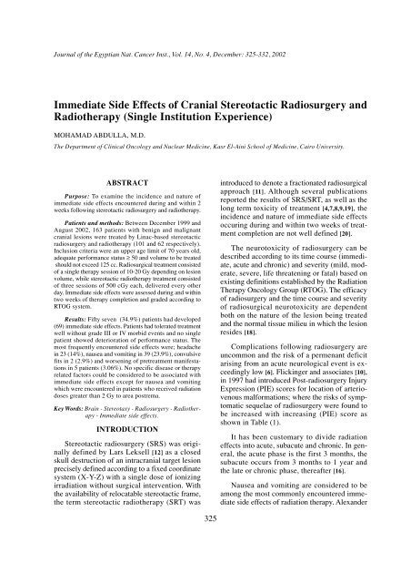
328 <strong>Immediate</strong> <strong>Side</strong> <strong>Effects</strong> <strong>of</strong> <strong>Cranial</strong> <strong>Stereotactic</strong> <strong>Radiosurgery</strong>experienced nausea <strong>and</strong> vomiting as immediateside effects <strong>of</strong> cranial streotactic irradiation, ahighly significant positive correlation with thedose delivered to the region harboring areapotrema (Vomiting Center) with cut <strong>of</strong>f point at2 Gy was found. After 2 Gy dose, all patientsTable (1): Post-radiosurgery injury expression (PIE) scoresfor location <strong>of</strong> arteriovenous malformation (Flickingeret al., 1997) [10].developed nausea <strong>and</strong> vomiting (Pearson’s ChiSquare) (p value < 0.0001) [3] as shown in Table(11). Poor correlation data were obtained onanalyzing other encountered immediate sideeffects in relation to any <strong>of</strong> the disease or therapyrelated parameters.Table (4): Karn<strong>of</strong>sky performance scale among 163 patientstreated by stereotactic radiosurgery <strong>and</strong> radiotherapy(NEMROCK 1999-2002).PIE scoreLocationPerformance statusNumber%1 (Low risk)234 (High risk)Frontal lobeCerebellum, temporal lobe, partietal lobeOccipital lobe or basal gangliaMedulla, thalamus, intraventricular, ponsor corpus callosum100-9080-7060-50Total65712716339.843.616.6100Table (2): Radiation therapy oncology group (RTOG)radiosurgery quality assurance guidelines [15].Per protocolMinor(acceptabledeviation)Major(unacceptableConformity≤ 2> 2 & < 2.5> 2.5Dosehomogenity1-2> 0.9 & < 1or> 2 & < 2.5< 0.9 & > 2.5Dosegradient≥ 0.3–Conformity: Prescription Isodose Surface Volume to Target Volume(TV) Ratio.Dose homogenity: Maximum Dose (MD) to Prescription Dose(PD) Ratio.Dose gradient: Target Volume (TV) to Volume Enclosed by 50%(V50%) Ratio.Table (3): Initial diagnosis among 163 patients treated bystereotactic radiosurgery <strong>and</strong> radiotherapy (NEM-ROCK 1999-2002).CatgeoryBenignDiseasesTumorsTotalDiagnosisMeningiomasArteriovenousMalformationsAcuosticNeuromasLow gradeGliomasHigh gradeGliomasPituitaryAdenomasMetastaticBrain diseaseOthers*Number2922*Others include; haemangiopericytoma in 2 patients (1%), chordoma<strong>of</strong> skull base in 4 patients (2%), ependymoma in 4 patients(2%), pineal body tumor in 3 patients (1.5%), glomus jugularein 3 patients (1.5%) <strong>and</strong> medulloblastoma in 3 patients (1.5%).2139208519163–1913132812.553%9.5100Table (5): Lesion volume, multiplicity <strong>and</strong> treatment scheduleamong 163 patients treated by stereotacticradiosurgery <strong>and</strong> radiotherapy (NEMROCK1999-2002).ItemVolume:Range (cc)Median volume (cc)St<strong>and</strong>ard deviationMultiplicity:One lesionTwo lesionsThree lesionsTreatment schedule:SRSSRTValues0.21-65.3112.5±13.6160 patients2 patients1 patient101 (61.9%)62 (38.1%)Table (6): Dose scheme adopted among (101) patientstreated by stereotactic radiosurgery (NEM-ROCK 1999-2002).Volume< 1 cc1-5 cc> 5-125 ccN113357% Dose (Gy)10.932.756.4201510Table (7): Dose contribution to risk structures <strong>and</strong> conformityindex among 163 patients treated by stereotacticradiosurgery <strong>and</strong> radiotherapy (NEMROCK1999-2002).RiskstructureLeft eyeRight eyeBrain stemChiasmaConformityIndexDose range(Gy)0-4.40-3.60-19.40-17.2Range1-2.95Mean dose(Gy)0.470.424.601.30Mean value1.44St<strong>and</strong>arddeviation±0.75±0.59±4.84±4.08St<strong>and</strong>ardDeviation±0.39
Mohamad Abdulla 329Table (8): Incidence <strong>of</strong> immediate side effects “<strong>of</strong> mild &moderate severity” reported among 163 patientstreated by stereotactic radiosurgery <strong>and</strong> radiotherapy(NEMROCK 1999-2002).<strong>Immediate</strong> sideeffectsNaudea <strong>and</strong> vomitingHeadacheWorsening <strong>of</strong> pretreat. DeficitsConvulsive fitsNumber <strong>of</strong> patientswith side effects392352% <strong>of</strong>total23.9143.061.2Table (9): Incidence <strong>of</strong> immediate side effects accordingto initial diagnosis among 163 patients treatedby stereotactic radiosurgery <strong>and</strong> radiotherapy(NEMROCK 1999-2002).DiagnosisAcuostic neuromaLow grade gliomaHigh grade gliomaArteriovenous malformationMeningiomaMetastatic brain diseasePituitary adenomaOthersNumber10/2113/3914/204/229/293/50/84/19%433370203160021.1Table (10): Distribution <strong>of</strong> immediate side effects (I.S.E.)within different disease entities among 163patients treated by stereotactic radiosurgery<strong>and</strong> radiotherapy (EMROCK 1999-2002).AcuosticNeuromaLow G.GliomaHigh G.GliomaAVMMeningiomaMetastasesOthersHeadache4553321Nausea &vomiting78101634Worsening <strong>of</strong>pretreatmentdeficits0120200FitsTable (11): Incidence to develop nausea <strong>and</strong> vomitingaccording to radiation dose delivered to areapostrema among 163 patients treated by stereotacticradiosurgery <strong>and</strong> radiotherapy (NEM-ROCK 1999-2002).DoseN/VNoYeTotal0-≤ 2 Gy124613095.44.6100> 2 GyNo. % No. %0333301001000010010p-value< 0.0001Fig. (1): Cut section in human brain showing area postrema“vomiting center”.Vol. (%)100% = 0.49 cm 3 LesionNormal Tissue140Min. dose = 15.80 GyMax. dose = 20.00 Gy12010080604020Dose (%)00 20 40 60 80 100 120 140 160 180 200100% = 20.00 GyNo dose delivery for:Rt eye mri (12.64 cm 3 ) Lt eye mri (11.94 cm 3 )Fig. (2): Dose volume histogram showing adequate lesionvolume coverage without dose contribution tosurrounding normal structures <strong>and</strong> conformityindex approaching value 1.Vol. (%)100% = 28.20 cm 3 Brain Stem MRIMin. dose = 0.00 Gy140Max. dose = 7.50 Gy12010080604020Dose (%)00 20 40 60 80 100 120 140 160 180 200Fig. (3): Dose volume histogram <strong>of</strong> brain stem denotingsparing <strong>of</strong> the major part <strong>of</strong> its volume, keepingthe maximum dose (7.50 Gy) to less than 1% <strong>of</strong>brain stem volume. The treated lesion is locatedat the cerebello-pontine angle.
Mohamad Abdulla 331receive a radiation dose ranging from 0-8.1 Gywith a median value <strong>of</strong> 1.21 Gy to the regionlocated at the floor <strong>of</strong> the 4th ventricle <strong>and</strong>harboring area postrema, but the onset <strong>of</strong> vomitingwas correlated only with doses greater than2 Gy, contrasting to data reported by Alex<strong>and</strong>eret al. [1].The discrepancy with data reported by Alex<strong>and</strong>er<strong>and</strong> co-workers [1] could be attributed todifferent disease entities treated in our study,where he treated only cases with acuostic neuromas.However, further analysis <strong>of</strong> our datarevealed that 33% (7/21 patients) with acuosticneuromas had developed nausea <strong>and</strong> vomitingbut at a lower threshold dose level (2 versus6.18 Gy) to area postrema, a finding whichrequires further clarification via future studiesenrolling higher number <strong>of</strong> patients to establishthe threshold radiation dose to area postremaabove which, nausea <strong>and</strong> vomiting are anticipatedas immediate post-stereotxay morbid events.Also, inspite <strong>of</strong> the relatively close results withthose <strong>of</strong> Bodis et al. [4]; (2 versus 2.75 Gy) asa threshold dose level to area postrema, none <strong>of</strong>our patients had received ondansetron as a premedication;only metclopramide <strong>and</strong> steroidswhich were effecive in alleviating such manifestations.The incidence <strong>of</strong> developing immediate sideeffects among patients with malignant craniallesions was clearly higher than those with benigndiseases, 70% versus 43% indicating the needto pay more attention to proper patient selection<strong>and</strong> adequate pre- as well a post-treatment medicationsfor patients with high grade gliomas<strong>and</strong> brain metastases.Only two patients had developed epilepticepisodes following stereotaxy. They had thediagnoses <strong>of</strong> high grade glioma <strong>and</strong> brainmetastasis, also they had a past history <strong>of</strong> convulsivefits during their disease course <strong>and</strong> coulddirect attention to the importance <strong>of</strong> employingmore potent anti-convulsive measures whenattempting to treat similar patients.Worsening <strong>of</strong> pre-therapy manifestations wasreported in one, two <strong>and</strong> two patients with diagnoses<strong>of</strong> low grade glioma, high garde glioma<strong>and</strong> meningioma respectively. All lesions werenoted to be located near the motor cortex whichcould be compromised by either direct radiationeffect or more likely due to cerebral oedemam<strong>and</strong>ating the adequate use <strong>of</strong> dehydrating measuresin both pre- <strong>and</strong> post-therapy periods.Moreover, It is difficult to conclude that patientswith pituitary adenomas are not anticipated todevelop immediate post-stereotaxy side effects,as this conclusion cannot be emphasized withthe small number <strong>of</strong> patients enrolled in our trial(8 Patients).The location <strong>of</strong> lesions <strong>and</strong> dose are criticalparameters related to the development <strong>of</strong> poststereotacticradiosurgery <strong>and</strong> radiotherapy edema.It is well known that parasagittal meningiomasare a risk group in this respect. Occlusion <strong>of</strong>bridging veins is a possible though unprovenmechanism for this phenomenon. However, thisedema did not occur within the time frame <strong>of</strong>the present study.Further clinical trials are clearly neededincluding higher numbers <strong>of</strong> patients with homogenouscharacteristics aiming at obtainingmore informative data about stereotaxy sideeffects <strong>and</strong> its proper management. Also, it shouldbe emphasized to avoid radiation doses greaterthan 2 Gy to the region in the floor <strong>of</strong> the 4thventricle harboring area potrema with the possibleuse <strong>of</strong> H3 antagonists if higher dose deliveryis an inevitable event.Acknowledgment: I am grateful to Pr<strong>of</strong>. KamalA. El-Ghamrawi for critical review <strong>of</strong> Themainscript. Also, I would like to express mydeep appreciation to Pr<strong>of</strong>. Ehsan El-Ghoneimy& Pr<strong>of</strong>. Magda Moustafa as well as NEMROCKradiosurgery team for devotion <strong>and</strong> help.REFERENCES1- Alex<strong>and</strong>er E. 3rd, Siddon R <strong>and</strong> Loeffler J.: The AcuteOnset <strong>of</strong> Nausea <strong>and</strong> Vomiting Following <strong>Stereotactic</strong><strong>Radiosurgery</strong>: Correlation with Total Dose to AreaPostrema. Surg. Neurol., 32 (1): 40-44, 1989.2- Alex<strong>and</strong>er E. III, Loeffler J.S., Lunsford L.D., et al.:<strong>Stereotactic</strong> <strong>Radiosurgery</strong>. New York: McGraw Hill,1993.3- Altman D.G.: Practical statistics for medical research.London, Chapman & Hall, 115-231, 1991.4- Bodis S., Alex<strong>and</strong>er E. III, Kooy H., et al.: The Prevention<strong>of</strong> <strong>Radiosurgery</strong>-Induced Nausea <strong>and</strong> Vomitingby Ondansetron: Evidence <strong>of</strong> a Direct Effect on TheCentral Nervous System Chemoreceptor Trigger Zone.Surg. Neurol., 42: 249-252, 1994.5- Brenner D.J., Martel M.K. <strong>and</strong> Hall E.J.: FractionatedRegimens For <strong>Stereotactic</strong> Radiotherapy <strong>of</strong> RecurrentTumors in The Brain. Int. J. Radiat. Oncol. Biol. Phys.,21: 819-824, 1991.
332 <strong>Immediate</strong> <strong>Side</strong> <strong>Effects</strong> <strong>of</strong> <strong>Cranial</strong> <strong>Stereotactic</strong> <strong>Radiosurgery</strong>6- Chin L.S., Lazio B.E., Biggins T. <strong>and</strong> Amin P.: AcuteComplications Following Gamma Knife <strong>Radiosurgery</strong>are Rare. Surg. Neurol., 53 (5): 498-502; Discussion502, 2000.7- Das I.J., Downs M.B., Corn B.W., et al: Characteristics<strong>of</strong> a Dedicated Linear Accelerator-Based <strong>Stereotactic</strong><strong>Radiosurgery</strong> - Radiotherapy Unit. Radiother. Oncol.,38: 61-68, 1996.8- Delannes M., Daly N.J., Bonnet J., et al: FractionatedRadiotherapy <strong>of</strong> Small Inoperable Lesions <strong>of</strong> The BrainUsing a Non-Invasive <strong>Stereotactic</strong> Frame. Int. J. Radiat.Oncol. Biol. Phys., 30: 531-539, 1991.9- Dunbar S.F., Tarbell N.J., Kooy H., et al: <strong>Stereotactic</strong>Radiotherapy for Pediatric <strong>and</strong> Adult Brain Tumors:Preliminary Report. Int. J. Radiat. Oncol. Biol. Phys.,30: 531-539, 1994.10- Flickinger J., Kondziolka D., Matiz A. <strong>and</strong> LunsfordL.: Analysis <strong>of</strong> Neurological Sequelae from <strong>Radiosurgery</strong><strong>of</strong> Arteriovenous Malformations: How LocationAffects Outcome. Int. J. Radiat. Oncol. Biol. Phys.,40: 273-278, 1997.11- Hall E.J. <strong>and</strong> Brenner D.J.: The Radiobiology <strong>of</strong> <strong>Radiosurgery</strong>:Rationale for Different Treatment Regimensfor AVMs <strong>and</strong> Malignancies. Int. J. Radiat. Oncol.Biol. Phys., 25: 381-385, 1993.12- Leksell L.: The <strong>Stereotactic</strong> Method <strong>and</strong> <strong>Radiosurgery</strong><strong>of</strong> The Brain. Acta Chir. Sc<strong>and</strong>, 102: 316-319, 1951.13- Linskey M.E., Lundsford L.D., Flickinger J.S., et al.:<strong>Stereotactic</strong> <strong>Radiosurgery</strong> for Acuostic Tumors. Neurosurg.Clin. North Am., 3 (1): 191-205, 1992.14- Loeffler J.S., Siddon R.L., Wen P.Y., et al.: <strong>Stereotactic</strong><strong>Radiosurgery</strong> <strong>of</strong> The Brain Using St<strong>and</strong>ard LinearAccelerator: A Study <strong>of</strong> Early <strong>and</strong> Late <strong>Effects</strong>. Radiother.Oncol., 17: 311-321, 1990.15- Modified from Rubin P. <strong>and</strong> Wasserman T.: InternationalClinical Trials in Oncology: International ClinicalTrials in Oncology: The Late <strong>Effects</strong> <strong>of</strong> Toxicity Scoring.Int. J. Radiat. Oncol. Biol. Phys.,14 (suppl):29-38, 1988. In Perez C. <strong>and</strong> Luther B.: Principles <strong>and</strong>Practice <strong>of</strong> Radiation Oncology, 3rd Ed Lippincott-Raven Publication p156, 1998.16- Sheline G.E., Wara W.M. <strong>and</strong> Smith V.: TherapeuticIrradiation <strong>and</strong> Brain Injury. Int. J. Radiat. Oncol. Biol.Phys., 6: 1215-1228, 1980.17- Show E.: RTOG: <strong>Radiosurgery</strong> Quality AssuranceGuideline. Int. J. Radiat. Oncol. Biol. Phys., 27: 1231-1241, 1993.18- Show E., C<strong>of</strong>fey R. <strong>and</strong> Dinapoli R.: Show E., C<strong>of</strong>feyR. <strong>and</strong> Dinapoli R.: Neurotoxicity <strong>of</strong> <strong>Radiosurgery</strong>.Semin Radiat. Oncol., 5 (3): 235-245, 1995.19- Souhami L., Olivier A., Podgorsak E.B., et al.: Fractionated<strong>Stereotactic</strong> Radiation Therapy for IntracranialTumors. Cancer, 68: 2101-2108, 1991.20- Wasik M.W., Rudoler S., Preston P., Lauck W.W.,Downes B.M., Leeper D., Andrews D., Corn B.W. <strong>and</strong>Curran W.J.: <strong>Immediate</strong> <strong>Side</strong> <strong>Effects</strong> <strong>of</strong> <strong>Stereotactic</strong>Radiotherapy <strong>and</strong> <strong>Radiosurgery</strong>. Int. J. Radiat. Oncol.Biol. Phys., 43 (2): 299-304, 1999.


