Immediate Side Effects of Cranial Stereotactic Radiosurgery and ...
Immediate Side Effects of Cranial Stereotactic Radiosurgery and ...
Immediate Side Effects of Cranial Stereotactic Radiosurgery and ...
You also want an ePaper? Increase the reach of your titles
YUMPU automatically turns print PDFs into web optimized ePapers that Google loves.
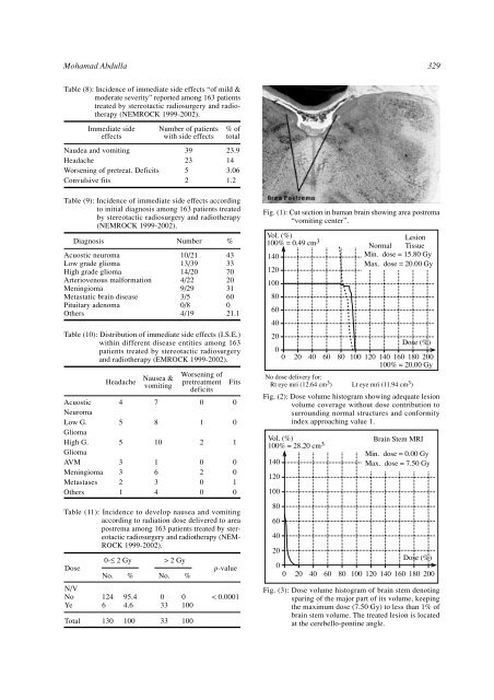
Mohamad Abdulla 329Table (8): Incidence <strong>of</strong> immediate side effects “<strong>of</strong> mild &moderate severity” reported among 163 patientstreated by stereotactic radiosurgery <strong>and</strong> radiotherapy(NEMROCK 1999-2002).<strong>Immediate</strong> sideeffectsNaudea <strong>and</strong> vomitingHeadacheWorsening <strong>of</strong> pretreat. DeficitsConvulsive fitsNumber <strong>of</strong> patientswith side effects392352% <strong>of</strong>total23.9143.061.2Table (9): Incidence <strong>of</strong> immediate side effects accordingto initial diagnosis among 163 patients treatedby stereotactic radiosurgery <strong>and</strong> radiotherapy(NEMROCK 1999-2002).DiagnosisAcuostic neuromaLow grade gliomaHigh grade gliomaArteriovenous malformationMeningiomaMetastatic brain diseasePituitary adenomaOthersNumber10/2113/3914/204/229/293/50/84/19%433370203160021.1Table (10): Distribution <strong>of</strong> immediate side effects (I.S.E.)within different disease entities among 163patients treated by stereotactic radiosurgery<strong>and</strong> radiotherapy (EMROCK 1999-2002).AcuosticNeuromaLow G.GliomaHigh G.GliomaAVMMeningiomaMetastasesOthersHeadache4553321Nausea &vomiting78101634Worsening <strong>of</strong>pretreatmentdeficits0120200FitsTable (11): Incidence to develop nausea <strong>and</strong> vomitingaccording to radiation dose delivered to areapostrema among 163 patients treated by stereotacticradiosurgery <strong>and</strong> radiotherapy (NEM-ROCK 1999-2002).DoseN/VNoYeTotal0-≤ 2 Gy124613095.44.6100> 2 GyNo. % No. %0333301001000010010p-value< 0.0001Fig. (1): Cut section in human brain showing area postrema“vomiting center”.Vol. (%)100% = 0.49 cm 3 LesionNormal Tissue140Min. dose = 15.80 GyMax. dose = 20.00 Gy12010080604020Dose (%)00 20 40 60 80 100 120 140 160 180 200100% = 20.00 GyNo dose delivery for:Rt eye mri (12.64 cm 3 ) Lt eye mri (11.94 cm 3 )Fig. (2): Dose volume histogram showing adequate lesionvolume coverage without dose contribution tosurrounding normal structures <strong>and</strong> conformityindex approaching value 1.Vol. (%)100% = 28.20 cm 3 Brain Stem MRIMin. dose = 0.00 Gy140Max. dose = 7.50 Gy12010080604020Dose (%)00 20 40 60 80 100 120 140 160 180 200Fig. (3): Dose volume histogram <strong>of</strong> brain stem denotingsparing <strong>of</strong> the major part <strong>of</strong> its volume, keepingthe maximum dose (7.50 Gy) to less than 1% <strong>of</strong>brain stem volume. The treated lesion is locatedat the cerebello-pontine angle.


