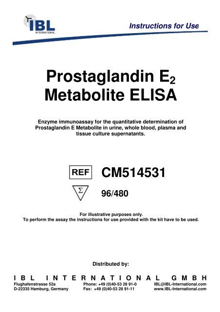
Prostaglandin E2 Metabolite ELISA - IBL International
Prostaglandin E2 Metabolite ELISA - IBL International
Prostaglandin E2 Metabolite ELISA - IBL International

Create successful ePaper yourself
Turn your PDF publications into a flip-book with our unique Google optimized e-Paper software.
<strong>Prostaglandin</strong> <strong>E2</strong><br />
<strong>Metabolite</strong> <strong>ELISA</strong><br />
Enzyme immunoassay for the quantitative determination of<br />
<strong>Prostaglandin</strong> E <strong>Metabolite</strong> in urine, whole blood, plasma and<br />
tissue culture supernatants.<br />
CM514531<br />
96/480<br />
For illustrative purposes only.<br />
To perform the assay the instructions for use provided with the kit have to be used.<br />
Distributed by:<br />
Instructions for Use<br />
I B L I N T E R N A T I O N A L G M B H<br />
Flughafenstrasse 52a Phone: +49 (0)40-53 28 91-0 <strong>IBL</strong>@<strong>IBL</strong>-<strong>International</strong>.com<br />
D-22335 Hamburg, Germany Fax: +49 (0)40-53 28 91-11 www.<strong>IBL</strong>-<strong>International</strong>.com
<strong>Prostaglandin</strong> E <strong>Metabolite</strong> EIA Kit<br />
Catalog No. 514531 (Strip Plate)<br />
Catalog No. 514531.1 (Solid Plate)
TABLE OF CONTENTS<br />
GENERAL INFORMATION 3 Materials Supplied<br />
4 Precautions<br />
4 If You Have Problems<br />
4 Storage and Stability<br />
INTRODUCTION 5 Background<br />
4 Materials Needed but Not Supplied<br />
5 About This Assay<br />
7 Description of ACE TM Competitive EIAs<br />
8 Biochemistry of Acetylcholinesterase<br />
10 Definition of Key Terms<br />
PRE-ASSAY PREPARATION 11 Buffer Preparation<br />
12 Sample Preparation<br />
13 Sample Purification<br />
ASSAY PROTOCOL 17 Derivatization of Standards and<br />
Samples to PGEM<br />
19 Preparation of Assay-Specific Reagents<br />
21 Plate Set Up<br />
22 Performing the Assay<br />
ANALYSIS 25 Calculations<br />
28 Performance Characteristics<br />
RESOURCES 32 Troubleshooting<br />
32 Additional Reading<br />
33 References<br />
33 Related Products<br />
34 Warranty and Limitation of Remedy<br />
35 Plate Template<br />
36 Notes<br />
Materials Supplied<br />
GENERAL INFORMATION<br />
Catalog Number Item 96 wells<br />
Quantity/Size<br />
GENERAL INFORMATION<br />
480 wells<br />
Quantity/Size<br />
414532 <strong>Prostaglandin</strong> E <strong>Metabolite</strong> EIA Antiserum 1 vial/100 dtn 1 vial/500 dtn<br />
414530 <strong>Prostaglandin</strong> E <strong>Metabolite</strong> AChE Tracer 1 vial/100 dtn 1 vial/500 dtn<br />
414534 <strong>Prostaglandin</strong> E <strong>Metabolite</strong> EIA Standard 1 vial 1 vial<br />
400060 EIA Buffer Concentrate (10X) 2 vials/10 ml 4 vials/10 ml<br />
400062 Wash Buffer Concentrate (400X) 1 vial/5 ml 1 vial/12.5 ml<br />
400035 Tween 20 1 vial/3 ml 1 vial/3 ml<br />
400005 Mouse Anti-rabbit IgG Coated Plate 1 plate 5 plates<br />
400012 Plate Cover 1 cover 5 covers<br />
400050 Ellman’s Reagent 3 vials/100 dtn 6 vials/250 dtn<br />
400032 Phosphate Buffer 1 vial/100 dtn 2 vials/250 dtn<br />
400027 Carbonate Buffer 1 vial/100 dtn 1 vial/500 dtn<br />
400040 EIA Tracer Dye 1 vial 1 vial<br />
400042 EIA Antiserum Dye 1 vial 1 vial<br />
If any of the items listed above are damaged or missing, please contact our Customer<br />
Service department at (800) 364-9897 or (734) 975-3999. We cannot accept any returns<br />
without prior authorization.<br />
!<br />
WARNING: Not for human or animal disease diagnosis or therapeutic<br />
drug use.<br />
3
Precautions<br />
Please read these instructions carefully before beginning this assay.<br />
The reagents in this kit have been tested and formulated to work exclusively with Cayman<br />
Chemical’s ACE TM EIA Kits. This kit may not perform as described if any reagent or<br />
procedure is replaced or modified.<br />
For research use only. Not for human or diagnostic use.<br />
If You Have Problems<br />
Technical Service Contact Information<br />
Phone: 888-526-5351 (USA and Canada only) or 734-975-3888<br />
Fax: 734-971-3641<br />
E-Mail: techserv@caymanchem.com<br />
Hours: M-F 8:00 AM to 5:30 PM EST<br />
In order for our staff to assist you quickly and efficiently, please be ready to supply the lot<br />
number of the kit (found on the outside of the box).<br />
Storage and Stability<br />
This kit will perform as specified if stored as directed at -20°C and used before the expiration<br />
date indicated on the outside of the box.<br />
Materials Needed But Not Supplied<br />
1. A plate reader capable of measuring absorbance between 405-420 nm.<br />
2. Adjustable pipettes and a repeat pipettor.<br />
3. A source of ‘UltraPure’ water. Water used to prepare all EIA reagents and buffers<br />
must be deionized and free of trace organic contaminants (‘UltraPure’). Use activated<br />
carbon filter cartridges or other organic scavengers. Glass distilled water (even if double<br />
distilled), HPLC-grade water, and sterile water (for injections) are not adequate for<br />
EIA. NOTE: UltraPure water is available for purchase from Cayman (Catalog No.<br />
400000).<br />
4. Materials used for Sample Preparation (see page 12).<br />
Background<br />
INTRODUCTION<br />
<strong>Prostaglandin</strong> E 2 (PGE 2 ) is produced by a variety of cell types which, in general, do not<br />
contain the enzymes required for metabolism of PGE 2 . Thus, cultured endothelial cells or<br />
osteoblasts will release PGE 2 into the culture medium where it will accumulate without<br />
appreciable metabolism. The direct assay of PGE 2 from the medium is a good way to<br />
measure PGE 2 production from these cells.<br />
PGE 2 is rapidly converted in vivo to its 13,14-dihydro-15-keto metabolite (see Figure 1,<br />
on page 6), with more than 90% of circulating PGE 2 cleared by a single passage through<br />
the lungs. Unfortunately, this metabolite is not chemically stable and undergoes a variable<br />
amount of degradation to PGA products. For this reason, blood, urine, or other samples<br />
from whole animals or humans often contain very little intact PGE 2 , and measurement of<br />
the metabolites is necessary to provide a reliable estimate of actual PGE 2 production.<br />
About This Assay<br />
Cayman’s <strong>Prostaglandin</strong> E <strong>Metabolite</strong> (PGEM) assay is a competitive assay that converts<br />
13,14-dihydro-15-keto PGA 2 and 13,14-dihydro-15-keto PGE 2 to a single, stable derivative<br />
that can be easily quantified. This assay is, therefore, the method of choice if the samples<br />
in question have undergone extensive metabolism prior to collection. The EIA typically<br />
displays an IC 50 (50% B/B 0 ) value of approximately 10 pg/ml and a detection limit (80%<br />
B/B 0 ) of approximately 2 pg/ml.<br />
4 GENERAL INFORMATION INTRODUCTION 5
HO<br />
t 1/2 - 3 minutes in plasma 15-OH PGDH<br />
HO<br />
HO<br />
O<br />
O<br />
OH<br />
<strong>Prostaglandin</strong> E 2<br />
15-keto <strong>Prostaglandin</strong> E 2<br />
O<br />
13,14,dihydro-15-keto<br />
<strong>Prostaglandin</strong> E 2<br />
(unstable)<br />
Figure 1. Metabolism of PGE 2<br />
O<br />
O<br />
COOH<br />
COOH<br />
15-oxoprostaglandin ∆ 13 -reductase<br />
COOH<br />
Non-enzymatic<br />
degradation<br />
O<br />
O<br />
13,14-dihydro-15-keto<br />
<strong>Prostaglandin</strong> A 2<br />
COOH<br />
Description of ACE TM Competitive EIAs 1,2<br />
This assay is based on the competition between Prostagladin E <strong>Metabolite</strong> (PGEM) and<br />
a PGEM-acetylcholinesterase (AChE) conjugate (PGEM Tracer) for a limited number of<br />
PGEM-specific rabbit antiserum binding sites. Because the concentration of the PGEM<br />
Tracer is held constant while the concentration of PGEM varies, the amount of PGEM<br />
Tracer that is able to bind to the rabbit antiserum will be inversely proportional to the<br />
concentration of PGEM in the well. This rabbit antiserum-PGEM (either free or tracer)<br />
complex binds to the mouse monoclonal anti-rabbit IgG that has been previously attached<br />
to the well. The plate is washed to remove any unbound reagents, and then Ellman’s Reagent<br />
(which contains the substrate to AChE) is added to the well. The product of this enzymatic<br />
reaction has a distinct yellow color and absorbs strongly at 412 nm. The intensity of this<br />
color, determined spectrophotometrically, is proportional to the amount of PGEM Tracer<br />
bound to the well, which is inversely proportional to the amount of free PGEM present in<br />
the well during the incubation; or<br />
Absorbance ∝ [Bound PGEM Tracer] ∝ 1/[PGEM]<br />
A schematic of this process is shown in Figure 2, below.<br />
Plates are pre-coated with<br />
mouse monoclonal anti-rabbit<br />
IgG and blocked with a<br />
proprietary formulation of<br />
proteins.<br />
2. Wash to remove all<br />
unbound reagents.<br />
1. Incubate with tracer,<br />
antiserum, and either<br />
standard or unknown<br />
sample.<br />
3. Develop the well with<br />
Ellman's Reagent.<br />
Figure 2. Schematic of the ACE TM EIA<br />
= Mouse Monoclonal Anti-rabbit IgG<br />
= Blocking proteins<br />
= Acetylcholinesterase linked<br />
to PGEM (Tracer)<br />
= Specific antiserum to PGEM<br />
= Free PGEM<br />
6 INTRODUCTION INTRODUCTION 7
Biochemistry of Acetylcholinesterase<br />
The electric organ of the electric eel, Electrophorus electricus, contains an avid<br />
acetylcholinesterase (AChE) capable of massive catalytic turnover during the generation of its<br />
electrochemical discharges. The electric eel AChE has a clover leaf-shaped tertiary structure<br />
consisting of a triad of tetramers attached to a collagen-like structural fibril. This stable<br />
enzyme is capable of high turnover (64,000 s -1 ) for the hydrolysis of acetylthiocholine.<br />
A molecule of the analyte covalently attached to a molecule of AChE serves as the tracer<br />
in ACE enzyme immunoassays. Quantification of the tracer is achieved by measuring<br />
its AChE activity with Ellman’s Reagent. This reagent consists of acetylthiocholine and<br />
5,5’-dithio-bis-(2-nitrobenzoic acid). Hydrolysis of acetylthiocholine by AChE produces<br />
thiocholine (see Figure 3, on page 9). The non-enzymatic reaction of thiocholine with 5,5’dithio-bis-(2-nitrobenzoic<br />
acid) produces 5-thio-2-nitrobenzoic acid, which has a strong<br />
absorbance at 412 nm (ε = 13,600).<br />
AChE has several advantages over other enzymes commonly used for enzyme immunoassays.<br />
Unlike horseradish peroxidase, AChE does not self-inactivate during turnover. This property<br />
of AChE also allows redevelopment of the assay if it is accidentally splashed or spilled. In<br />
addition, the enzyme is highly stable under the assay conditions, has a wide pH range (pH<br />
5-10), and is not inhibited by common buffer salts or preservatives. Since AChE is stable<br />
during the development step, it is unnecessary to use a ‘stop’ reagent, and the plate may be<br />
read whenever it is convenient.<br />
8 INTRODUCTION INTRODUCTION 9<br />
O 2 N<br />
- OOC<br />
S<br />
O 2 N<br />
S<br />
O<br />
- OOC<br />
O -<br />
N +<br />
O<br />
S<br />
N +<br />
- S<br />
S S NO 2<br />
Acetylthiocholine<br />
COO -<br />
Figure 3. Reaction catalyzed by acetylcholinesterase<br />
N + �iocholine<br />
- S<br />
5,5'-dithio-bis-<br />
(2-Nitrobenzoic Acid)<br />
COO -<br />
NO 2<br />
5-thio-2-Nitrobenzoic Acid<br />
λ max : 412 nm<br />
ε: 13,600
Definition of Key Terms<br />
Blank: background absorbance caused by Ellman’s Reagent. The blank absorbance should<br />
be subtracted from the absorbance readings of all the other wells.<br />
Total Activity: total enzymatic activity of the AChE-linked tracer. This is analogous to<br />
the specific activity of a radioactive tracer.<br />
NSB (Non-Specific Binding): non-immunological binding of the tracer to the well.<br />
Even in the absence of specific antibody a very small amount of tracer still binds to the<br />
well; the NSB is a measure of this low binding.<br />
B 0 (Maximum Binding): maximum amount of the tracer that the antibody can bind in<br />
the absence of free analyte.<br />
%B/B 0 (%Bound/Maximum Bound): ratio of the absorbance of a particular sample<br />
or standard well to that of the maximum binding (B 0 ) well.<br />
Standard Curve: a plot of the %B/B 0 values versus concentration of a series of wells<br />
containing various known amounts of analyte.<br />
Dtn: determination where one dtn is the amount of reagent used per well.<br />
PRE-ASSAY PREPARATION<br />
NOTE: Water used to prepare all EIA reagents and buffers must be deionized and free of trace<br />
organic contaminants (‘UltraPure’). Use activated carbon filter cartridges or other organic<br />
scavengers. Glass distilled water (even if double distilled), HPLC-grade water, and sterile<br />
water (for injections) are not adequate for EIA. UltraPure water may be purchased from<br />
Cayman (Catalog No. 400000).<br />
Buffer Preparation<br />
Store all buffers at 4°C; they will be stable for about two months.<br />
1. EIA Buffer Preparation<br />
Dilute the contents of one vial of EIA Buffer Concentrate (Catalog No. 400060)<br />
with 90 ml of UltraPure water. Be certain to rinse the vial to remove any salts that<br />
may have precipitated. NOTE: It is normal for the concentrated buffer to contain<br />
crystalline salts after thawing. These will completely dissolve upon dilution with water.<br />
2. Wash Buffer Preparation<br />
5 ml vial Wash Buffer (96-well kit; Catalog No. 400062): Dilute to a total volume<br />
of 2 liters with UltraPure water and add 1 ml of Tween 20 (Catalog No. 400035).<br />
10 INTRODUCTION PRE-ASSAY PREPARATION 11<br />
OR<br />
12.5 ml vial Wash Buffer (480-well kit; Catalog No. 400062): Dilute to a total<br />
volume of 5 liters with UltraPure water and add 2.5 ml of Tween 20 (Catalog No.<br />
400035).<br />
Smaller volumes of Wash Buffer can be prepared by diluting the Wash Buffer Concentrate<br />
1:400 and adding Tween 20 (0.5 ml/liter of Wash Buffer).<br />
NOTE: Tween 20 is a viscous liquid and cannot be measured by a regular pipette. A positive<br />
displacement pipette or a syringe should be used to deliver small quantities accurately.
3. Phosphate Buffer<br />
Prepare a 1 M Phosphate Buffer solution by dissolving the contents of the 100 dtn<br />
vial of Phosphate Buffer (Catalog No. 400032) in 30 ml UltraPure water, or dissolve<br />
the contents of one of the 250 dtn vials of Phosphate Buffer (Catalog No. 400032) in<br />
75 ml UltraPure water.<br />
4. Carbonate Buffer<br />
Prepare a 1 M Carbonate Buffer solution by dissolving the contents of the 100 dtn<br />
vial of Carbonate Buffer (Catalog No. 400027) in 25 ml UltraPure water, or dissolve<br />
the contents of the 500 dtn vial of Carbonate Buffer (Catalog No. 400027) in<br />
125 ml UltraPure water.<br />
5. PGEM Assay Buffer<br />
Prepare 20 ml of PGEM Assay Buffer by combining 13 ml EIA Buffer, 3 ml<br />
Carbonate Buffer, and 4 ml Phosphate Buffer. This quantity of buffer should be more<br />
than sufficient to complete one 96-well plate.<br />
Sample Preparation<br />
In general, after derivitizing urine and tissue culture supernatant samples may be diluted<br />
with PGEM Assay Buffer and added directly to the assay well. Plasma and whole blood,<br />
as well as other heterogeneous mixtures, such as lavage fluids and aspirates often contain<br />
contaminants which can interfere in the assay. It is best to check for interference before<br />
embarking on a large number of sample measurements. To test for interference, dilute<br />
one or two test samples to obtain at least two different dilutions of each sample between<br />
~2 and 50 pg/ml (i.e., between 20-80% B/B 0 ). If the two different dilutions of the sample<br />
show good correlation (differ by 20% or less) in the final calculated PGEM concentration,<br />
purification is not required. If you do not see good correlation of the different dilutions,<br />
purification is advised. The Purification Protocol, on page 13-16, is one such method.<br />
General Precautions<br />
• All samples must be free of organic solvents prior to assay.<br />
• Samples should be assayed immediately after collection; samples that cannot be<br />
assayed immediately should be stored at -80°C.<br />
Sample Purification<br />
The method of purification is by solid phase extraction (SPE) as described in the protocol<br />
below.<br />
Determination of Recovery<br />
Determination of percent recovery is recommended when any sample purification<br />
is performed. Detailed on page 15 are two methods that can be employed to monitor<br />
recovery. If the hot spike method (recommended) is used, 10,000 cpm of tritium-labeled<br />
PGE 2 is added directly to the sample and 10% is removed for scintillation counting after<br />
purification. If the cold spike method is used, the sample must be split prior to purification<br />
and an appropriate amount of 13,14-dihydro-15-keto PGE 2 added to one aliquot. The<br />
spiked sample is then assayed via EIA alongside the unspiked sample. Calculations for<br />
each method are found in the Analysis section on page 25.<br />
12 PRE-ASSAY PREPARATION PRE-ASSAY PREPARATION 13
C-18 SPE Purification Protocol<br />
Materials Needed<br />
1. Tritium-labeled PGE 2 to use as a hot spike or unlabeled 13,14-dihydro-15-keto PGE 2<br />
to use as a cold spike to allow determination of extraction efficiency.<br />
2. 1 M Acetic acid, deionized water, ethanol, methanol, hexane and ethyl acetate<br />
3. 200 mg C-18 solid phase extraction (SPE) columns (non end-capped)<br />
Sample<br />
Add ethanol Centrifuge Acidify<br />
1. Wash with H2O (very polar) 3. Elute with Ethyl acetate/<br />
1% Methanol<br />
(intermediate polarity)<br />
Water removes polar substances.<br />
PG is not highly water soluble and<br />
prefers to bind to the C18.<br />
SPE (C-18) Cartridge<br />
(non end-capped)<br />
2. Wash with Hexane<br />
(very non-polar)<br />
Hexane removes the water. PG<br />
prefers to bind to the silica OH<br />
backbone. Therefore PG stays on<br />
the column.<br />
Figure 5. Schematic of PGEM Purification by C-18 SPE<br />
PG is very soluble in this solvent<br />
and therefore elutes easily.<br />
Hot Spike Cold Spike<br />
1. Aliquot a known amount of each<br />
sample into a clean test tube<br />
(500 µl is recommended). If your<br />
samples need to be concentrated,<br />
a larger volume should be used<br />
(e.g., a 5 ml sample will be<br />
concentrated by a factor of 10, a<br />
10 ml sample will be concentrated<br />
by a factor of 20, etc.).<br />
2. Add 10,000 cpm of tritiumlabeled<br />
PGE 2 ([ 3 H]-PGE 2 ).<br />
Proceed to step 3 below<br />
1. Aliquot a known amount of each<br />
sample into each of two tubes.<br />
Label the first tube ‘sample #’<br />
and the second tube ‘sample #<br />
+ spike’. If your samples need<br />
to be concentrated, a larger<br />
volume should be used (e.g., a<br />
5 ml sample will be concentrated<br />
by a factor of 10, a 10 ml sample<br />
will be concentrated by a factor of<br />
20, etc.).<br />
2. Add a cold spike of 13,14dihydro-15-keto<br />
PGE 2 to the<br />
‘sample + spike’ tubes. Follow the<br />
procedure below for both spiked<br />
and unspiked samples.<br />
3. Precipitation of proteins using ethanol is optional and may not be needed if samples<br />
are clean enough to flow through the C-18 SPE cartridge. Body fluids such as<br />
plasma and urine can typically be applied directly to the C-18 cartridge after the<br />
acidification step (step 4) below. To precipitate proteins, add ethanol (approximately<br />
four times the sample volume) to each tube. Vortex to mix thoroughly. Incubate<br />
samples at 4°C for five minutes, then centrifuge a 3,000 x g for 10 minutes to remove<br />
precipitated proteins. Transfer the supernatant to a clean test tube. Evaporate the<br />
ethanol under nitrogen.<br />
4. Acidify the sample to ~pH 4 by the addition of 1 M acetate buffer. (To avoid having<br />
to measure the pH of each individual sample, adjust the pH of an equivalent volume<br />
of sample matrix to pH 4.0 using the 1M acetic acid. Add this volume of acetic acid<br />
to each sample. NOTE: For samples of different volumes, the amount of acid should be<br />
adjusted to maintain this ratio of acid to sample.). If the samples are cloudy or contain<br />
precipitate, either filter or centrifuge to remove the precipitate. Particulate matter in<br />
the sample may clog the SPE cartridge.<br />
14 PRE-ASSAY PREPARATION PRE-ASSAY PREPARATION 15
5. Prepare C-18 SPE columns by rinsing with 5 ml methanol followed by 5 ml deionized<br />
water. Do not allow the SPE cartridge to dry.<br />
6. Apply the sample to the SPE cartridge and allow the sample to completely enter the<br />
packing material.<br />
7. Wash the column with 5 ml deionized water followed by 5 ml HPLC grade hexane<br />
(allow the cartridge to become dry after this step). Discard both washes.<br />
8. Elute the PGE 2 and PGEM from the column with 5 ml ethyl acetate containing 1%<br />
methanol. Higher recovery and better reproducibility may be obtained if the sample<br />
is applied and eluted by gravity. The wash steps may be performed under vacuum or<br />
pressure.*<br />
9. Evaporate the ethyl acetate to dryness under a stream of nitrogen. It is very important<br />
that all of the organic solvent be removed as even small quantities will adversely affect<br />
the EIA.<br />
10. To resuspend the sample, add 500 µl EIA Buffer. Vortex. It is common for insoluble<br />
precipitate to remain in the sample after addition of EIA Buffer; this will not affect<br />
the assay. This sample is now ready for use in the EIA.<br />
Hot Spike (continued)<br />
11. Use 50 µl of the resuspended sample for scintillation counting.<br />
*If it is necessary to stop during this purification, samples may be stored in the ethyl acetate/<br />
methanol solution at -20°C or -80°C.<br />
ASSAY PROTOCOL<br />
Derivatization of Standards and Samples to PGEM<br />
Derivatization Hints<br />
• Allow the derivatization to proceed overnight to ensure that all the PGE 2<br />
metabolites derivatize completely.<br />
• Derizatize all standards and samples for the same amount of time.<br />
Derivatization of the PGEM EIA Standard<br />
Equilibrate a pipette tip in ethanol by repeatedly filling and expelling the tip with ethanol<br />
several times. Using the equilibrated pipette tip, transfer 100 µl of the PGEM Standard<br />
(Catalog No. 414534) into a clean test tube, then dilute with 900 µl UltraPure water. The<br />
concentration of this solution (the bulk standard) will be 40 ng/ml.<br />
Aliquot 50 µl of this solution into a clean tube and dilute to a total volume of 1 ml with<br />
EIA Buffer (i.e., add 950 µl). Add 300 µl of Carbonate Buffer and incubate at 37°C<br />
overnight. Then add 400 µl Phosphate Buffer and 300 µl EIA Buffer. This solution is<br />
1,000 pg/ml.<br />
Derivatization of the Samples<br />
Aliquot 500 µl of each sample into a clean test tube. Add 150 µl of Carbonate Buffer and<br />
incubate at 37°C overnight. Then add 200 µl Phosphate Buffer and 150 µl EIA Buffer. The<br />
samples are now ready to assay. If you need to dilute your samples after derivatization, be<br />
sure to use the PGEM Assay Buffer.<br />
16 PRE-ASSAY PREPARATION ASSAY PROTOCOL 17
Preparing the Standard Curve<br />
NOTE: Because of the high salt concentration in the 1,000 pg/ml solution, all the points of<br />
the standard curve must contain the same salt concentration. Thus, when performing the serial<br />
dilution, use the PGEM Assay Buffer.<br />
To prepare the standard for use in EIA: Obtain 8 clean test tubes and number them #1<br />
through #8. Aliquot 950 µl PGEM Assay Buffer to tube #1 and 500 µl PGEM Assay Buffer<br />
to tubes #2-8. Transfer 50 µl of the derivatized standard (1,000 pg/ml) to tube #1 and mix<br />
thoroughly. Serially dilute the standard by removing 500 µl from tube #1 and placing it in<br />
tube #2; mix thoroughly. Next, remove 500 µl from tube #2 and place it into tube #3; mix<br />
thoroughly. Repeat this process for tubes #4-8. These diluted standards should not be stored<br />
for more than 24 hours.<br />
1,000 pg/ml<br />
Standard<br />
50 µl 500 µl 500 µl 500 µl 500 µl 500 µl 500 µl 500 µl<br />
S1 S2 S3 S4 S5 S6 S7 S8<br />
50<br />
pg/ml<br />
950 µl<br />
PGEM<br />
Buffer<br />
25<br />
pg/ml<br />
500 µl<br />
PGEM<br />
Buffer<br />
12.5<br />
pg/ml<br />
500 µl<br />
PGEM<br />
Buffer<br />
Figure 4. Preparation of the PGEM standards<br />
6.25<br />
pg/ml<br />
500 µl<br />
PGEM<br />
Buffer<br />
3.13<br />
pg/ml<br />
500 µl<br />
PGEM<br />
Buffer<br />
1.56<br />
pg/ml<br />
500 µl<br />
PGEM<br />
Buffer<br />
0.78<br />
pg/ml<br />
500 µl<br />
PGEM<br />
Buffer<br />
0.39<br />
pg/ml<br />
Final<br />
500 µl<br />
PGEM<br />
Buffer<br />
Preparation of Assay-Specific Reagents<br />
PGEM AChE Tracer<br />
Reconstitute the PGEM Tracer as follows:<br />
100 dtn PGEM AChE Tracer (96-well kit; Catalog No. 414530): Reconstitute<br />
with 6 ml EIA Buffer.<br />
18 ASSAY PROTOCOL ASSAY PROTOCOL 19<br />
OR<br />
500 dtn PGEM AChE Tracer (480-well kit; Catalog No. 414530): Reconstitute<br />
with 30 ml EIA Buffer.<br />
Store the reconstituted PGEM Tracer at 4°C (do not freeze!) and use within four weeks. A<br />
20% surplus of PGEM Tracer has been included to account for any incidental losses.<br />
Tracer Dye Instructions (optional)<br />
This dye may be added to the tracer, if desired, to aid in visualization of tracercontaining<br />
wells. Add the dye to the reconstituted tracer at a final dilution of<br />
1:100 (add 60 µl of dye to 6 ml tracer or add 300 µl of dye to 30 ml of tracer).
PGEM EIA Antiserum<br />
Reconstitute the PGEM Antiserum as follows:<br />
100 dtn PGEM Antiserum (96-well kit; Catalog No. 414532): Reconstitute with<br />
6 ml EIA Buffer.<br />
OR<br />
500 dtn PGEM Antiserum (480-well kit; Catalog No. 414532): Reconstitute with<br />
30 ml EIA Buffer.<br />
Store the reconstituted PGEM Antiserum at 4°C. It will be stable for at least four weeks. A<br />
20% surplus of PGEM Antiserum has been included to account for any incidental losses.<br />
Antiserum Dye Instructions (optional)<br />
This dye may be added to the antiserum, if desired, to aid in visualization of<br />
antiserum-containing wells. Add the dye to the reconstituted antiserum at a final<br />
dilution of 1:100 (add 60 µl of dye to 6 ml antiserum or add 300 µl of dye to<br />
30 ml of antiserum).<br />
Plate Set Up<br />
The 96-well plate(s) included with this kit is supplied ready to use. It is not necessary to<br />
rinse the plate(s) prior to adding the reagents. NOTE: If you do not need to use all the strips<br />
at once, place the unused strips back in the plate packet and store at 2-4°C. Be sure the packet is<br />
sealed with the desiccant inside.<br />
Each plate or set of strips must contain a minimum of two blanks (Blk), two non-specific<br />
binding wells (NSB), two maximum binding wells (B 0 ), and an eight point standard curve<br />
run in duplicate. NOTE: Each assay must contain this minimum configuration in order to<br />
ensure accurate and reproducible results. Each sample should be assayed at two dilutions<br />
and each dilution should be assayed in duplicate. For statistical purposes, we recommend<br />
assaying samples in triplicate.<br />
A suggested plate format is shown in Figure 5, below. The user may vary the location and<br />
type of wells present as necessary for each particular experiment. The plate format provided<br />
below has been designed to allow for easy data analysis using a convenient spreadsheet<br />
offered by Cayman (see page 25, for more details). We suggest you record the contents of<br />
each well on the template sheet provided (see page 35).<br />
1 2 3 4 5 6 7 8 9 10 1112<br />
A Blk S1 S1 1 1 1 9 9 9 17 17 17<br />
B Blk S2 S2 2 2 2 10 10 10 18 18 18<br />
C NSB S3 S3 3 3 3 11 11 11 19 19 19<br />
D NSB S4 S4 4 4 4 12 12 12 20 20 20<br />
E B0 S5 S5 5 5 5 13 13 13 21 21 21<br />
F B0 S6 S6 6 6 6 14 14 14 22 22 22<br />
G B0 S7 S7 7 7 7 15 15 15 23 23 23<br />
H TA S8 S8 8 8 8 16 16 16 24 24 24<br />
Figure 5. Sample plate format<br />
Blk - Blank<br />
TA - Total Activity<br />
NSB - Non-Specific Binding<br />
B 0 - Maximum Binding<br />
S1-S8 - Standards 1-8<br />
1-24 - Samples<br />
20 ASSAY PROTOCOL ASSAY PROTOCOL 21
Performing the Assay<br />
Pipetting Hints<br />
• Use different tips to pipette the buffer, standard, sample, tracer, and<br />
antiserum.<br />
• Before pipetting each reagent, equilibrate the pipette tip in that reagent<br />
(i.e., slowly fill the tip and gently expel the contents, repeat several times).<br />
• Do not expose the pipette tip to the reagent(s) already in the well.<br />
Addition of the Reagents<br />
1. PGEM Buffer<br />
Add 50 µl EIA Buffer and 50 µl of PGEM Buffer to Non-Specific Binding (NSB)<br />
wells. Add 50 µl PGEM Buffer to Maximum Binding (B 0 ) wells.<br />
2. PGEM EIA Standard<br />
Add 50 µl from tube #8 to both of the lowest standard wells (S8). Add 50 µl from<br />
tube #7 to each of the next two standard wells (S7). Continue with this procedure<br />
until all the standards are aliquoted. The same pipette tip should be used to aliquot<br />
all the standards. Before pipetting each standard, be sure to equilibrate the pipette tip<br />
in that standard.<br />
3. Samples<br />
Add 50 µl of sample per well. Each sample should be assayed at a minimum of two<br />
dilutions. Each dilution should be assayed in duplicate (triplicate recommended).<br />
4. PGEM AChE Tracer<br />
Add 50 µl to each well except the Total Activity (TA) and the Blank (Blk) wells.<br />
5. PGEM EIA Antiserum<br />
Add 50 µl to each well except the Total Activity (TA), the Non-Specific Binding<br />
(NSB), and the Blank (Blk) wells.<br />
Well PGEM Buffer EIA Buffer Standard/<br />
Sample<br />
Tracer Antibody<br />
Blk - - - - -<br />
TA - - - 5 µl (at devl. step) -<br />
NSB 50 µl 50 µl - 50 µl -<br />
B 0 50 µl - - 50 µl 50 µl<br />
Std/Sample - - 50 µl 50 µl 50 µl<br />
Table 1. Pipetting Summary<br />
Incubate the Plate<br />
Cover each plate with plastic film (Catalog No. 400012) and incubate for 18 hours at room<br />
temperature.<br />
Develop the Plate<br />
1. Reconstitute Ellman’s Reagent immediately before use (20 ml of reagent is sufficient<br />
to develop 100 wells):<br />
100 dtn vial Ellman’s Reagent (96-well kit; Catalog No. 400050): Reconstitute<br />
with 20 ml of UltraPure water.<br />
22 ASSAY PROTOCOL ASSAY PROTOCOL 23<br />
OR<br />
250 dtn vial Ellman’s Reagent (480-well kit; Catalog No. 400050): Reconstitute<br />
with 50 ml of UltraPure water.<br />
NOTE: Reconstituted Ellman’s Reagent is unstable and should be used the same day it is prepared;<br />
protect the Ellman’s Reagent from light when not in use. Extra vials of the reagent have been<br />
provided should a plate need to be re-developed or multiple assays be run on different days.
2. Empty the wells and rinse five times with Wash Buffer.<br />
3. Add 200 µl of Ellman’s Reagent to each well<br />
4. Add 5 µl of tracer to the Total Activity wells.<br />
5. Cover the plate with plastic film. Optimum development is obtained by using an<br />
orbital shaker equipped with a large, flat cover to allow the plate(s) to develop in the<br />
dark. This assay typically develops (i.e., B 0 wells ≥0.3 A.U. (blank subtracted)) in<br />
60-90 minutes.<br />
Read the Plate<br />
1. Wipe the bottom of the plate with a clean tissue to remove fingerprints, dirt, etc.<br />
2. Remove the plate cover being careful to keep Ellman’s Reagent from splashing on the<br />
cover. NOTE: Any loss of Ellman’s Reagent will affect the absorbance readings. If Ellman’s<br />
Reagent is present on the cover, use a pipette to transfer the Ellman’s Reagent into the well.<br />
If too much Ellman’s Reagent has splashed on the cover to easily redistribute back into the<br />
wells, wash the plate three times with Wash Buffer and repeat the development with fresh<br />
Ellman’s Reagent.<br />
3. Read the plate at a wavelength between 405 and 420 nm. The absorbance may be<br />
checked periodically until the B 0 wells have reached a minimum of 0.3 A.U. (blank<br />
subtracted). The plate should be read when the absorbance of the B 0 wells in the range<br />
of 0.3-1.0 A.U. (blank subtracted). If the absorbance of the wells exceeds 1.5, wash the<br />
plate, add fresh Ellman’s Reagent and let it develop again.<br />
ANALYSIS<br />
Many plate readers come with data reduction software that plots data automatically.<br />
Alternatively a spreadsheet program can be used. The data should be plotted as %B/B 0<br />
versus log concentration using either a 4-parameter logistic or log-logit curve fit. NOTE:<br />
Cayman has a computer spreadsheet available for data anaylsis. Please contact Technical Service<br />
or visit our website (www.caymanchem.com/analysis/eia) to obtain a free copy of this convenient<br />
data analysis tool.<br />
Calculations<br />
Preparation of the Data<br />
The following procedure is recommended for preparation of the data prior to graphical<br />
analysis.<br />
NOTE: If the plate reader has not subtracted the absorbance readings of the blank wells from the<br />
absorbance readings of the rest of the plate, be sure to do that now.<br />
1. Average the absorbance readings from the NSB wells.<br />
2. Average the absorbance readings from the B 0 wells.<br />
3. Subtract the NSB average from the B 0 average. This is the corrected B 0 or corrected<br />
maximum binding.<br />
4. Calculate the %B/B 0 (% Sample or Standard Bound/Maximum Bound) for the<br />
remaining wells. To do this, subtract the average NSB absorbance from the S1<br />
absorbance and divide by the corrected B 0 (from Step 3). Multiply by 100 to obtain<br />
%B/B 0 . Repeat for S2-S8 and all sample wells.<br />
NOTE: The total activity (TA) values are not used in the standard curve calculations. Rather,<br />
they are used as a diagnostic tool; the corrected B 0 divided by the actual TA (10X measured<br />
absorbance) will give the % Bound. This value should closely approximate the % Bound that can<br />
be calculated from the Sample Data (see page 28). Erratic absorbance values and a low (or no)<br />
% Bound could indicate the presence of organic solvents in the buffer or other technical problems<br />
(see page 32 for Troubleshooting).<br />
24 ASSAY PROTOCOL Analysis<br />
25
Plot the Standard Curve<br />
Plot %B/B 0 for standards S1-S8 versus PGEM concentration using linear (y) and log (x) axis<br />
and fit the data to a 4-parameter logistic equation.<br />
Alternative Plot - The data can also be lineraized using a logit transformation. The equation<br />
for this conversion is as follows, NOTE: Do not use %B/B 0 in this calculation:<br />
logit (B/B 0 ) = ln [B/B 0 /(1 - B/B 0 )]<br />
Plot the data as logit (B/B 0 ) versus log concentrations and perform a linear regression fit.<br />
Determine the Sample Concentration<br />
Calculate the %B/B 0 value for each sample. Determine the concentration of each sample<br />
using the equation obtained from the standard curve. NOTE: Remember to account for any<br />
concentration or dilution of the sample prior to the addition to the well. Samples with %B/B 0<br />
values greater than 80% or less than 20% should be re-assayed as they generally fall out of<br />
the linear range of the standard curve. A 20% or greater disparity between the apparent<br />
concentration of two different dilutions of the same sample would indicate interference<br />
which could be eliminated by purification.<br />
Recovery Factor =<br />
Hot Spike Method<br />
10 x cpm of sample<br />
[ 3 H]-PGE 2 added to sample (cpm)<br />
PGEM (pg) in purified sample =<br />
Value from EIA (pg/ml)<br />
x 0.5 ml<br />
[ Recovery Factor ]<br />
PGEM (pg) in purified sample<br />
Total PGEM in sample (pg/ml) =<br />
Volume of sample used for purification (ml)<br />
Cold Spike Method<br />
The original concentration of the sample and recovery factor can be determined by the<br />
following method:<br />
V = EIA determined concentration of the unspiked sample (pg/ml)<br />
S = concentration of the spike (pg/ml)<br />
Y = EIA determined concentration of the spiked sample (pg/ml)<br />
Purification Recovery Factor =<br />
PGEM (pg) in purified sample =<br />
PGEM in original sample =<br />
V<br />
x 0.5 ml<br />
Recovery Factor]<br />
26 Analysis Analysis<br />
27<br />
[<br />
[<br />
Y - V<br />
S<br />
]<br />
PGEM (pg) in purified sample<br />
Volume of sample used for purification (ml)
Performance Characteristics<br />
Sample Data<br />
The standard curve presented here is an example of the data typically produced with this kit;<br />
however, your results will not be identical to these. You must run a new standard curve - do<br />
not use this one to determine the values of your samples. Depending on the development<br />
conditions and the purity of the water used, your results could differ substantially from the<br />
data presented below.<br />
Raw Data Average Corrected<br />
Total Activity 1.752 1.965 1.859<br />
NSB -0.001 -0.003 -0.002<br />
B 0 0.674 0.726<br />
0.684 0.719 0.701 0.703<br />
Dose (pg/ml) Raw Data Corrected %B/B 0<br />
50 0.138 0.120 0.140 0.122 19.88 17.44<br />
25 0.230 0.213 0.232 0.215 33.08 30.60<br />
12.5 0.335 0.307 0.337 0.309 47.96 44.01<br />
6.25 0.441 0.431 0.443 0.433 63.00 61.63<br />
3.13 0.518 0.521 0.520 0.523 74.00 74.37<br />
1.56 0.592 0.582 0.594 0.584 84.54 83.16<br />
0.78 0.640 0.618 0.642 0.620 91.35 88.20<br />
0.39 0.649 0.687 0.651 0.689 92.58 97.98<br />
Table 2. Typical results<br />
0.1 1 10 100<br />
28 Analysis Analysis<br />
29<br />
%B/B 0<br />
120<br />
100 100<br />
80<br />
60<br />
40<br />
20<br />
0<br />
PGEM Standard curve<br />
<strong>Prostaglandin</strong> E <strong>Metabolite</strong> (pg/ml)<br />
PGEM Intra-assay variation<br />
PGEM Inter-assay variation<br />
50% B/B 0 - 11 pg/ml<br />
Detection Limit (80% B/B 0 ) - 2 pg/ml<br />
Figure 6. Typical standard curve<br />
Evaluate data cautiously<br />
Use data with confidence<br />
120<br />
80<br />
60<br />
40<br />
20<br />
0<br />
%CV
Precision:<br />
The intra- and inter-assay CV’s have been determined at multiple points on the standard<br />
curve. These data are summarized in the graph on page 29.<br />
Dose (pg/ml)<br />
%CV*<br />
Intra-assay variation<br />
%CV*<br />
Inter-assay variation<br />
50 8.1 18.2<br />
25 5.4 7.2<br />
12.5 5.9 8.3<br />
6.25 5.5 11.2<br />
3.13 12.8 8.0<br />
1.56 25.1 13.4<br />
0.78 23.7 39.3<br />
0.39 N.D. 123<br />
Table 3. Intra- and inter-assay Variation<br />
*%CV represents the variation in concentration (not absorbance) of 40 repetitions<br />
of each point on the standard curve as determined using a reference standard curve.<br />
Specificity:<br />
Compound Cross-reactivity<br />
13,14-dihydro-15-keto PGE 1 (derivatized) 100%<br />
13,14-dihydro-15-keto PGE 2 (derivatized) 100%<br />
Bicyclo <strong>Prostaglandin</strong> E 1<br />
Leukotriene B 4<br />
30 Analysis Analysis<br />
31<br />
38%<br />
Troubleshooting<br />
RESOURCES<br />
Problem Possible Causes Recommended Solutions<br />
Erratic values; dispersion of<br />
duplicates<br />
A. Trace organic contaminants in the<br />
water source<br />
B. Poor pipetting/technique<br />
High NSB (>0.035) A. Poor washing<br />
B. Exposure of NSB wells to specific<br />
antibody<br />
Very low B 0<br />
Low sensitivity (shift in dose<br />
response curve)<br />
Analyses of two dilutions of a<br />
biological sample do not agree<br />
(i.e., more than 20% difference)<br />
Only Total Activity (TA) wells<br />
develop<br />
Additional Reading<br />
A. Contamination of water with<br />
organic solvents<br />
B. Plate requires additional<br />
development time<br />
C. Dilution error in preparing reagents<br />
A. Replace activated carbon<br />
filter or change source of<br />
UltraPure water<br />
A. Rewash plate and redevelop<br />
A. Replace activated carbon<br />
filter or change source of<br />
UltraPure water<br />
B. Return plate to shaker and<br />
reread later<br />
Standard is degraded Replace standard<br />
Interfering substances are present Purify sample prior to analysis<br />
by EIA 3<br />
Trace organic contaminants in the<br />
water source<br />
Replace activated carbon filter or<br />
change source of UltraPure water<br />
Go to www.caymanchem.com/514531/references for a list of publications citing the use<br />
of Cayman’s PGEM <strong>Metabolite</strong> EIA Kit.<br />
References<br />
1. Maclouf, J., Grassi, J., and Pradelles, P. Development of enzyme-immunoassay<br />
techniques for the measurement of eicosanoids, Chapter 5, in <strong>Prostaglandin</strong> and Lipid<br />
Metabolism in Radiation Injury. Walden, T.L., Jr. and Hughes, H.N., editors, Plenum<br />
Press, Rockville, 355-364 (1987).<br />
2. Pradelles, P., Grassi, J. and Maclouf, J.A. Enzyme immunoassays of eicosanoids<br />
using acetylcholinesterase as label: An alternative to radioimmunoassay. Anal.<br />
Chem. 57, 1170-1173 (1985).<br />
3. Maxey, K.M., Maddipati, K.R. and Birkmeier, J. Interference in enzyme<br />
immunoassays. J. Clin. Immunoassay 15,116-120 (1992).<br />
Related Products<br />
<strong>Prostaglandin</strong> E 2 EIA Kit - Monoclonal - Cat. No. 514010<br />
<strong>Prostaglandin</strong> E 2 EIA Kit - Monoclonal (Solid Plate) - Cat. No. 514010.1<br />
<strong>Prostaglandin</strong> Screening EIA Kit - Cat. No. 514012<br />
<strong>Prostaglandin</strong> Screening EIA Kit (Solid Plate) - Cat. No. 514012.1<br />
SPE Cartridges (C-18) - Cat. No. 400020<br />
STAT-<strong>Prostaglandin</strong> E 2 EIA Kit - Cat. No. 514131<br />
STAT-<strong>Prostaglandin</strong> E 2 EIA Kit (Solid Plate) - Cat. No. 514131.1<br />
UltraPure Water - Cat. No. 400000<br />
32 RESOURCES RESOURCES 33
Warranty and Limitation of Remedy<br />
Cayman Chemical Company makes no warranty or guarantee of any kind, whether<br />
written or oral, expressed or implied, including without limitation, any warranty of<br />
fitness for a particular purpose, suitability and merchantability, which extends beyond<br />
the description of the chemicals hereof. Cayman warrants only to the original customer<br />
that the material will meet our specifications at the time of delivery. Cayman will carry<br />
out its delivery obligations with due care and skill. Thus, in no event will Cayman have<br />
any obligation or liability, whether in tort (including negligence) or in contract, for any<br />
direct, indirect, incidental or consequential damages, even if Cayman is informed about<br />
their possible existence. This limitation of liability does not apply in the case of intentional<br />
acts or negligence of Cayman, its directors or its employees.<br />
Buyer’s exclusive remedy and Cayman’s sole liability hereunder shall be limited to a refund<br />
of the purchase price, or at Cayman’s option, the replacement, at no cost to Buyer, of all<br />
material that does not meet our specifications.<br />
Said refund or replacement is conditioned on Buyer giving written notice to Cayman<br />
within thirty (30) days after arrival of the material at its destination. Failure of Buyer to<br />
give said notice within thirty (30) days shall constitute a waiver by Buyer of all claims<br />
hereunder with respect to said material.<br />
For further details, please refer to our Warranty and Limitation of Remedy located on<br />
our website and in our catalog.<br />
A<br />
B<br />
C<br />
D<br />
E<br />
F<br />
G<br />
H<br />
34 RESOURCES RESOURCES 35<br />
1 2 3 4 5 6 7 8 9 10 11 12
36 RESOURCES<br />
NOTES<br />
This document is copyrighted. All rights are reserved. This document may not, in whole or<br />
part, be copied, photocopied, reproduced, translated, or reduced to any electronic medium<br />
or machine-readable form without prior consent, in writing, from Cayman Chemical<br />
Company.<br />
©06/18/2008, Cayman Chemical Company, Ann Arbor, MI, All rights reserved. Printed<br />
in U.S.A.
Symbols / Symbole / Symbôles / Símbolos / Símbolos / Σύµβολα<br />
REF Cat.-No.: / Kat.-Nr.: / No.- Cat.: / Cat.-No.: / N.º Cat.: / N.–Cat.: / Αριθµός-Κατ.:<br />
LOT Lot-No.: / Chargen-Bez.: / No. Lot: / Lot-No.: / Lote N.º: / Lotto n.: / Αριθµός -Παραγωγή:<br />
Use by: / Verwendbar bis: / Utiliser à: / Usado por: / Usar até: / Da utilizzare entro: /<br />
Χρησιµοποιείται από:<br />
No. of Tests: / Kitgröße: / Nb. de Tests: / No. de Determ.: / N.º de Testes: / Quantità dei tests: /<br />
Αριθµός εξετάσεων:<br />
CONC Concentrate / Konzentrat / Concentré / Concentrar / Concentrado / Concentrato / Συµπύκνωµα<br />
LYO Lyophilized / Lyophilisat / Lyophilisé / Liofilizado / Liofilizado / Liofilizzato / Λυοφιλιασµένο<br />
IVD<br />
In Vitro Diagnostic Medical Device. / In-vitro-Diagnostikum. / Appareil Médical pour Diagnostics In<br />
Vitro. / Dispositivo Médico para Diagnóstico In Vitro. / Equipamento Médico de Diagnóstico In<br />
Vitro. / Dispositivo Medico Diagnostico In vitro. / Ιατρική συσκευή για In-Vitro ∆ιάγνωση.<br />
Evaluation kit. / Nur für Leistungsbewertungszwecke. / Kit pour évaluation. / Juego de Reactivos<br />
para Evaluació. / Kit de avaliação. / Kit di evaluazione. / Κιτ Αξιολόγησης.<br />
Read instructions before use. / Arbeitsanleitung lesen. / Lire la fiche technique avant emploi. /<br />
Lea las instrucciones antes de usar. / Ler as instruções antes de usar. / Leggere le istruzioni<br />
prima dell’uso. / ∆ιαβάστε τις οδηγίες πριν την χρήση.<br />
Keep away from heat or direct sun light. / Vor Hitze und direkter Sonneneinstrahlung schützen. /<br />
Garder à l’abri de la chaleur et de toute exposition lumineuse. / Manténgase alejado del calor o la<br />
luz solar directa. / Manter longe do calor ou luz solar directa. / Non esporre ai raggi solari. / Να<br />
φυλάσσεται µακριά από θερµότητα και άµεση επαφή µε το φως του ηλίου.<br />
Store at: / Lagern bei: / Stocker à: / Almacene a: / Armazenar a: / Conservare a: / Αποθήκευση<br />
στους:<br />
Manufacturer: / Hersteller: / Fabricant: / Productor: / Fabricante: / Fabbricante: / Παραγωγός:<br />
Caution! / Vorsicht! / Attention! / ¡Precaución! / Cuidado! / Attenzione! / Προσοχή!<br />
Symbols of the kit components see MATERIALS SUPPLIED.<br />
Die Symbole der Komponenten sind im Kapitel KOMPONENTEN DES KITS beschrieben.<br />
Voir MATERIEL FOURNI pour les symbôles des composants du kit.<br />
Símbolos de los componentes del juego de reactivos, vea MATERIALES SUMINISTRADOS.<br />
Para símbolos dos componentes do kit ver MATERIAIS FORNECIDOS.<br />
Per i simboli dei componenti del kit si veda COMPONENTI DEL KIT.<br />
Για τα σύµβολα των συστατικών του κιτ συµβουλευτείτε το ΠΑΡΕΧΟΜΕΝΑ ΥΛΙΚΑ.<br />
<strong>IBL</strong> <strong>International</strong> GmbH<br />
Flughafenstr. 52A, 22335 Hamburg, Germany<br />
<strong>IBL</strong> AFFILIATES WORLDWIDE<br />
<strong>IBL</strong> <strong>International</strong> B.V.<br />
Zutphenseweg 55, 7418 AH Deventer, The Netherlands<br />
<strong>IBL</strong> <strong>International</strong> Corp.<br />
194 Wildcat Road, Toronto, Ontario M3J 2N5, Canada<br />
Tel.: + 49 (0) 40 532891 -0 Fax: -11<br />
E-MAIL: <strong>IBL</strong>@<strong>IBL</strong>-<strong>International</strong>.com<br />
WEB: http://www.<strong>IBL</strong>-<strong>International</strong>.com<br />
Tel.: + 49 (0) 40 532891 -0 Fax: -11<br />
E-MAIL: <strong>IBL</strong>@<strong>IBL</strong>-<strong>International</strong>.com<br />
WEB: http://www.<strong>IBL</strong>-<strong>International</strong>.com<br />
Tel.: +1 (416) 645 -1703 Fax: -1704<br />
E-MAIL: Sales@<strong>IBL</strong>-<strong>International</strong>.com<br />
WEB: http://www.<strong>IBL</strong>-<strong>International</strong>.com<br />
LIABILITY: Complaints will be accepted in each mode –written or vocal. Preferred is that the complaint is accompanied with the test performance<br />
and results. Any modification of the test procedure or exchange or mixing of components of different lots could negatively affect the results. These<br />
cases invalidate any claim for replacement. Regardless, in the event of any claim, the manufacturer’s liability is not to exceed the value of the test kit.<br />
Any damage caused to the kit during transportation is not subject to the liability of the manufacturer.<br />
Symbols Version 3.5 / 2011-07-01