

PhD thesis - Biologisk Institut - Københavns Universitet
PhD thesis - Biologisk Institut - Københavns Universitet
PhD thesis - Biologisk Institut - Københavns Universitet

Create successful ePaper yourself
Turn your PDF publications into a flip-book with our unique Google optimized e-Paper software.
2DEPARTMENT OF BIOLOGYFACULTY OF SCIENCEUNIVERSITY OF COPENHAGEN<strong>PhD</strong> <strong>thesis</strong>Alen KristofThe molecular and developmental basisof bodyplan patterning in Sipunculaand the evolution of segmentationPrincipal supervisorAssociate Prof. Dr. Andreas WanningerCo-supervisorProf. Dr. Pedro Martinez, University of BarcelonaApril, 2011
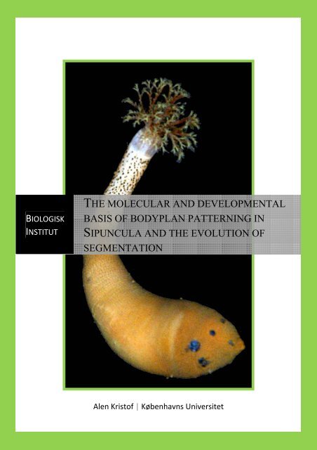
3Reviewed by:Assistant Professor Anja SchulzeDepartment of Marine Biology, Texas A&M University at GalvestonGalveston, USAProfessor Stefan RichterDepartment of Biological Sciences, University of RostockRostock, GermanyFaculty opponent:Associate Professor Danny Eibye-JacobsenNatural History Museum of Denmark, University of DenmarkCopenhagen, Denmark______________________________________________________________Cover illustration:Front: Frontal view of an adult specimen of the sipunculan Themiste pyroides with atotal length of 13 cm.Back: Confocal laserscanning micrograph of a Phascolosoma agassizii pelagospheralarva showing its musculature. Lateral view. Age of the specimen is 15 days and itstotal size approximately 300 µm in length.
4“In a world that keeps on pushin’ me around,but I’ll stand my ground,and I won’t back down.”Thomas Earl Petty, 1989
5 PrefacePrefaceThe content of this dissertation comprises three years of research at the University ofCopenhagen from May 1, 2008 to April 30, 2011. The <strong>PhD</strong> project on thedevelopment of Sipuncula was mainly carried out in the Research Group forComparative Zoology, Department of Biology, University of Copenhagen under thesupervision of Assoc. Prof. Dr. Andreas Wanninger. I spent nine months working onbody patterning genes in the lab of Prof. Dr. Pedro Martinez, Department of Genetics,University of Barcelona, Spain. Within these three years, research trips of altogether16 weeks were made to collect, rear, and fix different sipunculan species at the SvenLovén Center for Marine Sciences in Kristineberg, Sweden (6 weeks), at the EspelandMarine Biological Station in Bergen, Norway (4 weeks), and at the Vostok Station ofthe A. V. Zhirmunsky <strong>Institut</strong>e of Marine Biology in Vladivostok, Russia (6 weeks).Furthermore, <strong>PhD</strong> courses had a great impact on my scientific knowledge and skills.During my studies I attended the EMBO course Marine Animal Models in Evolutionand Development at the University of Gothenburg, Sweden, Comparative Embryologyof Marine Invertebrates at the Friday Harbor Laboratories, University of Washington,USA, and Scientific Writing at the University of Copenhagen, Denmark. This <strong>PhD</strong>project was funded by the European Research Council (MEST-CT-2005-020542) andthe Faculty of Science of the University of Copenhagen.This <strong>thesis</strong> is composed of four chapters. The first chapter gives a generalintroduction to the topics of the present study, its objectives, and the discussion of theresults in a broader perspective, leading to conclusions and perspectives for futureresearch. Chapters II to IV contain the major findings of this <strong>PhD</strong> project in threepublished papers.Copenhagen, April 2011Alen Kristof
Acknowledgements 6AcknowledgementsI am heavily indebted to my principal supervisor Andreas Wanninger for more thanthree years of support, guidance, education, motivation, and invaluable help. Hisoverall excitement for evolution, invertebrates, science, and finally life had thegreatest impact on me. Thank you Andi!Gratefully acknowledged is also my co-supervisor Pedro Martinez for a greattime in his lab in Barcelona. Many thanks are going to his lab members JohannesAchatz, Alex Alsen, Eduardo Moreno, Elena Perea, and especially to Marta Chiodinwho introduced me to molecular techniques as well as to the city of Barcelona.Furthermore, I am grateful to José María Martín-Durán from the “planarian group”also from the University of Barcelona for helping me establishing an in situ protocolfor my animals.I thank Prof. Dr. Danny Eibye-Jacobsen that he agreed to act as the head ofmy defence committee at the University of Copenhagen, as well as Prof. Dr. StefanRichter and Prof. Dr. Anja Schulze for kindly reviewing this <strong>thesis</strong>.Thanks to the working group for Comparative Zoology for a wonderful time inCopenhagen. All these years of the <strong>PhD</strong> project would not be the same without thesemembers and associates. Therefore, special thanks are going to my friends TimWollesen, Uwe Spremberg, Judith Fuchs, and my crazy office mate Jan Bielecki.Special thanks are also going to Anders Garm who helped me with the Danishabstract. Thanks man! In addition, I would like to thank Dany “Sahne” Zemeitat,Ricardo Neves, Nora Brinkmann, Lennie Rotvit, Andreas Altenburger, Birgit Meyer,Henrike Semmler, Christopher Grubb, Tommy Kristensen, Jens Høgh, LisbethHaukrogh, and Åse Jaspersen.A huge thank-you goes to Christiane Todt (University of Bergen, Norway) forhosting and taking care of me during my visit at the Marine Station of Bergen. Inaddition, I want to thank Anastassia S. Maiorova (Marine Biology <strong>Institut</strong>e,Vladivostok, Russia) for being helpful, reliable, and fun to work with during my timeat the Vostok Station. I am grateful to Andrey V. Adrianov (Marine Biology <strong>Institut</strong>e,Vladivostok, Russia) and the staff of the Vostok Station for their help and hospitality.For comments, important discussions and collaboration, I highly acknowledgeMary Rice (Smithsonian Research Center, Florida, USA) and Katrine Worsaae(University of Copenhagen, Denmark).
7 AcknowledgmentsI am deeply grateful for the never ending support from my family, in particularmy parents. Za sve što ste napravili da krenem ovim uspješnim putem, jednostavno sezahvaljulem vama tisuču puta. Puno hvala mama! Puno hvala tata i puno hvala momburazu Jožefu!Finally, thousand thanks to my lovely girlfriend Maria Eracleous for being theone. Maria mou, ise i zoi mou! Also, I want to thank all members of her family,especially Despina, Iraclis, and Georgia for treating me as one of them.For financial support I would like to thank Friday Harbor Laboratories,Washington, USA, the European Research Council (MEST-CT-2005-020542), andthe Faculty of Science, University of Copenhagen, Denmark.
Content 8ContentPreface…………………………………………………………..…………………......5Acknowledgements………………………………………………..……………....6 – 7Content……………………………………………………….………………………..8Chapter I……………………………………………………..…………………...9 – 46Danish abstract……………...…………………………………………………9Abstract……….…………….………………………………………………..10Short abstract………………....………………………………………………11General introduction.…………....……………………………………....12 – 15Sipuncula – a neglected taxon with annelid affinities..................13 – 15Thesis objectives..…………………………………………………....…15 – 16Material and methods…………………………………………………...16 – 22RNA isolation and cDNA syn<strong>thesis</strong>.....................................................17Degenerate PCR...........................................................................17 – 19Rapid amplification of cDNA ends (RACE)................................19 – 21Whole mount in situ hybridisation...............................................21 – 22Results..............................…………………………………………........22 – 31Cloning of homeobox genes in Themiste pyroides......................22 – 23Establishing of an in situ hybridisation protocol..........................23 – 24Serotonergic and FMRFamidergic nervous system development insipunculan worms (chapters I-III)................................................25 – 30Myogenesis and cell proliferation during sipunculan development(chapter IV)...................................................................................30 – 31General discussion....................................................................................31 – 36Early neurogenesis in Trochozoa..................................................31 – 32Establishment and loss of segmentation during sipunculanneurogenesis.................................................................................32 – 33Comparative trochozoan neurogenesis.........................................33 – 34Sipunculan growth zone(s)...........................................................34 – 35Myogenesis in sipunculans and annelids......................................35 – 36Conclusions and future perspectives…………………………………....37 – 38References…………………………………………………………........38 – 46Chapter IIKristof A, Wollesen T & Wanninger A. 2008. Segmental mode of neuralpatterning in Sipuncula. Current Biology 18: 1129-1132..........….….....47 – 51Chapter IIIWanninger A, Kristof A & Brinkmann N. 2009. Sipunculans andsegmentation. Communicative & Integrative Biology 2: 56-59.………..52 – 56Chapter IVKristof A, Wollesen T, Maiorova AS & Wanninger A. 2011. Cellular andmuscular growth patterns during sipunculan development. Journal ofExperimental Zoology Part B: Molecular and Developmental Evolution 316B:227-240.............................……..………………………………..............57 – 71
9 Chapter I – Danish abstractChapter IDanish abstractPølseorme (Sipuncula) har gennem tiderne været regnet som nære slægtninge tilmange forskellige dyregrupper, men for nyligt har molekylære data placeret demindenfor ledormene (Annelida) på trods af sipunculidernes ikke leddelte krop. For atundersøge om sipunculide orme har spor af en leddelt krop i deres ontogeni, blevnerve- og muskel-dannelsen sammen med fordelingen af celledelinger undersøgt hos3 arter af sipunculider. Resultaterne viser, at rudimenter af mange ringmuskler ikropsvæggen og den længdegående retraktormuskel dannes samtidigt i den tidligetrochophor-larve. Ydermere, gennem hele udviklingen dannes nye ringmuskler viaspaltning af allerede eksisterende muskelceller. I modsætning til det, så følgernervedannelsen et leddelt mønster gennem dannelsen af parrede serotonin-holdigecellekroppe langs den anteriøre-posteriøre akse. Cellekroppene er associeret med enparret ventral nervestreng og er arrangeret i fire stringent gentagne grupper. Detteleddelte mønster forsvinder inden metamorfosen, og den parrede ventrale nervestrengfusionerer til en enkelt nervestreng i det voksne dyr. Fordelingen af mitotiske cellerviser sammenstemmende en lighed med en annelid-ligende posteriør vækstzone, somogså forsvinder hos metamorfiserende pelagosphera larver. Disse udviklingsmæssigeog morfologiske data understøtter således de nye molekylære analyser og antyder, atsipunculider stammer fra en leddelt forfar. Tabet af en leddelt krop hos disse relativtstore og fritlevende dyr indikerer desuden, at leddeling forsvinder lettere gennemevolutionære processer end tidligere antaget. Initialiseringen og efterfølgende(sekundære) tab af leddeling gør Sipuncula ideel til evolutionære ogudviklingsmæssige studier af leddelingsprocesser indenfor Metazoa.
Abstract 10AbstractPeanut worms (Sipuncula) have been variously related to a number of animals in thepast, but, recently, and despite their non-segmented adult body, molecularphylogenetic analyses place them within Annelida. In order to contribute to thequestion whether or not sipunculan worms show traces of a segmental pattern in theirontogeny, neuro- and myogenesis as well as the distribution of proliferating cells wereanalysed in three different sipunculan species. The data show that the rudiments ofnumerous circular muscles of the body wall musculature and the longitudinal retractormuscles appear at the same time in the early trochophore larva. In addition,throughout development newly formed ring muscles emerge along the entire anteriorposterioraxis by fission from already existing myocytes. In contrast to that,neurogenesis does follow a segmental pattern by subsequently emerging pairs ofserotonergic perikarya along the anterior-posterior axis, which are associated with apaired ventral nerve cord and are arranged in four distinct repetitive units. Thismetameric pattern, however, disappears prior to metamorphosis and the ventral nervecords fuse to form the single ventral nerve of the adult. Congruently, the distributionpattern of mitotic cells show similarities to an annelid-like posterior growth zone thatalso disappears in metamorphic competent pelagosphera larvae. Accordingly, thesedevelopmental and morphological data corroborate recent molecular analyses andshow that sipunculans stem from a segmented ancestor. Furthermore, the loss ofsegmentation in these relatively large, free living animals indicates that bodysegmentation may easier be lost during evolution than previously assumed. Theontogenetic establishment and (secondary) loss of segmentation renders Sipunculaideal for developmental and evolutionary studies on the segmentation process inMetazoa.
11 Short abstractShort abstractIn this <strong>PhD</strong> <strong>thesis</strong>, three sipunculan species were investigated byimmunocytochemistry in conjunction with confocal laserscanning microscopy and 3Dreconstruction software, in order to clarify whether or not cryptic segmentation can befound during sipunculan ontogeny. The results show that sipunculan myogenesis doesnot follow a segmental manner, but for a short period of time neurogenesis and thedistribution of mitotic cells show transitional stages of segmentation duringsipunculan development, thus supporting a sipunculan/annelid affiliation. Moreover,the establishment of an in situ hybridisation protocol for the model sipunculanThemiste pyroides for gene expression analyses pave the way for future studies on themolecular processes underlying sipunculan segmentation that might give importantinsights into the evolution of segmentation.
General introduction 12General introductionAnnelids and arthropods are highly successful animals that have a “segmented” bodyplan. Therefore, understanding the origin of segmentation as well as the mechanismsof segment formation and diversification has been of scientific interest for decades.Segmented animals exhibit repeated units at the cellular, tissue or organ system levelalong the anterior-posterior axis (Wilmer 1990, Scholtz 2002). While many animalclades show serial repetition of organs along the anterior-posterior axis (i.g., seriallyarranged shell plates in polyplacophoran mollusks, ring muscles in platyhelminths, orcommissures in numerous worm-shaped invertebrates), only the above mentionedanimal clades display the definition of “true” segmentation, i.e., multiple organsystems that are generated successively along the anterior-posterior axis (Couso 2009,Chipman 2010). Recently, our view on animal interrelationships has changeddramatically, splitting them roughly into three mojor lineages (Deuterostomia,Ecdysozoa, and Lophotrochozoa) and placing the segmented animals all in separateclades (e.g., Aguinaldo et al. 1997, Halanych 2004, Dunn et al. 2008, Hejnol et al.2009). Consequently, the “new” animal tree of life has led to two equally possible andhotly debated scenaria: either, the common ancestor of the so-called Bilateria(bilaterally symmetrical animals) was very simple and segmentation evolvedindependently in each superclade; or, their last common ancestor was more complexand segmented, with segmentation having been lost in the vast majority of animalclades (Arendt 2005, De Robertis 2008, Couso 2009, Chipman 2010). Accordingly,the monophyly of segmentation is in conflict with the long-standing argument thatsegmentation is an evolutionary key invention in metazoan (multicellular animals)evolution that is unlikely to get lost (Seaver 2003). Interestingly, recent interpretationsof gene expression data on representatives of the segmented phyla have revealedremarkable similarities, proposing a segmented ancestor of all Bilateria (e. g., Arendt2005, De Robertis 2008, Couso 2009). Since these data largely come from a verylimited number of model system animals such as Drosophila (fly), Tribolium (beatle),Capitella (polychaete), Hirudo (leech), Cupiennius (spider), and so forth,developmental studies of non-model system taxa are needed in order to either verifyor falsify this hypo<strong>thesis</strong>. Moreover, comparative molecular, developmental, andmorphogenetic approaches, namely gene expression, cell proliferation, and organsystem formation, have proven useful in elucidating the evolution of body patterning
13 General introductionprocesses (e.g., Hessling and Westheide 2002, Denes et al. 2007, Hejnol andMartindale 2008, Maxmen 2008, Wanninger 2009, Boyle and Seaver 2010,Brinkmann and Wanninger 2010a, b).Sipuncula – a neglected taxon with annelid affinitiesSipuncula is a small, worm-shaped, exclusively marine lophotrochozoan taxon thatuniformly exhibits an unsegmented adult body, which is subdivided into a posteriortrunk and a retractable anterior introvert. Sipunculans are filter or deposit feeders thatlive in soft sediments, rock crevices, dead corals, or vacant mollusc shells, and arewidespread throughout the oceans (Rice 1975a). Their fossil record is generally sparsebut a description of six well preserved sipunculan specimens found in southwestChina dates their origin to more than 520 million years ago (Huang et al. 2004).Furthermore, the fossilised outer and inner morphology resembles extant sipunculanspecies, suggesting only limited changes since the Early Cambrium (Huang et al.2004). Cladograms based on morphological characters divide sipunculans into twoclasses, Sipunculidae and Phascolosomatidea, comprising four orders and six families(Aspidosiphonidae, Phascolosomatidae, Golfingiidae, Phascolionidae, Themistidae,and Sipunculidae) (Cutler and Gibbs 1985, Cutler 1994). Today, the use of DNAsequence data has changed this morphology-based view of the relationships amongthe currently recognised 147 species of Sipuncula (Maxmen et al. 2003, Schulze et al.2005, 2007). The two classes have not been recognised by Maxmen et al. (2003) andSchulze et al. (2005), whereas the class Phascolosomatidae was recovered in the byfar most comprehensive analysis on sipunculan phylogeny of Schulze et al. (2005).Congruently, all analyses strongly support Sipunculus nudus as the sister group to theremaining Sipuncula, while most of the previously recognised families and orderswere not recovered as monophyletic (Maxmen et al. 2003, Schulze et al. 2005, 2007).However, based on morphological characters and DNA sequence data, the monophylyof Sipuncula is strongly supported (Maxmen et al. 2003, Schulze et al. 2005, 2007).In general, sipunculans are dioecious (separate sexes), but there are a fewknown hermaphroditic and parthenogenetic species (asexual reproduction) (Åkesson1958, Rice 1970, Rajalu and Krishnan 1969, Pilger 1978, Cutler 1994). Four different,one direct, and three indirect developmental pathways have been described forSipuncula (Fig. 1; Rice 1975b, c). Since sipunculans share certain developmentalfeatures such as spiral cleavage, that gives rise to a trochophore larva with an apical
General introduction 14tuft and a ring of cilia used for locomotion, they have commonly been placed withinthe clade Spiralia, which, among others, also comprises annelids and mollusks(Jägersten 1972, Rice 1985, Cutler 1994, Rouse 1999).Figure 1. The four distinct developmental modes in Sipuncula. Development may be direct, wherebythe embryo emerges from the egg as a crawling worm, which eventually transforms into the juvenilestage without an intermediate larval form (black line and arrows). In some species the lecithotrophictrochophore, after a brief swimming period, transforms into a vermiform stage succeeded by thejuvenile (red line and arrows). In the lecithotrophic indirect developmental pathway the trochophorelarva starts to elongate and metamorphoses into a second larval stage, the pelagosphera, which swimsin the plankton for a short time, settles, and metamorphoses into a juvenile (blue line and arrows). Inthe planktotrophic indirect developmental mode the lecithotrophic trochophore transforms into apelagosphera, which remains in the plankton for up to several months before it settles andmetamorphoses into a juvenile (green line and arrows). Drawing modified from Rice 1975b, c.Until they were considered a distinct taxon among the spiralians, thephylogenetic position of Sipuncula has long been an enigma. Accordingly, they wereplaced close to sea cucumbers (holothurian echinoderms), as derived annelids, or asan in-group of Gephrea (sipunculans, echiurans, and priapulids), then as relatives ofphoronids, bryozoans, and brachiopods (Prosoygii), until developmental studiesproposed a close relationship to spiralians (reviewed in Rice 1985). From there on,
15 General introductionthey have been variously seen as either a sister taxon to Annelida or to Mollusca (e.g.,Åkesson 1958, Rice 1975b, c, Scheltema 1993, 1996, Cutler 1994, Zrzavy et al. 1998,Giribet et al. 2000). Nowadays, a growing number of morphological and molecularanalyses strongly suggest a close relationship to annelids, leaving the only questionwhether sipunculans cluster within Annelida or constitute their sister taxon (e.g.,Wanninger et al. 2005a, Tzetlin and Purschke 2006, Struck et al. 2007, Dunn et al.2008, Mwinyi et al. 2009, Shen et al. 2009, Sperling et al. 2009, Zrzavy et al. 2009,Dordel et al. 2010, Hausdorf et al. 2010, Struck et al. 2011). Accordingly, theinclusion of Sipuncula within Annelida suggests a secondary loss of a previouslysegmented body plan in Sipuncula rather than an initial evolutionary step towardssegmentation in these animals (Dordel et al. 2010, Struck et al. 2011).Thesis objectivesIn the light of the proposed sipunculan-annelid assemblage, it was the aim of thepresent <strong>PhD</strong> <strong>thesis</strong> to elucidate sipunculan body plan patterning and to deal with thefollowing scientific issues:1. Neuro- and myogenesis were described in three different sipunculan species,Phascolosoma agassizii, Themiste pyroides and Thysanocardia nigra (Fig. 2).Since nervous and muscle systems follow a segmental development inannelids, these experiments should clarify whether or not sipunculans showcryptic segmentation during their ontogeny.2. The distribution of proliferating cells in T. pyoides and T. nigra weredescribed throughout development to assess whether sipunculans possess aposterior growth zone similar to the segmented annelids.3. Comparative analysis of the acquired developmental data with those availablefor annelids and other lophotrochozoans (e.g., Mollusca) were carried out inorder to infer shared and possible ancestral neuromuscular features.4. The establishment and application of an in situ hybridisation protocol foranalyses of tempo-spatial expression patterns of selected candidate genesknown to be involved in body patterning (e.g., neural, muscular and so-called“segmentation” genes (hox1-9, even-skipped, hairy)).
Thesis objectives 16In the following, the main results are summerised and a general dicussion ispresented, dealing with these key issues of the present <strong>thesis</strong>. More detaileddiscussions are found in the chapters II to IV, which comprise three publishedmanuscripts. Finally, future perspectives of this project are proposed.Figure 2. Adults of investigated sipunculan species in the present <strong>PhD</strong> study. All in lateral view.Tentacles, which are at the top, mark the anterior end of animals. A. Phascolosoma agassizii, totallength approximately 15 cm. B. Themiste pyroides, total length approx. 10 cm. C. Thysanocardianigra, total length approx. 12 cm.Material and methodsIn the present <strong>PhD</strong> project, several sipunculan species were investigated bydevelopmental-morphological markers such as immunolabelling, confocalmicroscopy, and 3D reconstruction software (Fig. 2). In addition, developmentalstages of Themiste pyroides were used for cloning genes known to be involved in theformation of segmentation in arthropods and annelids, and for establishment of an insitu protocol for gene expression analyses (see below). An overview of the speciesinvestigated by morphological methods is given in Table 1. Further details of therespective techniques are provided in the chapters II to IV.
17 Material and methodsTable 1. List of species investigated in the course of the <strong>PhD</strong> project by fluorescence markers;Serotonin and FMRFamide – neurotransmitters/peptides; Phalloidin – F-actin of the musculature; Dapi(4’, 6-diamindino-2-phenylindole) – cell nuclei marker, EdU (5-ethynyl-2’-deoxyuridine) –proliferating cells.Species (Family) Neurogenesis Myogenesis Cell nuclei,Themiste pyroides(Themistidae)Thysanocardianigra (Golfingidae)Phascolosomaagassizii(Phascolosomatidae)Serotonin,FMRFamide(chapter I)Serotonin,FMRFamide(chapter I)Serotonin,FMRFamide(chapters I-III)F-actin(chapter IV)F-actin(chapter IV)F-actin(chapter IV)cell proliferationDapi, EdU(chapter IV)Dapi, EdU(chapter IV)-RNA isolation and cDNA syn<strong>thesis</strong>Total RNA was purified from Themiste pyroides embryos at 15, 28, 40, 48, 62, 72, 84,and 111 hours post fertilizaton (hpf) (miRCURY RNA isolation kit, Exiqon, Vedbaek,Denmark). cDNA samples were syn<strong>thesis</strong>ed by reverse transcription (RETROsript,Ambion, Woodward St. Austin, TX, USA), and stored at -20 °C until use.Degenerate PCRTo clone homeobox genes, a range of degenerate primers were designed referring toMartinez et al. (1997), Nederbragt et al. (2002), Seaver et al. (2006), and Paps et al.(2009). The sequences of the oligonucleotides used are given in Table 2.A touchdown PCR was performed with the degenerate primers given in Table2. The amplification parameters were: 3 min at 94 °C, 10 cycles of 45 sec at 94 °C, 45sec at 52 °C (every cycle -1 °C), 30 sec at 72 °C, and 30 cycles of 45 sec at 94 °C, 45sec at 42 °C, and final 30 sec extension at 72 °C. The PCR products were purified bya gel extraction kit (QIAquick, QIAGEN, Copenhagen, Denmark), and on 1 µl of thisreaction another PCR with the same parameters was performed. The samples weredisplayed by electrophoresis on a 1% agarose gel, purified, concentrated byspeedvacuum (GENEVAC EZ-2 plus , Ipswich, UK), resuspended in 10 µl distilledwater, and directly ligated into pGEM-T Easy vector using a ligation kit (Promega,
Material and methods 18Nacka, Sweden). The inserted DNA fragments were sequenced using BigDyeTerminator 3.1 Cycle Sequencing kit with an ABI Prism 377 DNA sequencer(Applied Biosystems, Carlsbad, CA, USA).Degenerate primers were also designed to clone a number of muscular andneural genes for establishment of a whole mount in situ hybridisation protocol (Table2). Subsequently, all recovered gene fragments were identified by the similarity oftheir nucleotide sequences to already known genes that are placed at the public onlinesource of the National Centre for Biotechnology Information (NCBI) using basic localalignement searches (BLASTs).Table 2. Nucleotide sequences of degenerate primers and their melting temperatures (Tm) used toclone homeobox, muscular and neural genes in Themiste pyroides.Primers Sequence TmHomeobox genes (Martinez et al. 1997)CT 77 5’-CGGATCCYTIGARYTIGARAARGARTCT 78 5’-GGAATTCATICKRTTYTGRAACCAIATTYEngrailed (Seaver et al. 2006, Nederbragt et al. 2002)EnEH2 5’-TGGCCTGCITGGGTNTAYTGYACen2-2 5’-TGRTTRTANARNCCYTGNGCCATEng1Eng45’-ATGGAATTCCNGCNTGGGTNTWYAC5’-TGGAAGCTTRTANARNCCYTSNGSCATMyosin heavy chain type II (Ruiz-Trillo et al. 2002)Mio7 (F)Mi6 (R)5’-TGYATCAAYTWYACYAAYGAG5’-CCYTCMARYACACCRTTRCATropomyosin (Paps et al. 2009)TropoF 5’-ATYRAGAAGAARATGNBKGVCATGTropoR5’-GTHYGRTCCARTTGNYCACTIntermediate filaments (Paps et al. 2009)FilF1 5’-TACATCGAGAAGGTGCGTTTCCTGGFilR1 5’-CCTCACCCTCCAGCAGCTTTCTGTAFilF3FilR35’-TACATCGAGAAGGTGCGTTTCCTGG5’-CYTCNCCYTCCAGCAGYTTYCTGTAβ-Actin (Matsuo and Shimizu 2006)42°C42 °C42 °C53 °C53 °C48 °C50 °C
19 Material and methodsβ-Actin F1 5’-TGGGAYGAYATGGARAARATβ-Actin R1 5’-GCCATYTCYTGYTCRAAα-Tubulin (Zheng et a. 1998)AT16-F 5’-ATHGGHAAYGCNTGYTGGGAT412-R 5’-RAAYTCDCCYTCYTCCATDCCβ-Tubulin (Garant and MacRea 2009)BT-F 5’-GGNCARTCNGGNGCNGGNAAYAAYTGGGCNBT-R 5’-NGGRAANGGNACCATRTTNACNGC50 °C50 °C55 °CRapid amplification of cDNA ends (RACE)Fragments of Themiste pyroides homeobox containing genes were isolated bydegenerate PCR using cDNA of mixed larval stages as template. In addition, 5’ and 3’cDNA were generated using the same template for rapid amplification of cDNA ends(RACE) with the SMARTer RACE cDNA amplification kit (Clonetech,Mountainview, CA, USA).For each of the recovered gene fragments, which ranged between 135 and 271base pairs (bp) (Table 3), gene specific primers (GSP) were designed and used forRACE (Advantage 2 PCR Kit; Clonetech). Touchdown PCRs were performed withfollowing amplification parameters: 5 min at 94 °C, 10 cycles of 45 sec at 94 °C, 45sec at 57 °C (every cycle -1 °C), 3 min at 72 °C, and 25 cycles of 45 sec at 94 °C, 45sec at 47 °C, and final 30 sec extension at 72 °C. Reactions were agarose-gelelectrophoresed, stained with SYBRsafe (Invitrogen, Taastrup, Denmark), andphotographed under UV light. Subsequently, 1 µl of RACE PCR products were usedas a template for another RACE PCR with nested GSPs (see Table 3). The parameterswere: 5 min at 94 °C, 35 cycles of 45 sec at 94 °C, 45 sec at 60 °C, 30 sec at 72 °C,and a final 10 min extension at 72 °C. Positive fragments were purified from theagarose gel, cloned into the vector pGEM-T easy, and sequenced as described above.However, I was able to amplify into the 3’ direction of two genes only, hox1 (47 bp)and hox8 (73 bp).
Material and methods 20Table 3. Isolated DNA fragments of Themiste pyroides mixed larval stages. Gene abbreviations: lab,labial; scr, sex combs reduced; ftz, fushi tarazu; abd-A, abdominal-A. Other abbreviations: bp, basepairs; e-value, expected value (the similarity between a given sequence and orthologues from thedatabase; high similarities are indicated by considerably low e-values, while high e-values indicate lowsimilarity). Red and underlined parts within the sequences indicate gene specific and nested primers,respectively.Gene Sequence bp/e-valuehox1(lab)hox3hox5(scr)hox5(ftz)hox8(abd-A)evenskippedlox2caudal(cdx)xloxnotCGGATCCCTGGAACTGGAGAAAGAATTCCACTTTAACAAATATCTCACAAGAGCCAGGCGGATAGAAATAGCTGCTGCACTTGGACTAAATGAAACACACAGGTTAAGATCTGGTTTCAGAACAGCCGCATGAATTCCGGATCCTTGGAACTGGAGAAGGAATTCCATTTTAACCGTTACTTGTGTCGGCCACGCCGTATTGAAATGGCAGCCATGCTCAACCTGACAGAAAGACAGATCAAGATCTGGTTTCAGAATAGCAGCATGAATTCCGGATCCCTGGAACTGGAGAAAGAATTTCATTACAACAGATACCTAACAAGACGAAGGAGAATAGAAATAGCACATGCTTTGGGACTCACGGAACGCCAAATTAAGATCTGGTTTCAGAATAGCAGCATGAATTCCGGATCCTTGGAATTGGAAAAAGAATTTCACTTCAACCGTTACTTGTCCAGCAAACGACGTACAGAGATAGCCGAGTCACTGGCTCTGACAGATAGACAAGTTAAGATCTGGTTTCAAAACAGCCGCATGAATTCCCGGATCCTTGGAATTGGAAAAAGAATTTCAGTTTAATCATTATTTAACCCGAAAAAGGAGAATAGAGATAGCGCACGCTTTGTGTCTAACAGAAAGACAGATAAAGATCTGGTTTCAGAATCGCAGCATGAATTCCCGGATCCTTGGAGTTGGAAAAAGAATTTTACAGAGAGAACTATGTTAGCAGACCAAGGAGGTGTGAACTGGCTGCGGAACTCAACCTGCCAGAATCAACAATCAAGGTAAGAAGAAAATAGTGCGACCATTTATCACAGTTTCAGAGCCAACATTTTCAAAGTCGATTTTTCTAATTTGGAATAAAATTGCAATAAATTAATGTTTTATTTTTTACTTTCAGATCTGGTTTCAGAACAGCAGCATGAATTCCCGGATCCTTGGAGCTGGAGAAAGAATTCAAATTCAACAGATACTTAACTCGCAGAAGACGAATAGAACTGTCTCATATGTTGTGCTTAACTGAGCGCCAAATCAAGATCTGGTTTCAGAACAGCAGCATGAATTCCGGAATTCATGCTGCTGTTCTGAAACCAGATTTTGACCTGTCTCTCAGACAGACTGAGTGACTGTGCTAGTTCTGCCTTTCTTCTGATTGTGATATAACGACTGTAATGGAATTCTTTCTCCAGTTCCAGGGATCCGCGGATCCCCTGGAATTGGAAAAAGAATTTCATTTTAATAAATATATCTCCAGACCAAGGAGAATAGAATTAGCTGCCATGTTAAATCTAACAGAGAGACATATAAAGATCTGGTTTCAGAATAGCAGCATGAATTCCCGGATCCTTGGAGTTGGAGAAAGAATTTCACTTCAACCGTTACTTGTCCAGCAAACGACGTACAGAGATAG136/10 -10135/10 -14135/10 -14135/10 -12136/10 -15252/10 -9136/10 -14136/10 -12137/10 -14271/10 -11
21 Material and methodsCCGAGTCACTGGCTCTGACAGATAGACAAGTTAAGATCTGGTTTCAGAACAGCAGCATGAATTCCGGATCCCTGGAATTGGAGAAGGAATTCGAAAGGCAACAATACATGGTTGGATCTGAAAGGTATTATCTGGCAGTATCGCTTAATTTATCCGAATCCCAAGTGAAGATCTGGTTTCAGAACCGCCGCATGAATTCCWhole mount in situ hybridisationEmbryos, larvae, and juveniles of Themiste pyroides were anesthetised by addingdrops of a 3.5 or 7% MgCl 2 solution to the seawater, and were subsequently fixed in4% paraformaldehyde (PFA) in 0.1 M phosphate-buffered saline (PBS) (pH 7.3) for20-30 min at room temperature. Subsequently, the fixative was removed by threewashes in 70% ethanol (15 min each), followed by the storage of the specimens at -20°C.Fixed specimens were rehydrated in PBS, washed four times for 5 min in PTw(phosphate-buffer containing 0.1% Tween), and permeabilised with proteinase K(Sigma; 10 µg/ml in PTw for 15 min at room temperature). Digestion was terminatedby two washes (5 min each) with PTw containing 2mg/ml of glycine and treated withtriethanolamine (TEA, 0,1M pH 7.6, Fluka; 3 x 5 min), following addition of 4µl/mland 8µl/ml of acetic anhydride without changing the TEA solution to block positivecharges. After two 5 min washes in PTw, specimens were post-fixed in 3.7% PFA for1 hr at room temperature. Subsequently, specimens were rinsed 5 x 5 min in PTw,incubated for 10 min in 50% pre-hybridisation buffer (50% formamide, 5x SSC,1mg/ml yeast RNA, 0.1 mg/ml heparin, 0.1% Tween 20, 10mM DTT) in PTw and anincubation in 100% pre-hybridisation buffer overnight at 65 °C. Antisense and sensedigoxigenin-labelled riboprobes were generated with a RNA labeling kit (SP6/T7;Roche; Copenhagen, Denmark), and used at a working concentration of 1 ng/µl forTp-mhc and Tp-actin (fragment sizes ca. 700 and 500 bp, respectively). Riboprobeswere warmed up in hybridisation buffer (50% formamide, 5x SSC, 0.1% Tween 20)for 10 min at 70 °C. Afterwards, specimens were added to that solution and incubatedfor 72 hr at 65 °C. After hybridisation, probes were recovered and specimens werewashed at 65 °C (30 min in 100% hybridisation buffer, in 75% hybridisation bufferand 75% 2x SSC, in 50% hybridisation buffer and 50% 2x SSC, in 25% hybridisationbuffer and 25% 2x SSC, 2 x 15 min in 2x SSC, and final 2 x 15 min washed in 0.2xSSC). Afterwards, specimens were washed two times 5 min in maleic buffer(MABTw; 100 mM maleic acid, 150 mM NaCl, 0.1% Tween 20, pH 7.5), blocked for
Material and methods 221 hr in 1% blocking solution (Roche) and incubated overnight with anti-digoxigeninalkaline phosphatase (AP) conjugated antibody (Roche; 1:200 dilution) in blockingsolution at 4 °C. Specimens were washed at least four times 15 min in MABTw andtwo times 5 min in AP buffer (0.1M NaCl, 0.1M Tris pH 9.5, 0.05M MgCl 2 , 0.5%Tween 20) and developed with NBT/BCIP (Roche) in the dark. The reactions werestopped with PTw, specimens were fixed in 3.7% PFA overnight at 4 °C and stored in80% glycerol in PBS at -20°C. Specimens were analysed and photographed usingDIC optics on a Zeiss Axiophot microscope (Zeiss, Jena, Germany) in conjunctionwith a Leica DFC300FX digital camera (Leica, Microsystems, Wetzlar, Germany).ResultsIn the course of the present <strong>PhD</strong> study, three different sipunculan species, whichcover three out of six currently recognised families, were investigated. Phascolosomaagassizii was collected and reared at the Friday Harbor Laboratories on the San JuanIsland (Washington, USA) whereas Themiste pyroides and Thysanocardia nigra wereacquired at the Vostok-Marine-Station (Vladivostok, Russia). The muscle andnervous system development is described for all investigated species, the distributionof proliferating cells for T. pyroides and T. nigra, and cloning of homeobox genes aswell as the establishment of an in situ hybridisation protocol for T. Pyroides. Themain findings are summarised in the following section. Further details are given in thechapters II – IV.Cloning of homeobox genes in Themiste pyroidesWith the pair of degenerate primers CT77 and CT78, which were designed to theconserved regions within the homeobox, the orthologues for the hox1 (labial), hox3,hox5 (sex combs reduced), hox5 (fushi tarazu), hox8 (abdominal-A), even-skipped,lox2, caudal, xlox, and not were isolated. The isolated gene fragments of Themistepyroides cDNA were generated from mixed embryonic and larval stages; their lengthswere between 135 and 271 bp (Table 3). Unfortunately, gene specific primers andRACE PCRs were not able to extent the recovered fragments neither in 5’ nor in 3’direction. The only exceptions were the genes hox1 (labial) (47 bp) and hox8(abdominal-A) (73 bp), which included the stop codon and a poly-A tail, but the
23 Resultsobtained fragments were too short to generate riboprobes for successful in situhybridisation experiments.Establishment of an in situ hybridisation protocolUsing a set of degenerate primers (Table 2), gene fragments of 700 bp of myosinheavy chain (Tp-mhc) and 500 bp of beta-actin (Tp-actin) were recovered by PCR.Tp-mhc is expressed from early trochophore stages (ca. 2 days afterfertilisation (dpf)) onwards in the four developing retractor muscles (Fig. 3A-C). FirstTp-actin expression is also in the early trochophore larvae in a prominent U-shapedband in the area of the forming retractor muscles (Fig. 3D). In pelagosphera larvae theTp-actin is expressed in distinct areas of the paired dorsal and ventral retractormuscles (Fig. 3E, F).
Results 24Figure 3. Expression of myosin heavy chain (Tp-mhc) (A-C) and beta-actin (Tp-actin) (D-F) duringlarval stages of Themiste pyroides. Anterior is to the top in all panels and scale bars equal 50 µm. Blackrepresents areas of gene expression – Tp-mhc in A-C and Tp-actin in D-F, respectively. (A) Dorsalview of an early trochophore larva showing Tp-mhc expression (arrowheads) in the anlagen of theretractor muscles. (B) Dorsal view of a late trochophore larva. Tp-mhc expression is restricted to theretractor muscles. Note that only three out of four are in focus; mt marks the ciliated metatroch. (C)Lateral view, ventral to the right, pelagosphera larva with Tp-mhc expression in the retractor muscles;pt marks the ciliated prototroch. (D) Dorsal view of an early trochophore larva with Tp-actinexpression in the area of the retractor muscles (arrows). (E) Dorsolateral view of an early pelagospheralarva, ventral to the left, showing Tp-actin expression in the retractor muscles. (F) Late pelagospheralarva, dorsal view with strong Tp-actin expression in the retractor muscles.
25 ResultsSerotonergic and FMRFamidergic nervous system development in sipunculanworms (chapters I – III)In the planktotrophic species Phascolosoma agassizii, the first detectable serotonergicand FMRFamidergic nervous structures are visible in the anterior region of thetrochophore larvae. A pair of neurites emerging from the apical organ contributes tothe ventral nerve cords. During further development, interconnecting commissuresand pairs of perikarya (four serotonergic and one pair of FMRFamidergic perikarya)appear subsequently along the ventral nerve cords from anterior to posterior, resultingin a rope-ladder like ventral nervous system. Due to migration of serotonergicperikarya and fusion of serotonergic ventral nerve cords this metameric arrangementis lost towards metamorphosis, and gives rise to the single non-metameric adultventral nervous system (chapters II and III).In the lecithotrophic species Themiste pyroides and Thysanocardia nigra,neurogenesis is similar to Phascolosoma agassizii (Figs. 4-6). The first serotonergicand FMRFamidergic cells are in the apical organ and a pair of neurites contributes tothe ventral nerve cords (Figs. 4A, 6A). In contrast to P. agassizii, a first, second, and athird pair of serotonergic perikarya appear already in the trochophore larvae of T.pyroides and T. nigra in an anterior to posterior progression, while later the fourthpair appears in the pelagosphera larva (Figs. 4A-D, 5E, H).
Results 26Figure 4. Confocal micrographs of the serotonergic nervous system in trochophore larvae of Themistepyroides. A and C are in ventrolateral left view, whereas B and D are in ventral view. Anterior facesupwards and scale bars equal 50 µm in all aspects except the insert in B where it is 10 µm. Age oflarvae is given in days post fertilisation (dpf). (A) Early trochophore larva (2 dpf) showing an apicalorgan comprising three cells (arrowheads), two neurites of the developing ventral nervous system (vn)that is interconnected by a commissure (arrow), and a pair of perikarya (asterisks). (B) Slighly later (2dpf), at the base of the apical organ (ao), the first cells of the adult cerebral organ are formed (cg), thetwo ventral neurites are stronger developed than in the stage before, and the first perikarya of thesecond pair appears. Insert is a magnification of the apical pole of B showing two flask-shaped and tworound cells in the apical organ. (C) Trochophore larva (2.5 dpf) with two pairs of perikarya associatedwith the ventral neurite bundles and the first perikarya of the third pair already formed. (D) Slightlylater (2.5 dpf) the second perikarya of the third pair is established posteriorly to the second pair. Notethat the cerebral ganglion is more elaborated than in the stage before.During subsequent development a second pair of FMRFamidergic perikaryaappears along the ventral nervous system in T. pyroides and T. nigra, while there isonly one in P. agassizii (Fig. 6D). All three species show four serotonergic and two tothree FMRFamidergic flask-shaped cells in their apical organ, as well as aserotonergic nerve ring underlying the ciliated metatroch (Figs. 4A, B, Insert, 5A, B,
27 ResultsE, F, I-L). It has to be noted that in T. pyroides and T. nigra the serotonergicmetatrochal nerve ring is considerebly weaker than that in P. agassizii (Fig. 5A, E, I-L).Figure 5. Confocal micrographs of the serotonergic nervous system in pelagosphera larvae of Themistepyroides. All aspects in ventral view except I, K, J and L, which are in ventro lateral left view. Anteriorfaces upwards and scale bars represent 50 µm in A, E, I, K, J and L, while they equal 10µm in B, C, D,F, G and H. A, E, I, and K are color-coded confocal micrographs, where blue and purple colors are
Results 28positioned to the ventral, colors of green and yellow in between, whereas orange and red colors are tothe dorsal side. Age of larvae is given in days post fertilisation (dpf). (A) Early pelagosphera larva (3dpf) showing a weakly stained metatrochal neurite (mtn), ventral neurite bundles (vn) that areinterconnected by commissures (arrows) always anteriorly positioned to a pair of perikarya (asterisks);cg marks the cerebral ganglion. (B) Dorsal portion of the same specimen as in A; magnification of theapical pole showing four cells (arrowheads) in the apical organ. Note that two cells are flask-shaped.(C) Ventral portion of the same specimen as in A; magnification of the apical pole showing numerouscells associated with the neuropile of the developing adult cerebral ganglion. (D) Magnification of theventral trunk area of an early pelagosphera larva (3 dpf) with three pairs of metamerically arrangedperikarya associated with the ventral neural bundles. (E) Slighly older pelagosphera (4 dpf) as in Awith stronger developed ventral neural bundles that bear four pairs of perikarya and a more elaboratecerebral ganglion. (F) Dorsal portion of the same specimen as in E; magnification of the apical poleshowing five cells in the apical organ. (G) Ventral portion of the same specimen as in E, magnificationof the apical pole showing numerous cells and a neuropile in the cerebral ganglion. (H) Same specimenas in E. Magnification of the ventral trunk area showing the ventral nervous system with four pairs ofperikarya and three commissures interconnecting the median with the two outer neural bundles. (I)Magnification of the anterior region of a pelagosphera larva (8 dpf) showing a complex central nervoussystem that comprises the cerebral ganglion with numerous cells, the apical organ with its cells andown neuropile (double arrows), peripheral cells (double arrowheads), metatrochal neurite, peripheralneurites (pn), and the ventral neural bundle. Note that the ventral neural bundle has retained its pairedcharacter only in the most anterior part. (J) Dorsal portion of the same specimen as in I showing theapical organ with its cells and neuropile. Note that two of the cells are flask-shaped. (K) Magnificationof the anterior region of a late pelagosphera larva (14 dpf) with a prominent cerebral ganglion, apicalorgan with two flask-shaped cells, peripheral cells and neurites, a prominent metatrochal neurite ring,and ventral neural bundles. (L) Dorsal portion of the same specimen as in K showing the apical organthat comprises less cells than in the previous stage. Three out of five cells disappeared while only thetwo flask-shaped cells and a part of the neuropile remain visible.In addition to the loss of the metameric arrangement in the ventral nervoussystem towards metamorphosis, less serotonergic and FMRFamidergic apical cells aredetectable in all of the investigated species, while the adult cerebral ganglion becomesmore elaborate (Figs. 5I-L, 6D).
29 ResultsFigure 6. Confocal micrographs of FMRFamidergic nervous system in pelagosphera larvae ofThysanocardia nigra. All aspects in ventral view except B and C which are in ventrolateral left view.Anterior faces upwards and scale bars represent 50 µm except the insert in A where it equals 30 µm.Age of larvae is given in days post fertilisation (dpf). (A) Early pelagosphera larva (3 dpf) with twoflask-shaped cells in the apical organ (arrowheads), the anlage of the adult cerebral ganglion (cg), anda pair of ventral neurites (vn). Insert shows a slightly older stage (3 dpf), with the first interconnectingcommissure (arrow) and pair of perikarya (asterisks) associated with the ventral neural bundles. Notethe presence of a median ventral neurite. (B) Pelagosphera larva (4 dpf) showing a more elaboratecerebral ganglion and ventral nervous system that now has a second commissure that interconnects theventral neurites; pn marks peripheral neurite. (C) Slighlty older pelagosphera larva (5 dpf) with a well
Results 30developed cerebral ganglion below the apical cells and ventral nervous system. (D) Late pelagospheralarva (11 dpf) with numerous peripheral neurites in the trunk area as well as in the ventral nervoussystem. Note the second pair of perikaya along the ventral neurites. No FMRF-positive cells appear inthe apical organ.Myogenesis and cell proliferation during sipunculan development (chapter IV)Myogenesis in all investigated species in the present <strong>PhD</strong> <strong>thesis</strong> display a similarmode of muscle system development.In Phascolosoma agassizii, Themiste pyroides and Thysanocardia nigra, therudiments of the four longitudinal retractor muscles appear at the same time as thefirst circular body wall muscles. Throughout subsequent development, novel circularbody wall muscles appear along the entire anterior-posterior axis and seem to beformed by fission from existing myocytes. In addition, all three sipunculans showloosely arranged longitudinal body wall muscles, except in the vicinity of theprominent retractor muscles. In contrast to P. agassizii, T. pyroides and T. nigra haveno terminal organ and, therefore, no respective retractor muscle. Furthermore, theanlagen of the buccal musculature appear much earlier in P. agassizii (trochophorelarva) than in T. pyroides and T. nigra (late pelagosphera, shortly before the onset ofmetamorphosis). However, in none of these species does myogenesis follow anannelid-like metameric development.Cell proliferation was described in Themiste pyroides and Thysanocardianigra during development from early embryos until early juvenile stages byvisualisation of the nucleotide analogue 5-ethynyl-2´-deoxyuridine (EdU) that isincorporated during the syn<strong>thesis</strong> phase (S-phase) of the cell cycle. There is a similardistribution of S-phase cells during the development of both investigated species.First, the proliferating cells are scattered throughout the ectoderm. At the beginning ofanterior-posterior axis elongation, proliferating cells are distributed in two lateralbands around the eyes, around the mouth, and the ventral trunk region. Slightly later,in the early pelagosphera larvae most S-phase cells appear in the ventral nerve cordand the rudiments of the digestive system. Interestingly, in the pelagosphera stage theproliferation is only present in the posterior tip of the ventral trunk area and in thevenral nerve cord. EdU-positive cells are arranged in units and by intensity loss itseems as if the direction of proliferation is from posterior to anterior. As the larvaegrow, more S-phase cells appear in the digestive system, around the anus, and in the
31 Resultsarea of the developing tentacles of post metamorphic stages. However, no S-phasecells can be detected in the posterior tip of the ventral trunk region anymore.General discussionImmunocytochemistry and F-actin labelling in conjunction with confocal microscopyand 3D reconstruction software has proven to be an excellent tool to characterise theneuromuscular system as well as the expression pattern of certain neurotransmitters,thus allowing for comparative analyses of neural and muscular characters (Wanninger2009, Richter et al. 2010). Using these methods, the main goal of the present <strong>PhD</strong><strong>thesis</strong> was to characterise nervous and muscle system formation as well as thedistribution of proliferating cells during sipunculan development. Additional studieson myo- and neurogenesis in other sipunculans supplement this comparative approach(Wanninger et al. 2005a, Schulze and Rice 2009). So far, eight species representingfour families and three different developmental modes have been investigated by theabove mentioned methods.Early neurogenesis in TrochozoaIn all investigated sipunculan species neurogenesis begins in the trochophore larvae atthe apical pole, from which two neurites grow posteriorly, forming a scaffold for thefuture ventral nervous system (Wanninger et al. 2005a; chapters I and II herein).Moreover, the investigated species herein, Phascolosoma agassizii, Themistepyroides, and Thysanocardia nigra, all show initially two and later up to fourserotonergic and FMRFamidergic flask-shaped cells within the apical organ (chaptersI-III), whereas Phascolion strombi lacks serotonergic apical cells (Wanninger et al.2005a). The most common developmental pathway in sipunculans is via alecithotrophic trochophore and a planktotrophic pelagosphera (Fig. 1), which mightremain pelagic for several weeks or even months (Scheltema and Hall 1975, Rice1981, 1985). Compared to other sipunculans, Phascolion strombi has a relativelyshort larval development (approx. 3 days) (Åkesson 1958, Wanninger et al. 2005a).Accordingly, the lack of serotonergic expression in the apical organ might be asecondary condition. However, the above described pattern of early neurogenesisfound in sipunculans is remarkably similar to that reported for the early trochophorelarvae of the polychaetes Polygordius lacteus, Pomatoceros lamarkckii, Sabellaria
General discussion 32alveolata, and Spirorbis cf. spirorbis (Hay-Schmidt 1995, McDougall et al. 2006,Brinkmann and Wanninger 2008, 2009). In sipunculans, congruently with annelids,mollusks, and nemerteans, the adult cerebral ganglion starts to form at the base of theapical organ (Croll and Dickinson 2004, Wanninger et al. 2005a, Brinkmann andWanninger 2008, 2009, 2010a, Chernyshev and Magarlamov 2010, Fischer et al.2010, Kristof and Klussmann-Kolb 2010; chapters I and II herein).Establishment and loss of segmentation during sipunculan neurogenesisAdult sipunculans exhibit a single ventral neurite bundle that lacks ganglia. However,in all investigated sipunculans to date, a paired serotonin- and FMRFamide-positiveventral neurite bundle with interconnecting commissures appears during ontogeny(Wanninger et al. 2005a; chapters I-III herein). Furthermore, four pairs ofserotonergic perikarya associated with the ventral neurite bundles and threeserotonergic and FMRFamidergic commissures appear subsequently in a strictanterior-posterior progression during sipunculan ontogeny, leading to a rope ladderlikeventral nervous system (chapters I-III). Interestingly, the FMRFamidergic ventralnervous system exhibits a median neurite bundle between the two ventral neurites andone (Phascolosoma agassizii) or two perikarya (Themiste pyroides, Thysanocardianigra), which appear in the same manner as the serotonergic ones subsequently on theouter ventral neurites (chapters I and II). A similar condition with a median neuritebundle in the ventral nervous system has been described for a number of polychaete,myzostomid, echiuran, as well as hirudinean larvae and adults (Sawyer 1986, Müllerand Westheide 2000, 2002, Eeckhaut et al. 2003, Orrhage and Müller 2005,McDougall et al. 2006, Hessling 2002, Hessling and Westheide 2002, Hessling 2003).Accordingly, the segmental mode of neurogenesis, which indicates the existence of aposterior growth zone, and a median neurite bundle clearly link sipunculans toannelids, thus strongly supporting a segmented ancestry of both taxa.Unique to sipunculans, the metameric arrangement of the ventral nervoussystem is lost towards metamorphosis (chapters I-III). The serotonergic ventralneurite bundles fuse, their commissures disappear, and the metameric arrangement ofthe perikarya is lost by the time a fifth pair appears (chapters I-III). The twoFMRFamidergic ventral neurite bundles that form the ventral nervous system come tolie closer to each other prior to metamorphosis, and more neurites appear betweenthem, resulting in a single, non-metameric ventral nerve cord in the adult stage
33 General discussion(chapters I-III). Accordingly, this is the first report of the ontogenetic establishmentand loss of a metamerically arranged organ system in the nervous system of ametazoan animal.Comparative trochozoan neurogenesisAn early neurogenesis that starts with a paired ventral neurite is common for allannelids (i.g., including myzostomids, sipunculans and echiurans sensu Mwinyi et al.2009 and Struck et al. 2011) as well as many lophotrochozoans, suggesting twoventral neurites to be part of the body plan of the last common ancestor ofLophotrochozoa (Wanninger 2009, Chernyshev and Magarlamov 2010, Fischer 2010;chapters I, and II herein).The sipunculan species investigated herein all have a serotonergic andFMRFamidergic neurite that underlies the metatrochal ciliary bands, which constitutethe primary locomotory organ of the larvae (chapters I-III). However, it has to benoted that the immunoreactivity against both neuronal markers of the metatrochalneurite in Themiste pyroides and Thysanocardia nigra is much weaker than inPhascolosoma agassizii, and even absent in Phascolion strombi (Wanninger et al.2005a; chapters I-III herein). This is probably due to the different life history patterns,since the pelagosphera larvae of P. agassizii are the ones that feed and have thelongest development in the plankton. This might explain why their locomotory organis well developed and innervated. T. pyroides and T. nigra both have lecithotrophic,short-lived pelagosphera stages (10-14 days at 17-19 °C), while this is even morereduced in P. strombi (pelagosphera stage for approx. 12 to 24 hours at 12-16 °C).However, a neurite underlying a ciliated locomotory organ is also found in othertrochozoan larvae including polychaetes (Hay-Schmidt 1995, Voronezhskaya et al.2003, McDougall et al. 2006, Brinkmann and Wanninger 2008, 2009, Fischer et al.2010), mollusks (Friedrich et al. 2002, Croll et al. 1997, Kempf et al. 1997),nemerteans (Lacalli and West 1985, Maslakova 2010), ectoprocts (Wanninger et al.2005b, Gruhl 2009), phoronids (Hay-Schmidt 1990a, b), and various deuterostomes(Nielsen and Hay-Schmidt 2007), although the expression of neural markers in thisneurite varies. Hence, this condition might be an adaptation of marine invertebratelarvae to swimming and feeding during their planktonic dispersal phase.A possible apomorphy of Lophotrochozoa might be an apical organ containingserotonergic and/or FMRFamidergic flask-shaped cells (Wanninger 2009). Similar to
General discussion 34the sipunculan species investigated herein, the polychaete Phyllodoce maculateexhibits four flask-shaped serotonergic and FMRFamidergic cells within the apicalorgan (Voronezhskaya et al. 2003; chapters I-III herein). In addition, some mollusks(but not the comparatively basal Polyplacophora with 8-10 flask-shaped apical cells),ectoprocts, nemerteans, and brachiopods share this feature of up to fourimmunoreactive flask-shaped cells within the apical organ (Hinman et al. 2003, Crolland Dickinson 2004, LaForge and Page 2007, Voronezhskaya et al. 2008, Dyachukand Odintsova 2009, Gruhl 2009, Wanninger 2009, Altenburger and Wanninger 2010,Chernyshev and Magarlamov 2010, Kristof and Klussmann-Kolb 2010), thusindicating that the last common lophotrochozoan ancestor probably had an apicalorgan comprising approximately four serotonergic flask-shaped apical cells.Taken together, sipunculan neurogenesis shares numerous features withannelid larvae. These include early neurogenesis with an apical organ from which twoneurite bundles are formed in posterior direction, giving rise to the ventral nervoussystem as well as the metatrochal and oral nerve ring (chapters I-III). Moreover, theformation of the ventral nerve cord follows a segmental pattern, whereby perikaryaand commissures associated with the ventral nerve cord are formed one after anotherin an anterior to posterior progression (chapters I-III). This supports recent molecularphylogenetic analyses and a segmental ancestry of Sipuncula (chapters I-III).Sipunculan growth zone(s)Annelid segment formation is defined by the presence of a posterior growth zonefrom which the segmentally arranged organs are progressively budded off, leading tothe anterior-posterior progression of developing organ systems such as the nervousand muscle system (Iwanonff 1928, Anderson 1966, 1973, Weisblatt et al. 1988).Interestingly, the sipunculans Themiste pyroides and Thysanocardia nigrashow a metameric pattern of mitotic cells in their development that originates fromthe ventral posterior trunk area just dorsal to the roots of the retractor muscles, andseem to progress from posterior to anterior (chapter IV). Congruently with sipunculanneurogenesis (chapters I-III), this metameric pattern disappears in metamorphiccompetent larvae (chapter IV). Furthermore, these findings are strikingly similar tothe description of a growth zone in sipunculan larvae that only remains for a shortperiod of time (Åkesson 1958).
35 General discussionWhile Åkesson (1958) described the embryology and neurogenesis inPhascolion strombi, he also reported a growth zone that is situated anterior to theretractor roots. In addition, he showed that with the exception of P. strombi thegrowth zone in sipunculan larvae remains in this anterior position for only a part oftheir life cycle. However, in P. strombi the introvert retractors are attached at the veryposterior end of the body. Moreover, during the juvenile stages the left nephridiumdegenerates and the ventral and dorsal retractors fuse, with the latter becoming moreprominent, while the ventral retractor often degenerates (Åkesson 1958). P. strombihas a short larval development as well as fusion and degeneration processes in thefirst juvenile stages. Accordingly, similar to Åkesson’s description, a putative growthzone in T. pyroides and T. nigra is only present in their pelagosphera larvae prior tometatmorphosis (chapter IV). However, the question remains whether and how longsuch a putative growth zone may exist during ontogeny of P. strombi orPhascolosoma agassizii, since both display different developmental pathways than T.pyroides and T. nigra.Recently, ontogenetic studies showed that segmentation in annelids is morevariable than previously thought (Seaver et al. 2005, Brinkmann and Wanninger 2008,2010b). For instance, the segments in the polychaetes Capitella and Hydroides areformed from a ventrolateral region of the body, while a posterior growth zone is onlyevident in post-metamorphic stages (Seaver et al. 2005). This corroborates the data onanother polychaete, Sabellaria alveolata, where no larval posterior growth zoneappears (Brinkmann and Wanninger 2010b). Moreover, this variability of segmentformation is also mirrored in the nervous and muscle system formation in Sabellariaalveolata, where certain parts develop synchronously and others subsequently in ananterior to posterior manner (Brinkmann and Wanninger 2008, 2010b). Clearly, moreontogenetic studies, preferebly on supposedly basal polychaetes such as Dinophilidae,Oweniidae, and Protodrilida, are needed to shed more light on segment formation inAnnelida (including Sipuncula).Myogenesis in sipunculans and annelidsIn all sipunculans investigated so far, myogenesis follows a similar pattern. Fourintrovert retractor muscles begin to develop together with a considerable number ofcircular muscles (Wanninger et al. 2005a, Schulze and Rice 2009; chapter IV herein).The planktotrophic species Phascolosoma agassizii and Nephasoma pellucidum
General discussion 36exhibit an additional retractor muscle for the terminal organ and the anlagen of thebuccal musculature (Schulze and Rice 2009; chapter IV herein). This is probably dueto the longer planktotrophic stage, since all other investigated species have eitherdirect or indirect lecithotrophic development, hence develop the buccal organconsiderably later (Wanninger et al. 2005a, Schulze and Rice 2009; chapter IVherein). However, the simultaneous establishment of circular body wall muscles alongthe entire anterior-posterior axis throughout sipunculan ontogeny seems to beindependent from the life cycle pathway, suggesting that this mode of myogenesismay be ancestral to sipunculans (Wanninger et al. 2005a, Schulze and Rice 2009,Wanninger 2009; chapter IV herein). By contrast, the circular body wall muscles inannelids develop in a segmental manner from anterior to posterior, although circularmuscles are not always present in polychaetes (Hill and Boyer 2001, Seaver et al.2005, Tzetlin and Filippova 2005, Purschke and Müller 2006, Bergter et al. 2007,Hunnekuhl et al. 2009, Wanninger 2009, Fillipova et al. 2010). Furthermore,comparative analyses on polychaete larvae suggest that the body wall of the lastcommon annelid ancestor probably had four longitudinal muscle strands that developfrom anterior to posterior (Purschke and Müller 2006, Bergter et al. 2008, Brinkmannand Wanninger 2010b).From early development onwards, sipunculans exhibit four longitudinalretractor muscles (Wanninger et al. 2005a, Schulze and Rice 2009; chapter IV herein),which in post-metamorphic stages might fuse (Åkesson 1958). Moreover, longitudinalbody wall muscles are always densely arranged in the areas of the retractor musclesand more loose towards the mid-body, thus suggesting that the retractor musclesevolved from fused longitudinal body wall muscles (chapter IV). Interestingly, onedorsal and one ventral pair of longitudinal body wall muscles appear first inpolychaetes and oligochaetes, which develop then in anterior to posterior direction(McDougall et al. 2006, Bergter et al. 2007, 2008, Brinkmann and Wanninger 2010b,Fischer et al. 2010). Accordingly, sipunculan and annelid larvae exhibit both fourseparate longitudinal muscle strands that develop from anterior to posterior, and areconsidered having been present in their last common ancestor (e.g., Cutler 1994,Schulze and Rice 2009, Brinkmann and Wanninger 2010b). This again supports therecent phylogenetic analyses that consider the sipunculans as in-group annelids (e.g.,Zrzavy et al. 2009, Struck et al. 2011).
37 Conclusions and future perspectivesConclusions and future perspectivesDespite the fact that circular body wall muscles do not develop in a segmentalmanner, the data presented herein document for the first time traits of segmentation inSipuncula. For a short period of time during sipunculan ontogeny, a repeated patternis generated from the posterior pole of the larvae in their nervous system, whichresembles the annelid-like mode of segmentation. In agreement with recent molecularphylogenetic analyses, this strongly argues for a segmented sipunculan and annelidlast common ancestor, which had four longitudinal body wall muscles that developedfrom anterior to posterior. However, the absence of circular body wall muscles in anumber of adult polychaetes and their larvae casts doubt on the assumption that ringmuscles were part of the annelid ground pattern, although multiple, independent lossof these muscles in the respective lineages are also possible. Accordingly, thedifferent pathways of body segmentation in annelids (including sipunculans,echiurans, and myzostomids) might be due to adaptations to different modes of life,thus indicating that segmentation is more complex as well as labile to evolutionarychanges than previously assumed. The fact that a segmented body is established andlater secondarily lost during ontogeny renders sipunculans an ideal group to study theontogeny and evolution of segmentation, in particular in comparison to the wellstudied annelid, arthropod and chordate model organisms. This might help to shedlight on the contradicting conclusions on morphological urbilaterian characteristics(i.e., a complex, segmented versus a simple, non-segmented species at the base of thebilaterian tree of life).In this context, one primary aim of future studies would be to deepen theknowledge on the “segmentation” process in Sipuncula from a molecular perspective.The way segments are specified in insects, especially Drosophila, is different from theway segments are controlled in vertebrates or annelids (Chipman 2010). Therefore,knowing which genes are involved in sipunculan segmentation, which unlike theabove mentioned model organisms looses its segmentation during ontogeny, can be agood objective for understanding the evolution of segmental specification and loss.Modern high-throughput sequencing technologies such as 454 reads orillumina are able to generate a large EST dataset of almost all genes expressed duringthe development of an animal. Accordingly, the analysis of the EST dataset and thegene expression of so-called “segmentation” genes (i.e., hairy, notch, delta, wingless,
Conclusions and future perspectives 38engrailed, even-skipped) will provide important insights into the molecular processesthat underlie the establishment and loss of segmentation during sipunculan ontogeny.The developmental-morphological study herein provides an anatomical frameworkthat enables detailed interpretation of gene expression patterns in sipunculans. Asshown in the present <strong>thesis</strong>, the sipunculan Themiste pyroides is ideally suited as amodel sipunculan for these kinds of studies in the future.ReferencesAguinaldo AMA, Tubeville JM, Linford LS, Rivera MC, Garey JR, Raff RA, LakeJA. 1997. Evidence for a clade of nematodes, arthropods and other moultinganimals. Nature 387: 489-493.Åkesson B. 1958. A study of the nervous system of the Sipunculoideae, with someremarks on the development of the two species Phascolion strombi Montaguand Golfingia minuta Keferstein. Undersökningar Over Öresund 38: 1-249.Altenburger A, Wanninger A. 2010. Neuromuscular development in Novocraniaanomala: evidence for the presence of serotonin and a spiralian-like apicalorgan in lecithotrophic brachiopod larvae. Evol Dev 12: 16-24.Anderson DT. 1966. The comparative embryology of the Polychaeta. Acta Zool 47:1-42.Anderson DT. 1973. Embryology and phylogeny in annelids and arthropods. Oxford:Pergamon Press.Arendt D. 2005. Genes and homology in nervous system evolution: comparing genefunctions, expression patterns, and cell type molecular fingerprints. TheoryBiosci 124: 185-197.Bergter A, Brubacher JL, Paululat A. 2008. Muscle formation during embryogenesisof the polychaete Ophryothrocha diadema (Dorvilleidae) – new insights intoannelid muscle patterns. Front Zool 5: 1.Bergter A, Hunnekuhl VS, Schniederjans M, Paululat A. 2007. Evolutionary aspectsof patterning formation during clitellate muscle development. Evol Dev 9: 602-617.Boyle MJ, Seaver EC. 2010. Expression of FoxA and GATA transcription factorscorrelates with regionalized gut development in two lophotrochozoan marine
39 Referencesworms: Chaetopterus (Annelida) and Themiste lageniformis (Sipuncula).EvoDevo 1: 2.Brinkmann N, Wanninger A. 2008. Larval neurogenesis in Sabellaria alveolatareveals plasticity in polychaete neural patterning. Evol Dev 10: 606-618.Brinkmann N, Wanninger A. 2009. Neurogenesis suggests independent evolution ofopercula in serpulid polychaetes. BMC Evo Biol 9: 270.Brinkmann N, Wanninger A. 2010a. Capitellid connections: contributions fromneuromuscular development of the maldanid polychaete Axiothella rubrocincta(Annelida). BMC Evol Biol 10: 168.Brinkmann N, Wanninger A. 2010b. Integrative analysis of polychaete ontogeny:cell proliferation patterns and myogenesis in trochophore larvae of Sabellariaalveolata. Evol Dev 12: 5-15.Chernyshev AV, Magarlamov TY. 2010. The first data on the nervous system ofhoplonemertean larvae (Nemertea, Hoplonemertea). Doklady BiologicalSciences 430: 48-50.Chipman AD. 2010. Parallel evolution of segmentation by co-option of ancestralgene regulatory networks. BioEssays 32: 60-70.Couso JP. 2009. Segmentation, metamerism and the Cambrian explosion. Int J DevBiol 53: 1305-1316.Croll RP, Dickinson AJG. 2004. Form and function of the larval nervous system inmolluscs. Inv Reprod Dev 46: 173-187.Croll RP, Jackson DL, Voronezhskaya EE. 1997. Catecholamine-containing cells inlarval and postlarval bivalve mollusks. Biol Bull 193: 116-124.Cutler EB. 1994. The Sipuncula. Their systematic, biology and evolution. Ithaca,New York: Cornell University Press.Cutler EB, Gibbs PE. 1985. A phylogenetic analysis of higher taxa in the phylumSipuncula. Syst Zool 34: 162-173.Denes AS, Jekely G, Steinmetz PRH, Raible F, Snyman H, Prud’homme B, FerrierDEK, Balavoine G, Arendt D. 2007. Molecular architecture of annelid nervecord supports common origin of nervous system centralization in Bilateria. Cell129: 277-288.De Robertis EM. 2008. Evo-Devo: variations on ancestral themes. Cell 132: 185-195.
References 40Dordel J, Fisse F, Purschke G, Struck TH. 2010. Phylogenetic position of Sipunculaderived from multi-gene and phylogenomic data and its implication for theevolution of segmentation. J Zool Syst Evol Res 48: 197-207.Dunn CW, Hejnol A, Matus DQ, Pang K, Browne WE, Smith SA, Seaver E, RouseGW, Obst M, Edgecombe GD, Sørensen MV, Haddock SHD, Schmidt-RhaesaA, Okusu A, Kristensen RM, Wheeler WC, Martindale MQ, Giribet G. 2008.Broad phylogenomic sampling improves resolution of the tree of life. Nature452: 754-749.Dyachuk V, Odintsova N. 2009. Development of the larval muscle system in themussel Mytilus trossulus (Mollusca, Bivalvia). Dev Growth Differ 51: 69-79.Eeckhaut I, Fievez L, Müller MCM. 2003. Larval development of Myzostomacirriferum. J Morphol 258: 269-283.Fischer AHL, Henrich T, Arendt D. 2010. The normal development of Platynereisdumerilii (Nereididae, Annelida). Front Zool 7: 31.Filippova A, Purschke G, Tzetlin AB, Müller MCM. 2010. Musculature inpolychaetes: comparison of Myrianida prolifera (Syllidae) and Spaerodoropsissp. (Spaerodoridae). Invert Biol 129: 184-198.Friedrich S, Wanninger A, Brückner M, Haszprunar G. 2002. Neurogenesis in themossy chiton, Mopalia muscosa (Gould) (Polyplacophora): Evidence againstmolluscan metamerism. J Morphol 253: 109-117.Garant KA, MacRea TH. 2009. Cloning and sequencing of tubulin cDNAs fromArtemia franciscana: evidence for differential expression of α- and β-tubulingenes. Biochem Cell Biol 87: 989-997.Giribet G, Distel DL, Polz M, Sterrer W, Wheeler WC. 2000. Triploblasticrelationships with emphasis on the acoelomates and the position ofGnatostomulida, Cycliophora, Plathelminthes, and Chaetognatha: a combinedapproach of 18S rDNA sequences and morphology. Syst Biol 49: 539-562.Gruhl A. 2009. Serotonergic and FMRFamidergic nervous system in gymnolaematebryozoan larvae. Zoomorphology 128: 135-156.Halanych KM. 2004. The new view of animal phylogeny. Annual Review of Ecology,Evolution and Systematics 35: 229-256.Hausdorf B, Helmkampf M, Nesnidal MP, Bruchhaus I. 2010. Phylogeneticrelationships within the lophophorate lineages (Entoprocta, Brachiopoda andPhoronida). Mol Phyl Evol 55: 1121-1127.
41 ReferencesHay-Schmidt A. 1990a. Distribution of catecholamine-containing, serotonin-like andneuropeptides FMRFamide-like immunoreactive neurons and processes in thenervous system of the actinotroch larva of Phoronis muelleri (Phoronida). CellTissue Res 259: 105-118.Hay-Schmidt A. 1990b. Catecholamine-containing, serotonin-like, and FMRFamidelikeimmunoreactive neurons and processes in the nervous system of the earlyactinotroch larva of Phoronis vancouverensis (Phoronida): distribution anddevelopment. Can J Zool 68: 1525-1536.Hay-Schmidt A. 1995. The larval nervous system of Polygordius lacteus Schneider,1868 (Polygordiidae, Polychaeta): Immunocytochemical data. Acta Zool(Stockholm) 76: 121-140.Hejnol A, Obst M, Stamatakis A, Ott M, Rouse GW, Edgecombe GD, Martinez P,Baguña J, Bailly X, Jondelius U, Wiens M, Müller WE, Seaver E, Wheeler WC,Martindale MQ, Giribet G, Dunn CW. 2009. Assessing the root of bilateriananimals with scalable phylogenomic methods. Proc R Soc Lond B 276: 4261-4270.Hejnol A, Martindale MQ. 2008. Acoel development indicates the indipendentevolution of the bilaterian mouth and anus. Nature 456: 382-386.Hessling R. 2002. Metameric organisation of the nervous system in developmentalstages of Urechis caupo (Echiura) and its phylogenetic implications.Zoomorphology 121: 221-234.Hessling R. 2003. Novel aspects of the nervous system of Bonellia viridis (Echiura)revealed by the combination of immunohistochemistry, confocal laser-scanningmicroscopy and three-dimensional reconstruction. Hydrobiologia 496: 225-239.Hessling R, Westheide W. 2002. Are Echiura derived from a segmental ancestor?Immunohistochemical analysis of the nervous system in developmental stagesof Bonellia viridis. J Morphol 252: 100-113.Hill SD, Boyer BC. 2001. Phalloidin labeling of developing muscle in embryos of thepolychaete Capitella sp. I. Biol Bull 201: 257-257.Hinman VF, O'Brien EK, Richards GS, Degnan BM. 2003. Expression of anteriorHox genes during larval development of the gastropod Haliotis asinina. EvolDev 5: 508-521.Huang D, Chen J, Vannier J, Saiz Salinas JI. 2004. Early Cambrian sipunculanworms from southwest China. Proc R Soc Lond B 271: 1671-1676.
References 42Hunnekuhl VS, Bergter A, Purschke G, Paululat A. 2009. Development andembryonic pattern of body wall musculature in the crassiclitellate Eiseniaandrei (Annelida, Clitellata). J Morphol 270: 1122-1136.Iwanoff PP. 1928. Die Entwicklung der Larvalsegmente bei den Anneliden. Z MorphÖkol Tiere 10: 62-161.Jägersten G. 1972. Evolution of the metazoan life cycle. A comprehensive theory.London: Academic Press.Kempf SC, Page LR, Pires A. 1997. Development of serotonin-like immunoreactivityin the embryos and larvae of nudibranch mollusks with emphasis on thestructure and possible function of the apical sensory organ. J Comp Neurol 386:507-528.Kristof A, Klussmann-Kolb A. 2010. Neuromuscular development of Aeolidiellastephanieae Valdéz, 2005 (Mollusca, Gastropoda, Nudibranchia). Front Zool7:5.Lacalli TC, West JE. 1985. The nervous system of a pilidium larva: evidence fromelectron-microscope reconstructions. Can J Zool 63: 1909-1916.LaForge NL, Page LR. 2007. Development in Berthella californica (Gastropoda:Opisthobranchia) with comparative observations on phylogenetically relevantlarval characters among nudipleuran opisthobranchs. Invertebr Biol 126: 318-334.Martinez P, Lee JC, Davidson EH. 1997. Complete sequence of SpHox8 and itslinkage in the single hox gene cluster of Stronglocentrotus purpuratus. J MolEvol 44: 371-377.Maslakova SA. 2010. Development to metamorphosis of the nemertean pilidiumlarva. Front Zool 7:30.Matsuo K, Shimizu T. 2006. Embryonic expression of a decapentaplegic gene in theoligochaete annelid Tubifex tubifex. Gene Expr Patterns 6: 800-806.Maxmen A. 2008. The sea spider’s contribution to T.H. Morgan’s (1866-1945)development. J Exp Zool (Mol Dev Evol) 310B: 203-215.Maxmen AB, King BF, Cutler EB, Giribet G. 2003. Evolutionary relationshipswithin the protostome phylum Sipuncula; a molecular analysis of ribosomalgenes and histone H3 sequence data. Mol Biol Evol 27: 489-503.
43 ReferencesMcDougall C, Chen W-C, Shimeld SM, Ferrier DEK. 2006. The development of thelarval nervous system, musculature and ciliary bands of Pomatoceros lamarckii(Annelida): heterochrony in polychaetes. Front Zool 3: 16.Müller MCM, Westheide W. 2000. Structure of the nervous system of Myzostomacirriferum (Annelida) as revealed by immunohistochemistry and cLSManalyses. J Morphol 245: 87-98.Müller MCM, Westheide W. 2002. Comparative analysis of the nervous system inpresumptive progenetic dinophilid and dorvilleid polychaetes (Annelida) byimmunohistochemistry and cLSM. Acta Zool (Stockholm) 83: 33-48.Mwinyi A, Meyer A, Bleidorn C, Lieb B, Bartolomaeus T, Podsiadlowski L. 2009.Mitochondrial genome sequence and gene order of Sipunculus nudus giveadditional support for an inclusion of Sipuncula into Annelida. BMC Genomics10: 27.Nederbragt AJ, van Loon AE, Dictus WJAG. 2002. Expression of Patella vulgataorthologs of engrailed and dpp-BMP2/4 in adjacent domains during molluscanshell development suggests a conserved compartment boundary mechanism.Dev Biol 246: 341-355.Nielsen C, Hay-Schmidt A. 2007. Development of the enteropneust Ptychodera flava:ciliary bands and nervous system. J Morphol 268: 551-570.Orrhage L, Müller MCM. 2005. Morphology of the nervous system of Polychaeta(Annelida). Hydrobiologia 535: 79-111.Paps J, Baguña J, Riutort M. 2009. Bilaterian phylogeny: a broad sampling of 13nuclear genes provides a new Lophotrochozoa phylogeny and supports aparaphyletic basal Acoelomorpha. Mol Biol Evol 26: 2397-2406.Pilger J. 1978. Reproduction of a parthenogenetic sipunculan. Am Zool 18: 663-663.Purschke G, Müller MCM. 2006. Evolution of body wall musculature. Integr CompBiol 46: 497-507.Rajalu GS, Krishnan N. 1969. Occurence of asexual reproduction by budding inSipunculida. Nature 223: 186-187.Rice ME. 1970. Asexual reproduction in a sipunculan worm. Science 167: 1618-1620.Rice ME. 1975a. Introduction. In Rice ME, Todorovic M (eds.). Proceedings of theInternational Symposium of Sipuncula and Echiura. Kotor: Naučno Delo Press,pp. XI-XIX.
References 44Rice ME. 1975b. Observations on the development of six species of CaribbeanSipuncula with a review of development in the phylum. In Rice ME, TodorovicM (eds.). Proceedings of the International Symposium of Sipuncula andEchiura. Kotor: Naučno Delo Press, pp. 141-160.Rice ME. 1975c. Sipuncula. In Giese AC, Pearse JS (eds.). Reproduction of MarineInvertebrates. New York: Academic Press, pp. 67-127.Rice ME. 1981. Larvae adrift: patterns and problems in life histories of sipunculans.Am Zool 21: 605-619.Rice ME. 1985. Sipuncula: developmental evidence for phylogenetic inference. InMorris SC, George JD, Gibson R, Platt HM (eds.). The Origins andRelationships of Lower Invertebrates. Oxford: Oxford University Press, pp.274-296.Richter S, Loesel R, Purschke G, Schmidt-Rhaesa A, Scholtz G, Stach T, Vogt L,Wanninger A, Brenneis G, Döring C, Faller S, Fritsch M, Grobe P, Heuer CM,Kaul S, Møller OS, Müller CHG, Rieger V, Rothe BH, Stegner MEJ, HarzschS. 2010. Invertebrate neurophylogeny: suggested terms and definitions for aneuroanatomical glossary. Front Zool 7: 29.Rouse GW. 1999. Trochophore concepts: ciliary bands and the evolution of larvae inspiralian Metazoa. Biol J Linn Soc 66: 411-464.Ruiz-Trillo I, Paps J, Loukota M, Ribera C, Jondelius U, Baguña J, Riutort M. 2002.A phylogenetic analysis of myosin heavy chain type II sequences corroboratesthat Acoela and Nemertodermatida are basal bilaterians. Proc Natl Acad SciUSA 99: 11246-11251.Sawyer RT. 1986. Leech Biology and Behaviour. Oxford: Clarendon Press, Vol. 1.Scheltema AH. 1993. Aplacophora as progenetic aculiferans and the coelomateorigin of mollusks as the sister taxon of Sipuncula. Biol Bull 184: 57-78.Scheltema AH. 1996. Phylogenetic position of Sipuncula, Mollusca and theprogenetic Aplacophora. In Taylor J (ed.). Origin and Evolutionary Radiation ofthe Mollusca. Oxford: Oxford University Press, pp. 53-58.Scheltema RS, Hall JR. 1975. The dispersal of pelagosphera larvae by ocean currentsand its relationship to geographical distribution of sipunculans. In Rice ME,Todorovic M (eds.). Proceedings of the International Symposium of Sipunculaand Echiura. Kotor: Naučno Delo Press , pp. 103-115.
45 ReferencesScholtz G. 2002. The Articulata hypo<strong>thesis</strong> – or what is a segment? Organisms,Diversity and Evolution 2: 197-215.Schulze A, Cutler EB, Giribet G. 2005. Reconstructing the phylogeny of theSipuncula. Hydrobiologia 535/536: 277-296.Schulze A, Cutler EB, Giribet G. 2007. Phylogeny of sipunculan worms: a combinedanalysis of for gene regions and morphology. Mol Phyl Evol 42: 171-192.Schulze A, Rice ME. 2009. Musculature in sipunculan worms: ontogeny andancestral states. Evol Dev 11: 97-108.Seaver EC. 2003. Segmentation: mono- or polyphyletic? Int J Dev Biol 47: 583-595.Seaver EC, Kaneshige LM. 2006. Expression of ‘segmentation’ genes during larvaland juvenile development in the polychaetes Capitella sp I and H. elegans. DevBiol 289: 179-194.Seaver EC, Thamm K, Hill SD. 2005. Growth patterns during segmentation in thetwo polychaete annelids, Capitella sp. I and Hydroides elegans: comparison atdistinct life history stages. Evol Dev 7: 312-326.Shen X, Ma X, Ren J, Zhao F. 2009. A close phylogenetic relationship betweenSipuncula and Annelida evidenced from the complete mitochondrial genomesequence of Phascolosoma esculenta. BMC Genomics 10: 136.Sperling EA, Vinther J, Moy VN, Wheeler BM, Sémon M, Briggs DEG, PetersonKJ. 2009. MicroRNAs resolve an apparent conflict between annelid systematicsand their fossil record. Proc R Soc B 276: 4315-4322.Struck TH, Paul C, Hill N, Hartmann S, Hösel C, Kube M, Lieb B, Meyer A,Tiedemann R, Purschke G, Bleidorn C. 2011. Phylogenomic analyses unravelannelid evolution. Nature 471: 95-100.Struck TH, Schult N, Kusen T, Hickman E, Bleidorn C, McHugh D, Halanych KM.2007. Annelid phylogeny and the status of Sipuncula and Echiura. BMC EvolBiol 7: 57.Tzetlin AB, Filippova AV. 2005. Muscular system in polychaetes (Annelida).Hydrobiologia 535/536: 113-126.Tzetlin AB, Purschke G. 2006. Fine structure of the pharyngeal apparatus of thepelagosphera larva in Phascolosoma agassizii (Sipuncula) and its phylogeneticsignificance. Zoomorphology 125: 109-117.
References 46Voronezhskaya EE, Nezlin LP, Nellia A, Plummer JT, Croll RP. 2008. Neuraldevelopment in larval mussel Mytilus trossulus (Mollusca: Bivalvia).Zoomorphology 127: 97-110.Voronezhskaya EE, Tsitrin EB, Nezlin LP. 2003. Neuronal development in larvalpolychaete Phyllodoce maculata (Phyllodocidae). J Comp Neurol 455: 299-309.Wanninger A. 2009. Shaping the things to come: ontogeny of lophotrochozoanneuromuscular systems and the Tetraneuralia concept. Biol Bull 216: 293-306.Wanninger A, Koop D, Bromham L, Noonan E, Degnan BM. 2005a. Nervous andmuscle system development in Phascolion strombus (Sipuncula). Dev GenesEvol 215: 509-518.Wanninger A, Koop D, Degnan BM. 2005b. Immunocytochemistry andmetamorphic fate of the larval nervous system of Triphyllozoon mucronatum(Ectoprocta: Gymnolaemata: Cheilostomata). Zoomorphology 124: 161-170.Weisblat DA, Price DJ, Wedeen CJ. 1988. Segmentation in leech development.Development 104: 161-168.Willmer P. 1990. Body divisions – metamerism and segmentation. In InvertebrateRelationships; Patterns in Animal Evolution, Cambridge University Press, pp.39-45.Zheng Y, Roy PJ, Liang P, MacRea TH. 1998. Cloning and sequencing of an α-tubulin cDNA from Artemia franciscana: evidence for translational regulationof α-tubulin syn<strong>thesis</strong>. Biochim Biophys Acta 1442: 419-426.Zrzavy J, Mihulka S, Kepka P, Bezdek A, Tietz D. 1998. Phylogeny of the Metazoabased on morphological and 18S ribosomal DNA evidence. Cladistics 14: 249-285.Zrzavy J, Riha P, Pialek L, Janouskovec J. 2009. Phylogeny of Annelida(Lophotrochozoa): total-evidence analysis of morphology and six genes. BMC EvolBiol 9:189.
47 Chapter IIChapter IIKristof A, Wollesen T & Wanninger A. 2008. Segmental mode of neuralpatterning in Sipuncula. Current Biology 18: 1129-1132
Current Biology 18, 1129–1132, August 5, 2008 ª2008 Elsevier Ltd All rights reservedDOI 10.1016/j.cub.2008.06.066Segmental Modeof Neural Patterning in SipunculaReportAlen Kristof, 1 Tim Wollesen, 1 and Andreas Wanninger 1, *1Department of BiologyResearch Group for Comparative ZoologyUniversity of CopenhagenCopenhagen DK-2100DenmarkSummaryRecent molecular phylogenetic analyses suggest a closerelationship between two worm-shaped phyla, the nonsegmentedSipuncula (peanut worms) and the segmented Annelida(e.g., earthworms and polychaetes) [1–5]. The strikingdifferences in their bodyplans are exemplified by the annelids’paired, ladder-like ventral nervous system, which containssegmentally arranged ganglia, and the sipunculans’single ventral nerve cord (VNC), which is devoid of any segmentalstructures [6, 7]. Investigating central nervous system(CNS) formation with serotonin and FMRFamide labelingin a representative sipunculan, Phascolosoma agassizii, wefound that neurogenesis initially follows a segmental patternsimilar to that of annelids. Starting out with paired FMRFamidergicand serotonergic axons, four pairs of associatedserotonergic perikarya and interconnecting commissuresform one after another in an anterior-posterior progression.In late-stage larvae, the two serotonergic axons of the VNCsfuse, the commissures disappear, and one additional pair ofperikarya is formed. These cells (ten in total) migrate towardone another, eventually forming two clusters of five cellseach. These neural-remodeling processes result in the singlenonmetameric CNS of the adult sipunculan. Our dataconfirm the segmental ancestry of Sipuncula and renderPhascolosoma a textbook example for the Haeckelian hypo<strong>thesis</strong>of ontogenetic recapitulation of the evolutionaryhistory of a species [8].Results and Discussion*Correspondence: awanninger@bio.ku.dkOntogenetic Establishment of the SegmentedSipunculan CNSDespite their proposed close phylogenetic relationship [1–5],annelids and sipunculans differ significantly from each otherin that most annelids exhibit a typical segmented body withmetamerically arranged appendages, commissures interconnectingtwo ventral nerve cords (VNCs), and paired sets ofganglia, whereas sipunculans lack any trace of metamerismand have only a single VNC. Using antibodies against theneurotransmitters serotonin and FMRFamide, we found thatneurogenesis in Phascolosoma starts in the anterior regionwith the larval apical organ and the subsequently developingpaired serotonergic and FMRFamidergic axons of the VNC.Slightly later, one pair of serotonergic perikarya associatedwith the serotonergic portion of the VNCs appears(Figure 1A). As development and growth along the anteriorposterioraxis of the animal proceeds, a second pair of ventralperikarya arises (Figure 1B). In the following stages, the entireneural architecture of Phascolosoma is elaborated, resulting ina complex anterior nervous system that shares numerous featureswith annelid larvae. These include a (larval) serotonergicprototroch nerve ring, a (larval) apical organ comprising severalserotonergic cells, the early anlage of the adult brain,and an oral nerve ring (Figures 1C and 1D). Most notable andimportant, however, is the successive formation of two additionalpairs of serotonergic perikarya associated with the serotonergicaxons of the VNCs, together with three serotonergicand FMRFamidergic commissures that interconnect therespective axons of the VNC (Figure 1B, inset, and Figures1C–1F). This results in a segmental central nervous system(CNS) comprising four metamerically arranged pairs of serotonergicperikarya along the VNCs of the late-stage Phascolosomalarva (Figure 2A and Figure 3). Interestingly, only onepair of FMRFamidergic perikarya is found to be associatedwith the FMRFamidergic VNC (Figure 1F).Annelid segmentation is not typified simply by the repetitivearrangement of organs along the longitudinal body axis but,more specifically, by their successive formation mode ina strict anterior-posterior progression; this indicates the presenceof a posterior growth zone from which the segmentallyarranged organs are budded off [9–13]. With the increasingmolecular evidence for an annelid-sipunculan assemblage[1–5], the question of whether the last common ancestor(LCA) of both taxa was segmented or not is still open becausewithin Lophotrochozoa (animals with a ciliated larval stage intheir life cycle), a segmented nervous system had been knownonly in annelids, including echiurans (spoon worms, a group ofmarine worms believed to nest within the annelid phylum) [1–5,12, 14, 15]. Our findings fill this gap in knowledge by providingthe hitherto-lacking proof of a segmented ancestry of the sipunculannervous system [16], thus strongly arguing in favorof a segmented annelid-echiuran-sipunculan LCA.Ontogenetic and Evolutionary Loss of the SegmentalSipunculan CNSThe fully developed segmented nervous system of the larva ofPhascolosoma agassizii comprises four pairs of serotonergicperikarya and three serotonergic and FMRFamidergic commissuresinterconnecting the VNCs (Figures 1D, IF, 2A, and3D). As the larva approaches metamorphic competence, thetwo serotonergic axons of the VNCs fuse, the serotonergiccommissures disappear, and a fifth pair of serotonergic perikaryais formed (Figure 2B). Together with subsequent cellularmigratory processes, this eventually results in a CNS consistingof a single serotonergic VNC, which only retains its pairedcharacter in the anterior-most region where it connects to thebrain, as well as two neural cell clusters comprising five perikaryaeach (Figures 2C and 3E). Accordingly, the segmentalserotonergic neural architecture is lost prior to the onset ofmetamorphosis of the Phascolosoma larva. At this stage, thethree FMRFamidergic commissures are still discernable, althoughthe two FMRFamidergic axons of the VNCs lie inmuch closer proximity to each other than in the previous larvalstages (Figure 1F).
Current Biology Vol 18 No 151130Figure 1. Ontogeny of the Serotonergic andFMRFamidergic Nervous System in the SipunculanPhascolosoma agassiziiAnterior faces upward and scale bars represent30 mm in all aspects.(A) Early larva expressing four cells in the apical organ(indicated by arrowheads), one pair of serotonergicaxons in the ventral nerve cord (Svnc) withone commissure (indicated by an arrow), and thefirst pair of perikarya (marked by asterisks), whichis associated with the ventral nerve cord.(B) Slightly older larva with two pairs of perikarya,three commissures, and a weak signal in the futureoral nerve ring (indicated by a double arrow). Notethat fusion of the two serotonergic axons of the ventralnerve cord has already begun in the posteriorpart of the animal. The large open arrow indicatesthe fusion point of the serotonergic axons. TheSvnc is fused in the entire region posterior to thispoint. The inset shows a close-up of a transitionalstage with two commissures (indicated by arrows).(C) Larva with three pairs of ventral perikarya andelaborated apical organ (ao) with underlying rudimentof the future cerebral ganglion (cg) (‘‘brain’’).The continuing fusion of the serotonergic axons oftheventralnervecordresultsinonlyoneremainingcommissure.(D) Late-stage larva with four pairs of metamericallyarranged perikarya along the almostcompletely-fusedserotonergic axons of the ventralnerve cord. As in (C), only the first commissurein the anterior part of the larva has been retained.‘‘ptr’’ indicates the serotonergic nerve ring underlyingthe prototroch.(E) Larva with paired FMRFamidergic axons of theventral nerve cord (Fvnc) emerging from the rudimentof the adult cerebral ganglion and expressingthe first commissure.(F) Late larva with three FMRFamidergic commissures (indicated by arrows) and one pair of FMRFamidergic perikarya (marked by asterisks). Note that the twoventral nerve cords have begun to migrate toward each other. ‘‘pn’’ indicates the peripheral nervous system.Initially paired ventral serotonergic nerves that fuse duringdevelopment have recently been described for another sipunculan,Phascolion strombi. Although this species did not showunambiguous signs of segmentation during its ontogeny (afact that is probably due to its shortened larval phase), thetransitional occurrence of three FMRFamidergic commissuresin late P. strombi larvae indicates cryptic remnants of a oncesegmentedCNS in this species as well [17].The findings of earlier morphological and developmentalstudies proposing a taxon comprising Sipuncula and Mollusca[18] have recently been rejected by a number of molecular analysesthat clearly argue for an annelid-echiuran-sipunculanclade [1–5, 16]. Accordingly, novel data on CNS formation inthese groups cast new light on the evolution of annelid segmentation.Although the architecture of the adult annelid CNSseems to be quite plastic, ranging from one to five VNCs [19],neurogenesis consistently starts with an anterior neuropil (thepredecessor of the brain) and one pair of serotonergic andFMRFamidergic axons in the VNCs, irrespective of the numberof VNCs that are present in the adult stage [20–23]. This is congruentwith the findings on echiuran and sipunculan neurogenesisand suggests that a paired ventral CNS was also present inthe LCA of these three groups. However, various kinds of independentfusion and duplication events have taken place duringevolution, accounting for the phenotypic plasticity of the adultCNS that is encountered in these worm-shaped animals today.In many annelids, ganglia and commissures are commonlyformed after the establishment of the first rudiments of thepaired VNC. Development of these commissures and gangliais strictly directional and follows an anterior-posterior progression,a fact that is typically assigned to the presence ofa posterior growth zone [9, 10, 24, 25], and that eventuallyleads to the typical ladder-like metameric annelid ventralCNS. Although rather obvious in ‘‘typical’’ (e.g., polychaete)annelids, indications for the segmental ancestry of theechiurans are much more subtle and were only revealed recentlyby studies of their neural development [12, 14, 15].As such, echiurans exhibit a double-stranded VNC in earlylarval stages similar to the annelids [12]. Although thesenerve cords fuse during subsequent development, the associatedperikarya form in anterior-posterior succession untilthe juvenile neural bodyplan is established [12, 14, 15].Thus, although modified, the segmental origin of the echiuranCNS can still be recognized in the adult stage by the metamericarrangement of the perikarya associated with theVNC. Moreover, cell-proliferation markers indicate an annelid-likeposterior growth zone in Bonellia viridis, thus furtherstrengthening the evidence for the segmented origin ofechiurans [15]. In sipunculans, reduction of neural segmentationtraits has progressed even further than in echiurans,resulting in a CNS that is devoid of any metameric structuresin the adult stage (Figures 2C and 3E).
Segmental Neurogenesis in Sipuncula1131Figure 2. Loss of the Metameric CNS Patternduring Ontogeny of the Serotonergic NervousSystem in Late Phascolosoma LarvaeAnterior faces upward and scale bars represent15 mm in all aspects. The total size of the specimensis approximately 300 mm in length.(A) Late larva with four pairs of metameric perikarya(marked by asterisks) and progressivelyfused serotonergic axons of the ventral nervecord (Svnc). The large open arrow indicates thefusion point of the serotonergic axons. TheSvnc is fused in the entire region posterior tothis point.(B) Appearance of two additional cell bodies andstart of disintegration of the paired metamericarrangement of the perikarya.(C) Disintegration of the metameric arrangementof the perikarya is completed, resultingin two clusters of five perikarya each (boxedareas).To our knowledge, this work for the first time documentsontogenetic establishment and loss of CNS segmentation inthe life cycle of a recent metazoan and demonstratesthe segmental origin of Sipuncula. Accordingly,Phascolosoma confirms theHaeckelian notion that cryptic characterstates, which have been lost during evolutionin the adult bodyplan, may havebeen conserved during ontogeny [8],thus stressing the importance of developmental studies forthe reconstruction of ancestral bodyplan features of metazoanclades.Supplemental DataSupplemental Data include Supplemental Experimental Procedures andtwo movies and can be found with this article online at http://www.current-biology.com/cgi/content/full/18/15/1129/DC1/.AcknowledgmentsWe thank Bernard M. Degnan, Danny Eibye-Jacobsen, Gerhard Haszprunar,Claus Nielsen, and Christiane Todt for comments on an earlier versionFigure 3. Summary Figure of Establishment and Secondary Loss of theSegmental Serotonergic Neural Bodyplan in Phascolosoma as Depictedby 3D Reconstructions of Selected Confocal Microscopy Data Sets fromFigures 1 and 2Anterior faces upward and scale bars represent 15 mm in all aspects exceptfor (A), in which it equals 7.5 mm. The total size of the specimens is approximately150 mm in (A), (C), and the inset, 250 mm in (B), and 300 mm in (D) and(E). ‘‘a-p’’ indicates the anterior-posterior axis of the animals.(A) Early larva with paired serotonergic axons of the ventral nerve cord(Svnc), the first pair of perikarya (marked by asterisks), and the first commissure(indicated by an arrow).(B) Larva with two pairs of perikarya. The large open arrow indicates the fusionpoint of the serotonergic axons. The Svnc is fused in the entire regionposterior to this point. The inset shows the transitional stage with two commissuresestablished.(C) Larva with three pairs of perikarya. Continued fusion of the serotonergicaxons of the ventral nerve cord has begun. Note the prominent commissurein the anterior region.(D) Fully established metameric nervous system with four pairs of perikaryapresent.(E) The metameric character of the nervous system has been lost, and theten perikarya are now arranged in two distinct clusters containing five cellseach (boxed areas).
Current Biology Vol 18 No 151132of the manuscript, as well as members of the Wanninger lab for discussion.The comments of three anonymous reviewers helped to improve the manuscript.A.K. is the recipient of an EU fellowship within the MOLMORPH networkunder the 6th Framework Programme, ‘‘Marie Curie Host Fellowshipsfor Early Stage Research Training (EST)’’ (contract number MEST-CT-2005 –020542). T.W. is funded by a <strong>PhD</strong> fellowship from the University of Copenhagen.The research of A.W. is supported by grants from the Danish ResearchAgency (21-04-0356 and 271-05-0174) and the Carlsberg Foundation(2005-1-249). Author contributions are as follows: A.K. performed research,T.W. acquired the study material, A.K. and A.W. analyzed data, and A.W.designed research and wrote the paper.Received: April 23, 2008Revised: May 29, 2008Accepted: June 18, 2008Published online: July 24, 2008bands of Pomatoceros lamarckii (Annelida): Heterochrony in polychaetes.Front. Zool. 3, 16.22. Denes, A.S., Jékely, G., Steinmetz, P.R.H., Raible, F., Snyman, H.,Prud’homme, B., Ferrier, D.E.K., Balavoine, G., and Arendt, D. (2007).Molecular architecture of annelid nerve cord supports common originof nervous system centralization in Bilateria. Cell 129, 277–288.23. Brinkmann, N., and Wanninger, A. (2008). Larval neurogenesis in Sabellariaalveolata reveals plasticity in polychaete neural patterning. Evol.Dev., in press.24. Anderson, D. (1966). The comparative morphology of the Polychaeta.Acta Zool. 47, 1–42.25. Anderson, D. (1973). Embryology and Phylogeny in Annelids and Arthropods(Oxford: Pergamon Press).References1. McHugh, D. (1997). Molecular evidence that echiurans and pogonophoransare derived annelids. Proc. Natl. Acad. Sci. USA 94, 8006–8009.2. Boore, J.L., and Staton, J.L. (2002). The mitochondrial genome of the sipunculidPhascolopsis gouldii supports its association with Annelidarather than Mollusca. Mol. Biol. Evol. 19, 127–137.3. Halanych, K.M., Dahlgren, T.G., and McHugh, D. (2002). Unsegmented annelids?Possible origins of four lophotrochozoan worm taxa. Integr. Comp.Biol. 42, 678–684.4. Struck, T.H., Schult, N., Kusen, T., Hickman, E., Bleidorn, C., McHugh,D., and Halanych, K.M. (2007). Annelid phylogeny and the status ofSipuncula and Echiura. BMC Evol. Biol. 7, 57.5. Dunn, C.W., Hejnol, A., Matus, D.Q., Pang, K., Browne, W.E., Smith, S.A.,Seaver, E., Rouse, G.W., Obst, M., Edgecombe, G.D., et al. (2008). Broadphylogenomic sampling improves resolution of the animal tree of life.Nature 452, 745–749.6. Brusca, R.C., and Brusca, G.J. (2003). Invertebrates, Second Edition(Sunderland, MA: Sinauer Associates).7. Ruppert, E.E., Fox, R.S., and Barnes, R.D. (2004). Invertebrate Zoology,Seventh Edition (Belmont, CA: Thomson, Brooks/Cole).8. Haeckel, E. (1874). Anthropogenie (Leipzig: Engelmann).9. Weisblat, D.A., Price, D.J., and Wedeen, C.J. (1988). Segmentation inleech development. Development. Suppl. 104, 161–168.10. Shankland, M. (1991). Leech segmentation: Cell lineage and the formationof complex body patterns. Dev. Biol. 144, 221–231.11. Davis, G.K., and Patel, N.H. (1999). The origin and evolution of segmentation.Trends Cell Biol. 12, M68–M72.12. Hessling, R., and Westheide, W. (2002). Are Echiura derived from a segmentedancestor? Immunohistochemical analysis of the nervous systemin developmental stages of Bonellia viridis. J. Morphol. 252, 100–113.13. De Rosa, R., Prud’homme, B., and Balavoine, G. (2005). Caudal andeven-skipped in the annelid Platynereis dumerilii and the ancestry ofposterior growth. Evol. Dev. 7, 574–587.14. Hessling, R. (2002). Metameric organisation of the nervous system indevelopmental stages of Urechis caupo (Echiura) and its phylogeneticimplications. Zoomorphology 121, 221–234.15. Hessling, R. (2003). Novel aspects of the nervous system of Bonelliaviridis (Echiura) revealed by the combination of immunohistochemistry,confocal laser-scanning microscopy and three-dimensional reconstruction.Hydrobiologia 496, 225–239.16. Chipman, A.D. (2008). Annelids step forward. Evol. Dev. 10, 141–142.17. Wanninger, A., Koop, D., Bromham, L., Noonan, E., and Degnan,B.M. (2005). Nervous and muscle system development in Phascolionstrombus (Sipuncula). Dev. Genes Evol. 215, 509–518.18. Scheltema, A.H. (1993). Aplacophora as progenetic aculiferans and thecoelomate origin of mollusks as the sister taxon of Sipuncula. Biol. Bull.184, 57–78.19. Müller, M.C.M. (2006). Polychaete nervous systems: Ground pattern andvariations – cLS microscopy and the importance of novel characteristicsin phylogenetic analysis. Integr. Comp. Biol. 46, 125–133.20. Voronezhskaya, E.E., Tsitrin, E.B., and Nezlin, L.P. (2003). Neuronal developmentin larval polychaete Phyllodoce maculata (Phyllodocidae). J.Comp. Neurol. 455, 299–309.21. McDougall, C., Chen, W.C., Shimeld, S.M., and Ferrier, D.E.K. (2006).The development of the larval nervous system, musculature and ciliary
Chapter III 52Chapter IIIWanninger A, Kristof A & Brinkmann N. 2009. Sipunculans andsegmentation. Communicative & Integrative Biology 2: 56-59
[Communicative & Integrative Biology 2:1, 56-59; January/February 2009]; ©2009 Sipunculans Landes Bioscience and segmentationMini-ReviewSipunculans and segmentationAndreas Wanninger,* Alen Kristof and Nora BrinkmannUniversity of Copenhagen; Department of Biology; Research Group for Comparative Zoology; Copenhagen, DenmarkKey words: Sipuncula, evolution, segmentation, seriality, annelid, Echiura, nervous system, development, phylogeny, bodyplanComparative molecular, developmental and morphogeneticanalyses show that the three major segmented animal groups—Lophotrochozoa, Ecdysozoa and Vertebrata—use a wide range ofontogenetic pathways to establish metameric body organization.Even in the life history of a single specimen, different mechanismsmay act on the level of gene expression, cell proliferation,tissue differentiation and organ system formation in individualsegments. Accordingly, in some polychaete annelids the first threepairs of segmental peripheral neurons arise synchronously, whilethe metameric commissures of the ventral nervous system form inanterior-posterior progression. Contrary to traditional belief, lossof segmentation may have occurred more often than commonlyassumed, as exemplified in the sipunculans, which show remnantsof segmentation in larval stages but are unsegmented as adults.The developmental plasticity and potential evolutionary lability ofsegmentation nourishes the controversy of a segmented bilaterianancestor versus multiple independent evolution of segmentation inrespective metazoan lineages.Ontogeny and Functional Implications of SegmentationThe evolution of a segmented bodyplan is often considered acrucial metazoan innovation because it allows the subdivision andspecialization of individual body regions along the anterior-posterioraxis of an animal. 1,2 This partitioning typically involves both theectodermal and the endodermal germ layers and often resultsin metameric ectodermal appendages (parapodia) and segmentallyarranged, paired mesodermal body cavities (coelomic sacs).Traditionally, a condition where all segments have the same typeof parapodia and house identical sets of internal organs such asganglia, nephridia, gonads and muscles has been regarded as basalfor annelids and arthropods (homonomic segmentation). 1,2 Fromthis basal (plesiomorphic) condition, concentration of individualorgan systems into segments of specific body regions combinedwith reductions of organs in other segments are thought to haveoccurred multiple times within various lineages, and eventually led tomorphologically distinct segments along the anterior-posterior bodyaxis (heteronomic segmentation). 1,2*Correspondence to: Andreas Wanninger; University of Copenhagen; Departmentof Biology; Research Group for Comparative Zoology; <strong>Universitet</strong>sparken 15;Copenhagen DK-2100 Denmark; Email: awanninger@bio.ku.dkSubmitted: 11/26/08; Accepted: 12/01/08Previously published online as a Communicative & Integrative Biology E-publication:http://www.landesbioscience.com/journals/cib/article/7505While often considered an important hint towards ancestralsegmentation of a species, serial repetition of organs along the anterior-posterioraxis alone is not decisive for a segmental evolutionaryhistory (cf., e.g., the multiple ring muscles in the non-segmentedplatyhelminths). On the cellular and organ system level, segmentationcan only be proven with the aid of developmental studies,because segmented animals typically exhibit a posterior growth zonefrom which all segments are progressively budded off. 3-7 Accordingly,ontogenetically older segments—and thus also the organs associatedwith them—are found anterior to the younger segments, a fact thatis illustrated by the gradual decrease of the degree of organ systemdifferentiation from anterior to posterior (Fig. 1). 8 This makes thepattern of organogenesis an ideal marker to test for the segmentalancestry of worm-shaped lophotrochozoan taxa. 8-12Coelomic compartmentalization of a cylindrical body hasfrequently been proposed to be of selective advantage due to the factthat these animals are able to regulate the hemolymphic pressure ineach compartment (segment) individually. The interplay of coelomicpressure and the contractile ring and longitudinal muscles of thebody wall enable direct and independent control over the diameterof the body in each individual segment, thus allowing for a diversityof complex movement patterns. 2 However, while coelomic segmentationhas often (but not always) been retained in large, burrowingannelids (e.g., earthworms), secondary loss is often observed in nonbenthicfree-living (e.g., leeches), interstitial (e.g., Protodrilus), orsessile forms (e.g., tube worms).Loss of SegmentationDespite the loss of segmentation in various annelid taxa, ontogeneticremnants of their segmented ancestry are present in a numberof annelids that do not show obvious segmental features in the adultbody (e.g., leeches). 3,4 Recent developmental studies have shownthat this holds also true for representatives of the Sipuncula (peanutworms), unsegmented lophotrochozoans that are regarded eitheras derived ingroup annelids or as a direct annelid sister clade. 13-17Interestingly, however, segmental traits in the sipunculans arerestricted to the nervous system but have been completely lost on thelevel of coelom organisation. 18 Accordingly, larvae of Phascolosomaagassizii develop four pairs of perikarya that are associated with thepaired ventral nerve cord and express the common neurotransmitterserotonin (Fig. 2A). These paired perikarya form, together withcommissures that interconnect the ventral nerve cords, in a typicalannelid-like anterior-posterior progression, thus demonstratingthe segmental ancestry of Sipuncula. During subsequent larval56 Communicative & Integrative Biology 2009; Vol. 2 Issue 1
Sipunculans and segmentationdevelopment, the ventral nerve cords fuseand the paired perikarya migrate towardseach other, resulting in two cell clusters offive cells each, which are grouped aroundthe now single ventral nerve cord (Fig. 2B).This results in loss of the metameric neuralpattern and eventually in the establishmentof the non-segmented ventral nervoussystem with only one single nerve cord. 18Noticeably, within Sipuncula the degreeof preservation of neural segmentationappears to be dependent on the duration ofthe larval phase and is thus directly correlatedwith the basal versus the derived modeof sipunculan development. The segmentednervous system in Phascolosoma is expressedin the so-called pelagosphera larva. Thislarval stage is a defining (apomorphic)character for Sipuncula and is thus consideredpart of the ancestral sipunculan lifecycle. 19 Neurogenesis in another sipunculanspecies that lacks the pelagospherastage, Phascolion strombi, shows that theremnants of the metameric nervous systemhave been reduced even further. As such,while also having a primarily paired ventralnerve cord, this species lacks the associatedperikarya and only has retained threetransitional ventral commissures as the soleremnants of the ancestral segmented neuralbodyplan. 20Cryptic segmentation in taxa withclose annelid affinities seems to be morecommon than previously assumed. Theechiurans (spoon worms), now consideredas clustering within Annelida, 13,16,17 donot show any segmental traits in their adultgross morphology. However, neurogenesisrevealed the same ontogenetic mechanismsas found in annelids and sipunculans, namely that paired sets ofperikarya are formed in a discrete anterior-posterior progression. 9-11In contrast to the sipunculans and similar to the condition foundin “typical” annelids, this metameric organization of the nervoussystem persists in the adult echiurans. Accordingly, the annelid-echiuran-sipunculanlineage shows a gradual decrease of preservationof nervous system segmentation, a notion that is further supportedby patterns of myogenesis. Hereby, annelids exhibit the typicalanterior-posterior progression of ring and dorsoventral muscleformation, while sipunculan myogenesis starts with synchronousformation of early ring muscle rudiments, followed by the emergenceof additional ring muscles along the entire anterior-posterioraxis by fission from already existing myocytes. 8,20 Accordingly,Sipuncula represents a developmental mosaic of segmental andnon-segmental bodyplan patterning mechanisms, whereby theectoderm-derived nervous system has to some degree maintainedits segmental ancestry while the mesodermal musculature is formedentirely non-metamerically.Figure 1. Schematic representation of neurogenesis in the polychaete annelid Sabellaria alveolata basedon serotonin immunoreactivity, revealing differences in the mode of establishment of metamery in theperipheral segmental neurons and the ventral commissures, respectively. Both aspects are ventral viewswith anterior facing upwards. Total length of the specimens is approximately 280 μm in (A) and 330μm in (B). (A) Late larva with synchronously established peripheral segmental neurons (yellow). Ventralcommissures and perikarya along the paired ventral nerve cord (vnc) are still lacking. The prototrochnerve ring (pnr) and the nerve ring underlying the telotroch (ttn) constitute subsets of the larval nervoussystem, while the circumoesophageal commissures (cc) and the longitudinal trunk neurons (ltn) are partsof the adult neural bodyplan. (B) Larva prior to metamorphosis. The ventral commissures (asterisks) ofthe first five segments have been established progressively, together with the paired, metameric sets ofperikarya (red dots) along the ventral nerve cords (vnc). The six pairs of peripheral segmental neurons(yellow) correspond to the segments II–VII, because development of segment I is retarded in this species,resulting in development of the paired peripheral segmental neuron of this segment at a later stage. Notethat ontogeny of the peripheral segmental neurons precedes development of the ventral commissures insegments VI and VII. pns – the nerves of the peripheral nervous system.Plasticity of SegmentationDespite the long standing definitions concerning the characteristicsof a segmented bodyplan (see above), recent data haveshown that the ontogenetic establishment of annelid segmentationmay follow quite different developmental pathways. Traditionally,it had been proposed that the first three (larval) segments formmore or less synchronously by schizocoely from the paired lateralmesodermal band, while the following (adult) segments developfrom a pre-anal growth zone. 21 Accordingly, one would assume thatthe organ systems associated with the first three segments also arisesynchronously, while only the subsequent segmental organs followthe anterior-posterior differentiation gradient. However, this is onlypartly true for the polychaete Sabellaria alveolata. While the threelarval segments indeed arise synchronously in this species, only thecorresponding pairs of peripheral segmental neurons form synchronously,while the ventral commissures develop subsequently oneafter another (Fig. 1). 22 While this may be interpreted as (secondary)www.landesbioscience.com Communicative & Integrative Biology 57
Sipunculans and segmentationFigure 2. Expression and loss of the segmental pattern of the ventral CNS in the sipunculan Phascolosomaagassizii, as depicted by 3D reconstruction of serotonin immunoreactivity. Both aspects are ventral viewswith anterior facing upwards. Total length of the specimens is approximately 150 μm. (A) Late larvawith four pairs of metameric perikarya (red; boxed area) associated with the ventral nerve cord (vnc).The latter is already fused along the entire anterior-posterior axis except in the anterior-most region.Additional neural elements include the cells of the larval apical organ (dark blue) overlying the neuropilmass (np) of the adult brain, the first cell bodies of the developing adult brain (light blue), two cells ofthe peripheral nervous system (orange), and the larval prototroch nerve ring (green). (B) Larva prior tometamorphosis in which the metameric arrangement of the ventral perikarya (red) has been lost in favorof two cell clusters (boxed areas) comprising five cells each. Note the increased number of cells belongingto the adult brain (light blue).chronological dissociation of larval segment formation and thedevelopment of the ventral commissures, it could alternatively beexplained as the result of a heterochronic shift of the first threesegments, which were originally derived from a posterior growthzone, into the larval stage of the animal. This notion is supportedby reports that describe an—albeit rapid—progressive formation ofthese first three pairs of coelomic sacs in several polychaete taxa. 23Whatever alternative holds true, this example demonstrates that thedevelopmental mechanisms that underlie annelid segmentation aremuch more complex than previously assumed. This is confirmed byrecent cell proliferation pattern analyses, which suggest that the locationof the growth zone might have shifted from a posterior-medianposition to both lateral sides in some species, indicating positionalvariability of the annelid growth zone. 24 However, despite the highmorphogenetic plasticity of segmentation, some molecular mechanismsappear similar even between distant phylogenetic entities suchas arthropods and vertebrates. 25,26Ancestry of SegmentationComparative analyses of the developmentalmechanisms that form metamericorgans in segmented lophotrochozoanworms demonstrate a high plasticityof ontogenetic patterns that lead to asegmented bodyplan and show thatsegmentation may be lost during evolution.This raises the question as to whatextent such an evolutionary loss has yetremained unrecognized in other phyla, thusreviving the discussion about a possiblesegmented ancestor of Lophotrochozoa andBilateria as a whole. Such a scenario hasbeen repeatedly proposed by the advocatesof a conserved molecular pathwaythat is thought to underlie the ontogenyof metazoan segmentation. 7,26 However,despite some similarities on the molecularlevel, there are also significant differencesin the way typical “segmentation genes” areexpressed, and cellular and tissue differentiationpathways that eventually give rise toindividual segments vary between lophotrochozoans,vertebrates and ecdysozoans. 25To complicate matters further, segmentformation is highly variable even betweenphyla within the respective “superclades”Ecdysozoa and Annelida. 25 Moreover,morphologically similar segments mayfollow different ontogenetic pathways evenwithin the same individual, and distinctmetamerically arranged subunits of thenervous system may form differently inindividual segments of the same animal(e.g., the ventral commissures versus theperipheral segmental neurons in polychaetes;see above). Lastly, a mosaic ofsegmental and non-segmental modes oforganogenesis may occur within an individual, indicating the occurrenceof dissociation of organogenesis from the segmentation processin some species (e.g., muscle formation in sipunculans; see above).Given the incongruencies and the plasticity of the processesinvolved in the ontogeny of segmentation in the various major bilateriansubgroups, no final statement as to whether or not Urbilateriawas segmented can yet be made. In any case, assuming a segmentedurbilaterian would imply a wide range of evolutionary modificationsof the ancestral segmentation pathway on the molecular, cellularand morphogenetic level, as well as secondary loss of a segmentedbody in a number of lineages. Both, experimental developmentalgenetics employing RNAi experiments and comparative morphogeneticanalyses provide exciting tools that enable us to directly test forevolutionary hypotheses concerning shared molecular segmentationpathways on the one hand and for cryptic remnants of a segmentedbodyplan in seemingly non-segmented recent phyla on the other.This should eventually lead to a sound reconstruction of the ancestry58 Communicative & Integrative Biology 2009; Vol. 2 Issue 1
Sipunculans and segmentationof one of the key innovations of metazoan evolution: the origin ofsegmented bodies.AcknowledgementsResearch in the lab of Andreas Wanninger is funded by the EUEarly Stage Research Training Network MOLMORPH under the6 th Framework Programme (contract number MEST-CT-2005—020542). Both Alen Kristof and Nora Brinkmann are recipients of afellowship within the MOLMORPH programme.References1. Brusca RC, Brusca GJ. Invertebrates. Sunderland: Sinauer Associates 2003.2. Ruppert EE, Fox RS, Barnes RD. Invertebrate Zoology. Belmont: Thomson, Brooks/Cole2004.3. Weisblat DA, Price DJ, Wedeen CJ. Segmentation in leech development. Development1988; 104:161-8.4. Shankland M. Leech segmentation: cell lineage and the formation of complex body patterns.Dev Biol 1991; 144:221-31.5. Davis GK, Patel NH. The origin and evolution of segmentation. Trends Biochem Sci 1999;12:68-72.6. Nielsen C. Animal Evolution. Interrelationships of the Living Phyla. Oxford: OxfordUniversity Press 2001.7. De Rosa R, Prud’homme B, Balavoine G. Caudal and even-skipped in the annelid Platynereisdumerilii and the ancestry of posterior growth. Evol Dev 2005; 7:574-87.8. Wanninger A. Shaping the things to come: Ontogeny of lophotrochozoan neuromuscularsystems and the Tetraneuralia concept. Biol Bull 2009; In press.9. Hessling R. Metameric organization of the nervous system in developmental stages of Urechiscaupo (Echiura) and its phylogenetic implications. Zoomorphology 2002; 121:221-34.10. Hessling R. Novel aspects of the nervous system of Bonellia viridis (Echiura) revealed bythe combination of immunohistochemistry, confocal laser-scanning microscopy and threedimensionalreconstruction. Hydrobiologia 2003; 496:225-39.11. Hessling R, Westheide W. Are Echiura derived from a segmented ancestor?Immunohistochemical analysis of the nervous system in developmental stages of Bonelliaviridis. J Morphol 2002; 252:100-13.12. Wanninger A, Haszprunar G. Chiton myogenesis: Perspectives for the development andevolution of larval and adult muscle systems in molluscs. J Morphol 2002; 251:103-13.13. McHugh D. Molecular evidence that echiurans and pogonophorans are derived annelids.Proc Natl Acad Sci USA 1997; 94:8006-9.14. Boore JL, Staton JL. The mitochondrial genome of the sipunculid Phascolopsis gouldii supportsits association with Annelida rather than Mollusca. Mol Biol Evol 2002; 19:127-37.15. Halanych KM, Dahlgren TG, McHugh D. Unsegmented annelids? Possible origins of fourlophotrochozoan worm taxa. Int Comp Biol 2002; 42:678-84.16. Struck TH, Schult N, Kusen T, Hickman E, Bleidorn C, McHugh D, Halanych KM.Annelid phylogeny and the status of Sipuncula and Echiura. BMC Evol Biol 2007; 7:57.17. Dunn CW, Hejnol A, Matus DQ, Pang K, Browne WE, Smith SA, et al. Broadphylogenomic sampling improves resolution of the animal tree of life. Nature 2008;452:745-9.18. Kristof A, Wollesen T, Wanninger A. Segmental mode of neural patterning in Sipuncula.Curr Biol 2008; 18:1129-32.19. Jaeckle WB, Rice ME. In: Atlas of Marine Invertebrate Larvae. Young CM, ed. San Diego:Academic Press 2002; 375-96.20. Wanninger A, Koop D, Bromham L, Noonan E, Degnan BM. Nervous and muscle systemdevelopment in Phascolion strombus (Sipuncula). Dev Genes Evol 2005; 215:509-18.21. Iwanoff PP. Die Entwicklung der Larvalsegmente bei den Anneliden. Z Morph Ökol Tiere1928; 10:161-62.22. Brinkmann N, Wanninger A. Larval neurogenesis in Sabellaria alveolata reveals plasticity inpolychaete neural patterning. Evol Dev 2008; 10:606-18.23. Anderson DT. Embryology and phylogeny in annelids and arthropods. Oxford: PergamonPress 1973.24. Seaver EC, Thamm K, Hill SD. Growth patterns during segmentation in the two polychaeteannelids, Capitella sp. I and Hydroides elegans: comparisons at distinct life historystages. Evol Dev 2005; 7:312-26.25. Blair SS. Segmentation in animals. Curr Biol 2008; 18:991-5.26. De Robertis EM. The molecular ancestry of segmentation mechanisms. Proc Natl Acad SciUSA 2008; 105:16411-2.www.landesbioscience.com Communicative & Integrative Biology 59
57 Chapter IVChapter IVKristof A, Wollesen T, Maiorova AS & Wanninger A. 2011. Cellular andmuscular growth patterns during sipunculan development. Journal ofExperimental Zoology Part B: Molecular and Developmental Evolution 316B:227-240
RESEARCH ARTICLECellular and Muscular GrowthPatterns During SipunculanDevelopmentALEN KRISTOF 1 , TIM WOLLESEN 1 , ANASTASSYA S. MAIOROVA 2 ,AND ANDREAS WANNINGER 11 Department of Biology, Research Group for Comparative Zoology, University of Copenhagen,Copenhagen, Denmark2 A. V. Zhirmunsky <strong>Institut</strong>e of Marine Biology, Vladivostok, RussiaABSTRACTJ. Exp. Zool.(Mol. Dev. Evol.)314B, 2011Sipuncula is a lophotrochozoan taxon with annelid affinities, albeit lacking segmentation of theadult body. Here, we present data on cell proliferation and myogenesis during development ofthree sipunculan species, Phascolosoma agassizii, Thysanocardia nigra, andThemiste pyroides. Thefirst anlagen of the circular body wall muscles appear simultaneously and not subsequently as inthe annelids. At the same time, the rudiments of four longitudinal retractor muscles appear. Thissupports the notion that four introvert retractors were part of the ancestral sipunculan bodyplan.The longitudinal muscle fibers form a pattern of densely arranged fibers around the retractormuscles, indicating that the latter evolved from modified longitudinal body wall muscles. For ashort time interval, the distribution of S-phase mitotic cells shows a metameric pattern in thedeveloping ventral nerve cord during the pelagosphera stage. This pattern disappears close tometamorphic competence. Our findings are congruent with data on sipunculan neurogenesis, aswell as with recent molecular analyses that place Sipuncula within Annelida, and thus stronglysupport a segmental ancestry of Sipuncula. J. Exp. Zool. (Mol. Dev. Evol.) 314B, 2011. & 2011Wiley-Liss, Inc.How to cite this article: Kristof A, Wollesen T, Maiorova AS, Wanninger A. 2011. Cellular andmuscular growth patterns during sipunculan development. J. Exp. Zool. (Mol. Dev. Evol.)314B:[page range].The phylogenetic position and evolutionary origin of thesipunculans, a small and exclusively marine group of coelomate,vermiform animals that show no obvious segmental organizationin the adult stage, has been controversial for decades. They havebeen related to taxa as diverse as holothurians, echiurids,priapulids, phoronids, mollusks, or annelids (e.g., Åkesson, ’58;Hyman, ’59; Rice ’85; Scheltema, ’93; Cutler, ’94). Recently, anumber of independent molecular phylogenetic analyses havesuggested a close relationship to Annelida (including Echiura) oreven a nested position within this phylum (Boore and Staton,2002; Staton, 2003; Jennings and Halanych, 2005; Bleidornet al., 2006; Struck et al., 2007; Dunn et al., 2008; Hejnol et al.,2009; Mwinyi et al., 2009; Shen et al., 2009; Sperling et al., 2009;Zrzavy et al., 2009). The latter scenario has received significantsupport by a recent study, whereby topology tests significantlyreject the sistergroup relationship of Sipuncula and Annelida(Dordel et al., 2010). Moreover, ultrastructural similarities havebeen found in the foregut of certain sipunculans and polychaetesas well as in their collagenous cuticle (Tzetlin and Purschke,2006). This notion is further supported by recent developmentalstudies on sipunculans and echiurans that have revealedsegmental traits during neurogenesis (Hessling, 2002, 2003;Hessling and Westheide, 2002; Kristof et al., 2008; Wanningeret al., 2009). Given their proposed inclusion within Annelida iscorrect, it seems plausible to assume secondary loss of a onceGrant Sponsor: European Research Council; Grant number: MEST-CT-2005-020542; Grant Sponsors: Faculty of Science, University of Copenhagen;FEBRAS; Grant numbers: 10-III-B-06-089; 09-III-A-06-190; Grant Sponsor:RFFI; Grant numbers: 08-04-01001-a; 09-04-98584_r_vostok_a; 10-04-10062_k. Correspondence to: Andreas Wanninger, Department of Biology, ResearchGroup for Comparative Zoology, University of Copenhagen, <strong>Universitet</strong>sparken15, DK-2100 Copenhagen, Denmark. E-mail: awanninger@bio.ku.dkReceived 7 July 2010; Revised 4 October 2010; Accepted 1 December 2010Published online in Wiley Online Library (wileyonlinelibrary.com).DOI: 10.1002/jez.b.21394& 2011 WILEY-LISS, INC.
2KRISTOF ET AL.segmented body plan rather than an initial evolutionary steptoward segmentation in these animals. Interestingly, the lack ofcertain annelid key features, such as segmentation, coelomiccavities, nuchal organs, and chaetae, is also known for a varietyof interstitial, parasitic, and sessile annelid representatives, but toa much lesser extent for large burrowing forms, such asearthworms (reviewed in Bleidorn, 2007).Segmentation is usually considered a concerted repetition oforgans or organ systems that form subsequently from a posteriorgrowth zone along the anterior–posterior axis of an animal(Willmer, ’90). Studies on the cell proliferation patterns and theontogeny of organ systems typically associated with annelidsegments (e.g., subsequently emerging sets of paired perikaryaassociated with the ventral nerve cords, body wall ring muscles,nephridia) have proven to be ideal markers to assess whether ornot a taxon has derived from a segmented ancestor (Müller andWestheide, 2000; Hessling, 2002, 2003; Hessling and Westheide,2002; de Rosa et al., 2005; Seaver et al., 2005; Bergter et al.,2007; Brinkmann and Wanninger, 2008; Kristof et al., 2008;Wanninger, 2009). Furthermore, the growing number ofimmunocytochemical studies on neuro- and myogenesis of anumber of lophotrochozoan taxa allows for a comparison ofnervous and muscle system patterning pathways in putativelysegmented and nonsegmented clades (e.g., Hay-Schmidt, 2000;Croll and Dickinson, 2004; McDougall et al., 2006; Bergter et al.,2007; Wanninger, 2008; Wanninger et al., 2008, 2009). Althoughsome variation in annelid segment formation has been reported(Seaver et al., 2005; Brinkmann and Wanninger, 2010), thesegmentation process driven from a posterior growth zone isconsidered to be the ancestral condition for Annelida (Anderson,’66; de Rosa et al., 2005; Seaver et al., 2005; Wanninger et al.,2009). Herein, we compare the tempo–spatial distribution ofproliferating cells in Thysanocardia nigra and Themiste pyroideswith growth patterns reported for annelids. We supplement thiswork with data on myogenesis in Phascolosoma agassizii,T. nigra, and T. pyroides, and thus provide insights into theevolution of the myogenic bodyplans within the Lophotrochozoa.MATERIAL AND METHODSAnimalsAdult P. agassizii were collected in the vicinity of the FridayHarbor Laboratories (Washington) and were kept in thelaboratory until gametes were released. After fertilization, thedeveloping larvae were maintained in natural seawater atambient temperature (101C). Development was followed until15 days post-fertilization (dpf) when animals had reached the latepelagosphera stage. Adult specimens of T. nigra and T. pyroideswere obtained from Crenomytilus grayanus mussel beds. Musselaggregations were collected by scuba divers at depths of 4–8 mfrom the Vostok Bay, Sea of Japan (Russia). Adults were placed insmall tanks (15–30 specimens each) with ambient seawater(20–221C) until spawning occurred. In addition, fertilizationexperiments were performed. Adult specimens were cut open andgametes were transferred into glass jars. The eggs were fertilizedwith a few drops of a diluted sperm suspension. Embryos, larvae,and juveniles were reared in Petri dishes and glass vessels at17–191C. Elongation of the anterior–posterior axis started at3 dpf in both species. Metamorphosis occurred at 10 dpf inT. nigra and at 15 dpf in T. pyroides. Development was followedin both species until the first juvenile stages, which alreadyshowed the anlagen of the primary tentacles (i.e., at 18 dpf inT. pyroides and 15 dpf in T. nigra).EdU Labeling, F-Actin Staining, Confocal Laserscanning Microscopy,and 3D ReconstructionProliferating cells were visualized by in vivo labeling with thenucleotide analogue 5-ethynyl-2 0 -deoxyuridine (EdU) that isincorporated during the syn<strong>thesis</strong> phase (S-phase) of the cell cycle.Larvae were incubated in EdU (Invitrogen, Taastrup, Denmark),diluted in filtered seawater in the following concentrations andtime intervals at 17–191C: 250 mMfor1hr,8mMfor6hr,and5mMfor 24 hr. After EdU treatment and before F-actin staining, larvaewere anesthetized by adding drops of a 3.5 or 7% MgCl 2 solution tothe seawater and were subsequently fixed in 4% paraformaldehydein 0.1 M phosphate-buffered saline (PBS) (pH 7.3) for 1.5 hr at roomtemperature or overnight at 41C. This procedure was followed bythree washes (15 min each) in 0.1 M PBS (pH 7.3) with 0.1% sodiumazide (NaN 3 ). Until further processing, samples were stored in 0.1 MPBSwith0.1%NaN 3 at 41C.After storage, larvae were rinsed in 0.1 M PBS (pH 7.3) formore than 6 hr, followed by incubation in a blocking andpermeabilization solution (saponin-based permeabilization andwash reagent with 1% BSA) overnight at 41C. Incorporated EdUwas detected with a Click-iT EdU Kit (Cat] C3005, Invitrogen,Taastrup, Denmark). The larvae were incubated with the reactioncocktail provided by the supplier (7,5 mL Alexa Flour 488 azide,30 mLCuSO 4 ,1313mL EdU reaction buffer, and 150 mL EdU bufferadditive) for 24 hr at 41C. Some specimens were additionallyincubated for 24 hr at 41C in a polyclonal rabbit anti-serotoninprimary antibody (Calbiochem, Cambridge; dilution 1:200). Thespecimens were rinsed three times for 15 min in PBS. This wasfollowed by incubation with a goat anti-rabbit Alexa 594secondary antibody (dilution 1:300; Invitrogen, Taastrup, Denmark)in 0.1 M PBS for 24 hr at 41C. Finally, the specimens werewashed three times for 15 min each in the saponin-based washreagent with 1% BSA, incubated with the nucleic acid stain4 0 , 6-diamidino-2-phenylindole (DAPI; 1:10 dilution; Invitrogen,Taastrup, Denmark), and mounted in Fluoromount G (Southern-Biotech, Birmingham, Alabama) on glass slides.For F-actin labeling, the stored larvae were washed threetimes for 15 min in 0.1 M PBS, permeabilized for 6 hr in 0.1 MPBS containing 4% Triton X-100 at 41C, and incubated in a 1:40dilution of Alexa Flour 488 phalloidin (Invitrogen, Taastrup,J. Exp. Zool. (Mol. Dev. Evol.)
SIPUNCULAN GROWTH PATTERNS 3Denmark) in 0.1 M PBS in the dark for 24 hr at room temperature.Thereafter, the samples were washed three times for 15 min andmounted in Fluoromount G on glass slides. Some specimens weredouble labeled for DAPI and F-actin.Analysis and digital image acquisition was performed on aLeica DM IRBE microscope equipped with a Leica TCS SP2confocal unit (Leica Microsystems, Wetzlar, Germany). Opticalsections taken at intervals of 0.5–0.2 mm were digitally mergedinto maximum projection images. Images were further processedwith Photoshop 9.0.2 (Adobe Systems, San Jose, CA) to adjustcontrast and brightness. 3D reconstructions were created fromselected confocal stacks using isosurface algorithms of theimaging software Imaris v. 4.1 (Bitplane, Zürich, Switzerland).Digital line drawings were created with Corel Draw 11.0 (CorelCorporation, Ottawa, Ontario, Canada).RESULTSCell Proliferation During Larval Development of T. pyroides and T. nigraDividing cells were labeled with the thymidine analogue EdUduring development of T. pyroides and T. nigra. Combination ofEdU and DAPI labeling enabled identification of proliferatingcells and their location in the specimens (Fig. 1). Subsequentclones of S-phase cells that have incorporated EdU showprogressive loss of staining intensity in the respective daughtercells, which allows following the direction of proliferation(Fig. 1B, D, E; cf. Chehrehasa et al., 2009).In the trochophore stages, mitotic cells are scattered throughoutthe ectoderm (not shown). At the beginning of elongation(‘‘teardrop stage’’; age: 3 dpf), proliferating cells are denselyarranged around the eyes, mouth, metatroch, ventral trunk region,and along the lateral and dorsal epidermis (Figs. 1A, 2A and B).Moreover, the S-phase cells are symmetrically arranged in twolateral stripes in the introvert (Figs. 1A and 2A). Slightly later(3.5 dpf), proliferating cells appear in the area of the developingdigestive system (Figs. 1B, 2C and D). The highest number ofproliferating cells is found along the developing ventral nervecord (Figs. 1B, 2C and D), which at this stage has three pairs ofserotonergic perikarya associated with the serotonergic nervecords (Fig. 1C). Moreover, intensity loss toward the posterior poleof the ventral trunk region indicates an anterior to posteriordirection of cell proliferation (Figs. 1B, 2C and D). As in the 1 hrincubated pelagosphera larvae aged 8 dpf, few units of proliferatingcells appear in the posterior tip of the ventral nerve cord. Thesignal intensity of these cells decreases from posterior to anterior(Figs. 1D, 2E and F). In addition, few weakly stained mitotic cellsappear in the developing digestive system and in the posteriortrunk region (Figs. 1D, 2E and F). As expected, 8 dpf oldpelagosphera larvae that had been exposed to EdU for 6 hr showa higher number of proliferating cells in the ventral nerve cord,the developing digestive system, around the mouth, and in twobilateral stripes in the introvert (Figs. 1E, 2G and H). Moreover, thenumber of S-phase cells has increased in the posterior trunkregion, and more units of proliferating cells appear in theposterior part of the ventral nerve cord (Figs. 1E, 2G, H). Lateral tothe ventral nerve cord, bilateral patches of stained nuclei seem tomigrate into the developing digestive system (Figs. 1E, 2G and H).As in the 1 hr incubated specimens, the 6 hr incubated pelagospheralarvae show the same pattern of intensity decrease fromposterior to anterior (Figs. 1E, 2G and H). As the larvae grow, mostproliferating cells occur in the developing digestive system,around the anus, and in the anterior part of the ventral trunk area(Figs. 1F, 2F, G). We found no units of S-phase cells in theposterior ventral trunk region (Figs. 1F, G, 2I, J). In metamorphiccompetent larvae (18 dpf), the majority of proliferating cells isdetected in the anterior trunk, the introvert, and the digestivesystem (Fig. 1H). Post-metamorphic stages show proliferating cellsmostly in the introvert and tentacle anlagen (not shown). Overall,the distribution pattern of proliferating cells during ontogeny ofT. pyroides and T. nigra does not unambiguously reveal atextbook-like posterior growth zone. However, a distinct tissueformation zone appears temporarily in the posterior ventral trunkregion in the pelagosphera stage.Myogenesis in P. agassiziiIn the early trochophore larva of P. agassizii (i.e., at 6 dpf),numerous circular body wall muscles appear simultaneously,together with the anlagen of the longitudinal retractor muscles(Figs. 3A inset and 4A). Slightly later, the longitudinal retractorscan be distinguished as one pair of ventral, one pair of dorsal,and one single unpaired terminal organ retractor muscle(Fig. 3B). In addition, the mouth opening is visible and situatedanterior to the anlage of the buccal musculature (Fig. 3B).The ventral and dorsal retractor muscles and the terminal organretractor muscle are well developed in the late trochophore larvabefore the onset of longitudinal growth at 7 dpf (Figs. 3C and 4B).At the same time, numerous circular muscles of the future buccalmusculature emerge posteriorly to the mouth opening (Figs. 3Cand 4B). In addition, the entire length of the larval trunk iscovered by circular body wall muscles (Figs. 3C and 4B). Theirnumber remains constant during the early phases of elongation(7–8 dpf) (Fig. 3D and E). By contrast, additional longitudinalbody wall and introvert retractor muscle fibers start to form(Fig. 3D and E). At the same time, the esophageal ring, intestinalring, and longitudinal muscle fibers, together with the anal ringmuscles of the digestive system, are established (Figs. 3D, E and4C). As the pelagosphera larva grows (9–12 dpf), new circularbody wall muscles are synchronously added along the entireanterior–posterior axis by fission from existing myocytes(Figs. 3F and 4D). Accordingly, the formation of new circularbody wall muscle fibers does not occur in a directionalanterior–posterior process. The ventral longitudinal retractormuscles contribute to the muscles that encircle the mouthopening and surround the musculature of the buccal organ fromJ. Exp. Zool. (Mol. Dev. Evol.)
4KRISTOF ET AL.Figure 1. Confocal micrographs of cell nuclei (blue), proliferating cells (red), and the serotonergic nervous system (green) during ontogeny ofThemiste pyroides. Incubation with EdU was 1 hr in A and D and 6 hr in B, E, F, G, and H. All aspects are ventral views except B, C and G, whichare lateral right views. Anterior faces upwards and scale bars equal 50 mm in all aspects except for C, in which it equals 10 mm. The stippledarrow in D and E shows the posterior to anterior decrease of the staining intensity of proliferating cells. Age of larvae is given in days postfertilization(dpf). (A) Larva at the beginning of elongation (3 dpf) showing densely arranged proliferating cells throughout the ectoderm,around the eyes (e), the mouth (asterisk), the metatroch (mt), the ventral trunk region, and along the lateral and dorsal epidermis. (B) Earlypelagosphera larva (3.5 dpf) showing the majority of proliferating cells along the ventral trunk area, the mouth, and the developing digestivesystem (ds). Along the ventral nerve cord, the intensity of proliferating cells decreases from anterior to posterior; svnc marks the serotonergicportion of the ventral nerve cord (svnc). (C) Same specimen as in B showing metamerically arranged perikarya (double arrowheads) associatedwith the serotonergic ventral nerve cords. (D) EdU-stained (1 hr) pelagosphera larva (8 dpf) with numerous proliferating cells in the area of theventral nerve cord. The highest density of S-phase cells is in the posterior tip of the ventral nerve cord (arrows). Two weakly stained S-phasecells appear in the posterior end of the trunk (arrowheads). (E) Same stage as in D, incubated with EdU for 6 hr, showing the highest densityand strongest signal of proliferating cells (arrows) at the posterior end of the ventral nerve cord (vnc). Note the stained cells at the posteriorend of the trunk (arrowheads), in the developing digestive system, and around the mouth. (F) Late pelagosphera larva (15 dpf) with numerousproliferating cells in the developing gut and the ventral nerve cord. Note that the posterior part of the ventral nerve cord shows only fewS-phase cells, whereas no proliferation appears in the posterior trunk cells (arrowheads). (G) Late stage larva (17 dpf) showing a decrease inmitotic activity in the ventral nerve cord, including the posterior trunk area (arrowhead). The majority of the proliferating cells are found inthe digestive system. (H) Metamorphic competent larva (18 dpf) showing proliferation in the digestive system, around the mouth, and in theanterior part of the ventral trunk area. Note that the cells in the posterior trunk area (arrowheads) are not mitotic.J. Exp. Zool. (Mol. Dev. Evol.)
SIPUNCULAN GROWTH PATTERNS 5either side (Figs. 3F, 4C and D). Similarly, the dorsal pair oflongitudinal retractor muscle bands passes the prominentesophageal ring muscle fibers, which are situated dorsally tothe buccal organ (Figs. 3F, 4C and D). In addition, the twobranches of the terminal organ retractor muscle, which insert atthe body wall dorsal to the anal ring muscles, can bedistinguished (Figs. 3F, 4C and D). Close to metamorphosis(15 dpf), the myoanatomy of P. agassizii is composed of foursolid longitudinal introvert retractors, which persist into adulthood,a prominent longitudinal terminal organ retractor musclewith two distinct roots, and a strongly developed buccal andesophageal musculature (Figs. 4E and 5). In addition, the dorsalretractors intersect in the anterior introvert region (Figs. 4E and5B). The body wall consists of numerous longitudinal musclefibers which are loosely arranged in the mid-body region and inbands near the retractors, together with homogeneously arrangedcircular body wall muscles along the entire anterior–posterioraxis (Figs. 4F, 5A, C and D). Moreover, new helicoid muscles,which probably connect the gut to the body wall, appearclose to the insertion areas of the longitudinal retractor musclesFigure 2. Schematic representation of cell proliferation patternsduring Themiste pyroides development. Anterior faces upwards andscale bars represent 50 mm in all aspects. Relative staining intensityis indicated from high (h) to low (l). Ventral views in A, C, E, G, and Iand lateral right views in B, D, F, H, and J. Incubation with EdU was1hrinA,B,E,andFand6hrinC,D,G,H,I,andJ.Ageoflarvaeisgiven in days post-fertilization (dpf). (A) Tear-drop stage larva(3 dpf) with proliferating cells around the mouth (asterisk), alongthe metatroch (mt), the ventral nerve cord (vnc), the trunkepidermis, and the introvert. Note the bilateral band of proliferatingcells in the introvert, laterally to the eyes (e). at marks the apicaltuft, pt the prototroch, and ds the anlage of the digestivesystem. (B) Lateral right view of the same specimen as in A,Figure 2. (Continued ) illustrating that the majority of theproliferating cells is found in the area of the ventral nerve cord,the mouth, and the metatroch. (C) Early pelagosphera larva(3.5 dpf), showing numerous S-phase cells in the developingdigestive system, around the mouth, the introvert, and the ventralnerve cord. Note that the staining intensity in the ventral nervecord decreases in anterior to posterior direction, and that theproliferating cells are clustered in units. (D) Lateral right view ofthe same specimen as in C. (E) Pelagosphera larva (8 dpf) showingthe majority of the proliferating cells in the posterior portion of theventral nerve cord arranged in several units (arrows). The intensityof the proliferating cells along the ventral nerve cord decreasesfrom posterior to anterior (stippled arrow). Note the few weaklystained cells in the introvert and the posterior trunk area(arrowheads). (F) Lateral right view of the same specimen as in E.(G) Same stage as in D, with an increase of cell proliferation after6 hr of incubation with EdU compared with the 1 hr incubatedspecimens. The number of units as well as proliferating cells perunit has increased (arrows) and their intensity decreases fromposterior to anterior (stippled arrow). The number of proliferatingcells has also increased in the posterior trunk area (arrowheads),the introvert, and the developing digestive system. Note the clusterof S-phase cells in the anterior tip of the ventral nerve cord.(H) Lateral right view of the same specimen as in G. (I) Latestagelarva (17 dpf) showing most mitotic cells in the digestive systemand some proliferation in the ventral nerve cord, around the mouth,and the anus (a). Note that the proliferating cells in the ventralnerve cord are not arranged in units and that no mitotic cells arefound in the posterior trunk area. (J) Lateral right view of the samespecimen as in I.J. Exp. Zool. (Mol. Dev. Evol.)
6KRISTOF ET AL.Figure 3. Confocal micrographs of myogenesis from the trochophore (A–C) to the pelagosphera stage (D–E) ofPhascolosoma agassizii.Anterior faces upwards and scale bars represent 30 mm in all aspects except for the inset in F, in which it equals 10 mm. A and C are in ventralview, whereas B, D, E, and F are in lateral left view. Age of larvae is given in days post-fertilization (dpf). (A) Early trochophore larva (6 dpf)showing the synchronous appearance of the first circular body wall muscles (cm: see inset) and the anlage of the ventral (vrm) and dorsal(drm) longitudinal retractor muscles as well as the terminal organ retractor muscle (trm). (B) Trochophore (6 dpf) with well establishedretractor muscles and the anlage of the buccal musculature (bm); asterisk marks the mouth opening. (C) Late trochophore (7 dpf) with welldeveloped buccal musculature. Note that the entire trunk region is covered by circular body wall muscles. (D) Early pelagosphera larva (7 dpf)at the beginning of anterior–posterior elongation, showing the same number of circular muscles as in C, as well as the anal ring muscles (am).Note the establishment of additional longitudinal retractor muscle fibers (arrowheads). lm marks the longitudinal body wall muscles. (E) Larva(8 dpf), slightly more elongated than in D, but still retaining the number of circular muscles. The musculature of the esophagus (esm) and theintestine (inm) has been established. (F) Elongated pelagosphera larva (12 dpf) with new ring muscles being added. Strongly developed ventraland dorsal longitudinal retractor muscles, a terminal organ retractor muscle with two roots close to the anal ring muscles, and numerous welldeveloped longitudinal body wall muscles (arrow) are present. The inset is a magnification of the boxed area in F. Note that new circularmuscles are formed by fission (open triangles) from already existing myocytes (double arrow).J. Exp. Zool. (Mol. Dev. Evol.)
SIPUNCULAN GROWTH PATTERNS 7Figure 4. Schematic representation of myogenesis in Phascolosoma agassizii. Anterior faces upwards and the total size of the specimen isapproximately 150 mminAandB,190mminC,270mminD,and480mm in E and F. a-p indicates the anterior–posterior axis of the animals.Location of the apical tuft and the prototroch is indicated (purple). Eyes are omitted for clarity. Age of larvae is given in days post-fertilization(dpf). (A) Ventral view of a trochophore larva (6 dpf) with the anlagen of the paired ventral (yellow) and dorsal (turquoise) longitudinalretractor muscles, terminal organ retractor muscle (dark blue), buccal musculature (bm), and synchronously developing early anlagen of thecircular body wall musculature (red). (B) Ventral view of a slightly later stage (6 dpf) with well developed buccal musculature and retractormuscles as well as circular body wall muscles along the entire trunk. Note the anlage of the digestive tract with a straight-through hindgut(hg). (C) Lateral left view of a longitudinal section through an early pelagosphera larva (8 dpf), showing the well developed U-shapeddigestive tract with the dorsal anus (light green) and the retracted terminal organ (to). Note that the buccal musculature and the mouthopening (green) are enclosed by the ventral retractor muscles. The esophagus with its strong musculature (esm) lies between the dorsalretractor muscles, whereas the upper part of the intestine (in) lies between the two arms of the terminal organ retractor muscle. (D) Lateralleft view of a pelagosphera larva (12 dpf) with newly formed circular muscle fibers. Note that new circular muscle fibers are formed along theentire anterior–posterior axis by fission from existing myocytes (double arrows). (E) Lateral left view of a metamorphic competent larva(15 dpf), showing strong and solid longitudinal retractor muscles. During anterior–posterior elongation of the body, the intestine establishesits typical U-shape with newly formed gut-fixing muscles (double arrowheads). Circular muscles omitted for clarity. (F) Samespecimenasin E highlighting the two-layered body wall musculature with outer circular and inner longitudinal muscle fibers (black). Note thatthe longitudinal body wall muscle fibers are densely arranged close to the laterally positioned retractor muscles and loosely toward themidbody region.J. Exp. Zool. (Mol. Dev. Evol.)
8KRISTOF ET AL.Figure 5. Confocal micrographs of the myoanatomy of a metamorphic competent Phascolosoma agassizii larva aged 15 days post-fertilization(dpf). All aspects are in lateral left view except for B which is a dorsal view. Anterior faces upwards and scale bars represent 30 mminAand10mmin B, C, and D. (A) Larva with the prominent paired ventral (vrm), dorsal (drm), and the unpaired terminal organ retractor muscle (trm), longitudinal(lm) and circular body wall (cm) as well as buccal musculature (bm), helicoid gut-fixing muscles (double arrowheads), and the attachment sites ofthe longitudinal body wall muscles (asterisks). Left and right boxed areas are enlarged in C and D, respectively. (B) Dorsal view of the partly retractedintrovert of the larva showing the intersection of the dorsal retractor muscles (open arrowheads) above the buccal musculature. (C)Samespecimenas in A, with focal depth being restricted to the trunk region, showing the attachment site of a ventral retractor muscle and the longitudinal bodywall muscle fibers (arrows), as well as the attachment sites of the retractor muscle fibers at the body wall (asterisks). (D) SamespecimenasinA,with focal depth being restricted to the dorsal trunk region, showing the helicoid gut-fixing muscles (double arrowheads), which underlie the dorsalretractor muscle fibers, numerous longitudinal body wall muscle fibers (arrows), and the upended anal opening. am marks the anal ring muscles.in the late pelagosphera larva (Figs. 4E, 5A and D). No obliquemuscles were found in the body wall during myogenesis ofP. agassizii.Myogenesis in T. pyroides and T. nigraBecause myogenesis in T. pyroides and T. nigra is strikinglysimilar, a detailed description is provided for T. pyroides only.J. Exp. Zool. (Mol. Dev. Evol.)
SIPUNCULAN GROWTH PATTERNS 9Development from fertilization to the crawling juvenile is 11 dpfat 17–191C inT. nigra and 18 dpf in T. pyroides. In contrast toP. agassizii, T. pyroides and T. nigra have a lecithotrophicdevelopment. Interestingly, an earlier report mentions directdevelopment in T. pyroides populations from the Pacific Northeast(Rice, ’67), whereas indirect development is known for thisspecies from the Sea of Japan, i.e., the West Pacific (Adrianovet al., 2008; Adrianov and Maiorova, 2010).In T. pyroides, the first muscles appear simultaneously in 2 dpfold larvae as numerous circular body wall muscles, together withthe anlagen of two pairs of longitudinal retractor muscles (Fig. 6Aand F). In the tear-drop stage, where anterior–posterior axiselongation begins (2.5 dpf), the two pairs of longitudinal retractormuscles can be distinguished (Fig. 6B). In addition, the ventralretractor muscles encircle the mouth opening and more circularbody wall muscles appear (Fig. 6B). Slightly later (at approximately3 dpf), new circular body wall muscles form by fissionfrom existing myocytes along the entire anterior–posterior axis(Fig. 6C and G). In addition, the longitudinal retractor muscles arewell developed and numerous longitudinal body wall musclescan be distinguished as bands near the retractor muscles (Fig. 6Cand G). During elongation of the anterior–posterior axis in thepelagosphera larva (3.5 dpf), the retractor muscles become denserand new circular and longitudinal body wall muscles form(Fig. 6D). In late pelagosphera larvae (8 dpf), the retractor musclesbecome prominent and the body wall musculature consists ofhomogeneously arranged circular muscles along the entireanterior–posterior axis, as well as numerous longitudinal musclesaround the retractors and few in the mid-body region (Fig. 6Eand H). The body wall musculature of early juveniles (18 dpf)comprises ring muscles and underlying longitudinal muscles,which are densely arranged around the strongly developedpaired ventral and dorsal retractor muscles (not shown). As inP. agassizii, no anterior to posterior development of newlyformed circular body wall muscles and no oblique body wallmuscle fibers were found during myogenesis of either T. pyroidesor T. nigra.DISCUSSIONProliferation Zones and SegmentationTraditional descriptions of annelid segment formation havemainly dealt with clitellates (leeches and oligocheates). In thesegroups, segments are formed in anterior–posterior progressionfrom a posterior growth zone that contains both mesodermalteloblasts and ectoblasts (e.g., Weisblat et al., ’88; Shankland, ’91;de Rosa et al., 2005; Seaver et al., 2005; Brinkmann andWanninger, 2010). Divisions of the mesoteloblasts result inmesodermal bands that extend anteriorly and then formsegmentally iterated muscle units. In the ectoderm, a ring ofectoteloblasts localized in the growth zone generates thesegmental ectoderm (Anderson, ’66, ’73). In polychaetes,segments are added sequentially from proliferating cells locatedin one posterior or two lateral growth zone(s), and these cellsmight be homologous to the clitellate teloblasts (Shankland andSeaver, 2000; Seaver et al., 2005). Interestingly, a distinctposterior region of increased cell proliferation comparable to anannelid-like growth zone seems to be present in certainechiurans, a derived polychaete taxon (Hessling, 2003).In T. pyroides and T. nigra, proliferating cells were constantlypresent in tissues of developing organ systems, such as thedigestive system, the ventral nerve cord, and the post-metamorphictentacles. First, S-phase cells were scattered throughoutthe ectoderm, then they appeared in the endodermal tissue of thedeveloping gut before they formed metameric clusters (units)along the posterior ventral nerve cord during the pelagospherastage. The intensity of these EdU-labeled cells decreased in theirrespective daughter cells in anterior direction, which alsoindicates the direction of proliferation (Chehrehasa et al., 2009).However, toward metamorphosis and in post-metamorphicstages, these posterior clusters of mitotic cells disappear.Interestingly, in regenerating segments of Platynereis dumeriliijuveniles, BrdU experiments showed many stained cells on theventral side and around the ventral nerve cord. Subsequently,these cells migrate laterally as the segments become older(intensity loss in respective daughter cells; de Rosa et al., 2005).However, BrdU pulse (15 min incubation with BrdU) and chaseexperiments (fixation after 24 hr and 48 hr, respectively) inregenerating segments of P. dumerilii juveniles revealed adistinct band of proliferating cells between the pygidium andthe newly formed segment (de Rosa et al., 2005). A recent studyon the polychaetes Capitella and Hydroides showed that larvalsegments in these distantly related polychaetes are formed from afield of dividing cells from the ventrolateral region of the body,and that only post-metamorphic segments arise from theposterior growth zone (Seaver et al., 2005). Likewise, neither anobvious posterior band of proliferating cells were detected in thelarval segments of metatrochophores nor in later larval stages ofSabellaria alveolata (Brinkmann and Wanninger, 2010). Thesedata illustrate the ontogenetic plasticity of segment formation inannelids on a cellular level, a fact that is also reflected in thehighly variable mode of establishment of the various parts of thesegmental nervous and muscle system in some polychaeteannelids (Brinkmann and Wanninger, 2008, 2010).Our findings on T. pyroides and T. nigra show differentdistribution patterns of proliferating cells during development.Only after the metameric arrangement of the nervous system hasbeen established, proliferation increases in the ventral posteriortrunk area and seems to progress from posterior to anterior.Accordingly, the distribution of mitotic cells in the sipunculanpelagosphera larva—but not in the earlier stages—corroborates thedata on neurogenesis, which follows a segmental pattern(Wanninger et al., 2005, 2009; Kristof et al., 2008). Interestingly,a similar situation has recently been described for the polychaetesJ. Exp. Zool. (Mol. Dev. Evol.)
10KRISTOF ET AL.Figure 6. Myogenesis from the early trochophore (A) to the pelagosphera stage (D-E) ofThemiste pyroides. Anterior faces upwards andscale bars represent 50 mm in all aspects. A-E are confocal micrographs whereas F-H are schematic reconstructions. Color code in F, G, and His as follows: circular body wall muscles in red, paired ventral and dorsal retractor muscles in yellow and turquoise, respectively, and longitudinalbody wall muscles in black. Ventral views in all aspects except E, which is a lateral right view. Age of larvae is given in days post-fertilization (dpf).(A) Early trochophore larva (2 dpf) showing the anlage of the paired ventral (vrm) and dorsal (drm) longitudinal retractor muscles as well asnumerous circular body wall muscles (cm); asterisk marks the mouth opening. (B) Slightly later stage (2.5 dpf) at the onset of elongation of theanterior–posterior axis. Elaborated retractor muscles and synchronously developing circular body wall muscles are present. Note that the ventralretractor muscles encircle the mouth opening. (C) Early pelagosphera larva (approximately 3 dpf), showing body wall musculature with outer circularand inner longitudinal muscles (lm). Note that the entire trunk is covered by circular body wall muscles and that the retractor muscles become moreprominent. (D) Pelagosphera larva (3.5 dpf) with strong retractor muscles as well as circular and longitudinal body wall muscles. Note thatlongitudinal body wall muscles are more numerous near the retractors than in the mid-body region. (E) Late pelagosphera larva (8 dpf) with trunkcovered by homogeneously arranged outer circular and inner longitudinal body wall muscles that are arranged in bands near the well establishedretractor muscles. (F) Schematic reconstruction of A. Note the early anlage of the circular body wall muscles and the paired ventral and dorsalretractor muscles. at marks the apical tuft, cb the ciliary bands, e the eye, eg the egg envelope, and m the mouth opening. (G) Schematicreconstruction of C. Note that new circular muscle fibers are formed along the entire anterior–posterior axis by fission from existing myocytes(double arrows), and that longitudinal body wall muscles are numerous close to the retractor muscles. pt marks the prototroch. (H) Schematicreconstruction of (E). Note the strong and solid retractor muscles of the pelagosphera larva.J. Exp. Zool. (Mol. Dev. Evol.)
SIPUNCULAN GROWTH PATTERNS 11Hydroides and Capitella, whereby the posterior growth zone wasestablished only in later larval stages after the first segments hadalready been formed (Seaver et al., 2005).In T. pyroides and T. nigra, this metameric pattern ofectodermal S-phase cells is lost toward metamorphosis, as isthe case for the segmental arrangement of the nervous system inP. agassizii (Kristof et al., 2008) and T. pyroides (unpublisheddata). Future studies including expression patterns of genesknown to be involved in the establishment of segmentation inannelids and arthropods, such as caudal, engrailed, brachyury,and even-skipped, should shed further light on the evolution ofsegmentation loss in Sipuncula.Comparative Aspects of Myogenesis in Sipunculans and AnnelidsThe anlagen of the longitudinal retractor muscles and aconsiderable number of circular muscles appear simultaneouslyin the early trochophore larvae of P. agassizii, T. nigra, andT. pyroides. Slightly later, the entire trunk is covered by circularmuscle fibers. Their number remains constant and increases onlyduring the later larval stages. Thereby, newly formed circularmuscle fibers of the body wall appear to be formed by fissionfrom already existing myocytes along the entire length of thebody axis. In Phascolion strombi, which has a shortened larvalphase and an entirely lecithotrophic mode of development, adifferent pattern of muscle formation is found. Here, new circularbody wall muscle fibers are added between the already existingcircular muscles along the entire anterior–posterior axis, wherebysplitting of existing myocytes was not observed (Wanningeret al., 2005). Accordingly, the process of simultaneous ringmuscle differentiation along the entire anterior–posterior axisseems to be independent of a planktrotrophic or a lecithotrophiclife style, and thus probably ancestral to sipunculans (Wanninger,2009). This is in striking contrast to the situation found in thesegmented annelids, where ring muscles, if present, are typicallyformed in an anterior–posterior progression, probably owing tothe existence of the posterior growth zone (e.g., Hill and Boyer,2001; Seaver et al., 2005; Bergter et al., 2007; Hunnekuhl et al.,2009; Wanninger, 2009).In all sipunculans investigated so far, the longitudinal bodywall musculature develops later than the circular muscles, andbecause metamorphosis occurs without major dramatic grossmorphological changes, these larval muscles form the scaffold ofthe adult musculature (Jaeckle and Rice, 2002; Wanninger et al.,2005; Schulze and Rice, 2009). A body wall comprising an outerlayer of circular and an inner layer of longitudinal muscle fibersas expressed in a number of oligochaetes is often considered asbasal for Annelida (Dales, ’63; Lanzavecchia et al., ’88; Gardiner,’92), although ring muscles are absent in various polychaeteclades (Purschke and Müller, 2006). In contrast to the uniformmuscle layers found in clitellates, polychaetes exhibit a highdiversity of complex muscle patterns of distinct longitudinalmuscle bands and sometimes absent, incomplete, or only weaklydeveloped circular muscles (Tzetlin and Filippova, 2005;Purschke and Müller, 2006). This may be correlated with thelifestyle of many polychaete taxa that use their parapodia forwalking and swimming, thus probably positively selecting for apronounced longitudinal musculature rather than a prominentring musculature. However, irrespective of their developmentalmodes, one ventral and one dorsal pair of longitudinal musclesform the first rudiments of the longitudinal musculature inpolychaetes and oligochaetes, whereas additional muscles, ifpresent, arise considerably later (McDougall et al., 2006; Bergteret al., 2007, 2008; Brinkmann and Wanninger, 2010). In contrastto circular body wall muscles, which are absent in the majority ofpolychaetes, it seems likely that these two pairs of primarylongitudinal body wall muscles are part of the annelid musculargroundpattern.Several recent phylogenetic analyses suggest that the lastcommon annelid ancestor had a polychaete-like morphologywith parapodia and an aquatic, epibenthic lifestyle (Rousset et al.,2007; Struck et al., 2007; Zrzavy et al., 2009). Because body wallring muscles are common in most spiralians, it must be assumedthat these are part of the spiralian groundplan. Accordingly, twoscenarios seem possible: (1) circular body wall muscles were lostat the base of Annelida (including the echiurans and sipunculans)and independently regained in some polychaetes, oligochaetes,echiurans, and sipunculans or (2) circular body wall muscles werepresent in the last common ancestor of Annelida and lost severaltimes independently within several subtaxa of the phylum.Interestingly, a study by Bergter et al. (2007) showed that in thehirudinean Erpobdella octoculata the circular muscles do notshow a segmental appearance from anterior to posterior, but areformed simultaneously along the anterior–posterior body axis, asis the case in the sipunculans. Because Hirudinea (and maybe alsoSipuncula; see Struck et al., 2007; Dordel et al., 2010) isconsidered a derived annelid taxon that does not show coelomicsegmentation in the adults, the nonsegmental mode of myogenesisin these two clades is most likely a secondary condition thatevolved independently in these taxa (Erseus and Källersjö, 2004;Erseus, 2005; Jamieson and Ferraguti, 2006; Siddall et al., 2006;Bergter et al., 2007).P. agassizii, T. nigra, andT. pyroides exhibit two pairs ofintrovert retractor muscles, thus corroborating earlier and recentdata on sipunculan pelagosphera larvae and early juveniles(Åkesson, ’58; Hall and Scheltema, ’75; Wanninger et al., 2005;Schulze and Rice, 2009). These findings support the view of ahypothetical ancestral sipunculan with four separate introvertretractor muscles (Cutler and Gibbs, ’85; Cutler, ’94; Schulze andRice, 2009). In adult sipunculans, however, the number andarrangement of the introvert retractors varies between four (e.g.,Phascolosoma) and one (e.g., Phascolion), and is thereforeconsidered of taxonomic relevance (Gibbs, ’77; Cutler, ’94).The number of longitudinal body wall fibers, which underliethe circular body wall muscles, increases markedly during theJ. Exp. Zool. (Mol. Dev. Evol.)
12KRISTOF ET AL.pelagosphera stages of P. agassizii, T. nigra, andT. pyroides, andthese muscles form a pattern of densely arranged fibers in thearea of the retractor muscles while they are looser toward themid-body region. Because all sipunculans investigated so farexhibit this pattern of longitudinal body wall muscles, it seemslikely that the introvert retractor muscles evolved from fusedlongitudinal body wall muscles. The only sipunculans withintermediate oblique body wall muscle fibers are considerablylarge representatives of the Sipunculidae (Cutler, ’94). However,such muscles have neither been found in larvae or juveniles ofP. strombi (Wanninger et al., 2005) nor in late-stage larvae ofP. agassizii or juveniles of T. nigra and T. pyroides (this study).This may be owing to secondary loss of such muscles, because abody wall musculature containing multilayered grids, includingoblique muscle fibers, is present in numerous vermiform softbodiedlophotrochozoans, such as annelids, platyhelminthes,nemertines, or basal mollusks (Solenogastres, Caudofoveata,larval Polyplacophora), and was thus likely a feature of thespiralian stem species (Westheide and Rieger, ’96; Haszprunarand Wanninger, 2000; Ladurner and Rieger, 2000; Wanningerand Haszprunar, 2002; Filippova et al., 2005; Sørensen, 2005).A similar arrangement in the muscular bodyplan of Acoela,supposedly the most basal bilaterian offshoot, increases theprobability that such a multilayered body wall musculature wasalso present in the last common bilaterian ancestor (Hooge andTyler, 2006; Semmler et al., 2008).CONCLUSIONSDevelopment of Phascolosoma expresses a mosaic of segmentaland nonsegmental features. Although the ontogeny of the seriallyrepeated circular body wall muscles does not follow ananterior–posterior pattern, neurogenesis and the distribution ofproliferating cells exhibit transitional stages that resemble asegmental (i.e., annelid-like) mode of formation. This stronglysupports a sipunculan–annelid clade, as suggested by recentmolecular phylogenetic analyses, and argues in favour of asegmented sipunculan stem species. The obvious loss ofsegmentation in adult sipunculans indicates that segmentedbodyplans may be more prone to evolutionary loss thanpreviously thought and raises the question as to what extent thismay have also occurred in other ‘‘nonsegmented’’ metazoan taxa.ACKNOWLEDGMENTSThe authors acknowledge the support, hospitality, and help ofAndrey V. Adrianov and the staff of the Vostok Marine BiologicalStation of the A. V. Zhirmunsky <strong>Institut</strong>e of Marine Biology(Vladivostok, Russia). AK has been a recipient of an EU fellowshipwithin the MOLMORPH network under the 6th FrameworkProgram ‘‘Marie Curie Host Fellowships for Early Stage ResearchTraining,’’ which is coordinated by AW. AK and TW are fundedby a Ph.D. fellowship from the University of Copenhagen. ASM issupported by FEBRAS and RFFI.AUTHORS’ CERTIFICATIONThe authors declare that they agreed to be listed as such, and thatthey have read and approved the final version of the manuscript.LITERATURE CITEDAdrianov AV, Maiorova AS. 2010. Reproduction and development ofcommon species of peanut worms (Sipuncula) from the Sea ofJapan. Russ J Mar Biol 36:1–15.Adrianov AV, Maiorova AS, Malakhov VV. 2008. Embryonic and larvaldevelopment of the peanut worm Themiste pyroides (Sipuncula:Sipunculoidea) from the Sea of Japan. Invert Reprod Dev 52:143–151.Åkesson B. 1958. A study of the nervous system of theSipunculoideae, with some remarks on the development of thetwo species Phascolion strombi Montagu and Golfingia minutaKeferstein. Undersökningar Over Öresund 38:1–249.Anderson DT. 1966. The comparative embryology of the Polychaeta.Acta Zool 47:1–42.Anderson DT. 1973. Embryology and phylogeny in annelids andarthropods. Oxford: Pergamon Press.Bergter A, Hunnekuhl VS, Schniederjans M, Paululat A. 2007.Evolutionary aspects of patterning formation during clitellatemuscle development. Evol Dev 9:602–617.Bergter A, Brubacher JL, Paululat A. 2008. Muscle formation duringembryogenesis of the polychaete Ophryothrocha diadema (Dorvilleidae)—newinsights into annelid muscle patterns. Front Zool 5:1.Bleidorn C. 2007. The role of character loss in phylogeneticreconstruction as exemplified for the Annelida. J Zool Syst EvolRes 45:285–387.Bleidorn C, Podsiadlowski L, Bartolomaeus T. 2006. The completemitochondrial genome of the orbiniid polychaete Orbinia latreillii(Annelida, Orbiniidae)—a novel gene order for Annelida andimplications for annelid phylogeny. Gene 370:96–103.Boore JL, Staton JL. 2002. The mitochondrial genome of the sipunculidPhascolopsis gouldii supports its association with Annelida ratherthan with Mollusca. Mol Biol Evol 19:127–137.Brinkmann N, Wanninger A. 2008. Larval neurogenesis in Sabellariaalveolata reveals plasticity in polychaete neural patterning. EvolDev 10:606–618.Brinkmann N, Wanninger A. 2010. Integrative analysis of polychaeteontogeny: cell proliferation patterns and myogenesis in trochophorelarvae of Sabellaria alveolata. Evol Dev 12:5–15.Chehrehasa F, Meedeniya ACB, Dwyer P, Abrahamsen G, Mackay-Sim A.2009. EdU, a new thymidine analogue for labelling proliferating cellsin the nervous system. J Neurosci Meth 177:122–130.Croll RP, Dickinson AJG. 2004. Form and function of the larval nervoussystem in molluscs. Inv Reprod Dev 46:173–187.Cutler EB. 1994. The Sipuncula. Their systematics, biology andevolution. Ithaca, New York: Cornell University Press.Cutler EB, Gibbs PE. 1985. A phylogenetic analysis of higher taxa inthe phylum Sipuncula. Syst Zool 34:162–173.Dales RP. 1963. Annelids, 2nd edition. London: Hutchinson & Co.J. Exp. Zool. (Mol. Dev. Evol.)
SIPUNCULAN GROWTH PATTERNS 13de Rosa R, Prud’homme B, Balavoine G. 2005. Caudal and evenskippedin the annelid Platynereis dumerilii and the ancestry ofposterior growth. Evol Dev 7:574–587.Dordel J, Fisse F, Purschke G, Struck TH. 2010. Phylogenetic position ofSipuncula derived from multi-gene and phylogenomic data and itsimplication for the evolution of segmentation. J Zool Syst Evol Res48:197–207.Dunn CW, Hejnol A, Matus DQ, Pang K, Browne WE, Smith SA, Seaver E,Rouse GW, Obst M, Edgecombe GD, Sørensen MV, Haddock SHD,Schmidt-Rhaesa A, Okusu A, Kristensen RM, Wheeler WC,Martindale MQ, Giribet G. 2008. Broad phylogenomic samplingimproves resolution of the tree of life. Nature 452:754–749.Erseus C. 2005. Phylogeny of oligochaetous Clitellata. Hydrobiologia535:357–372.Erseus C, Källersjö M. 2004. 18S rDNA phylogeny of Clitellata(Annelida). Zool Scripta 33:187–196.Filippova A, Purschke G, Tzetlin AB, Müller MCM. 2005. Reconstructionof the musculature of Magelona cf. mirabilis (Magelonidae)and Prionospio cirrifera (Spionidae) (Polychaeta, Annelida) byphalloidin labeling and cLSM. Zoomorphology 124:1–8.Gardiner SL. 1992. Polychaeta: general organization. In: Harrison FW,Gardiner SL, editors. Microscopic anatomy of invertebrates.New York: Wiley-Liss. p 19–52.Gibbs PE. 1977. A synopsis of the British sipunculans. Synopsis ofthe British fauna (new series): British sipunculans. London:Academic Press.Hall JR, Scheltema RS. 1975. Comparative morphology of open-oceanpelagosphera. In: Rice ME, Todorovic M, editors. Proceedings of theInternational Symposium on the Biology of the Sipuncula andEchiura. Kotor: Naucno Delo Press. p 183–197.Haszprunar G, Wanninger A. 2000. Molluscan muscle systems indevelopment and evolution. J Zool Syst Evol Res 38:157–163.Hay-Schmidt A. 2000. The evolution of the serotonergic nervoussystem. Proc R Soc Lond B 267:1071–1079.Hejnol A, Obst M, Stamatakis A, Ott M, Rouse GW, Edgecombe GD,Martinez P, Baguña J, Bailly X, Jondelius U, Wiens M, Müller WE,Seaver E, Wheeler WC, Martindale MQ, Giribet G, Dunn CW. 2009.Assessing the root of bilaterian animals with scalable phylogenomicmethods. Proc R Soc Lond B 276:4261–4270.Hessling R. 2002. Metameric organisation of the nervous system indevelopmental stages of Urechis caupo (Echiura) and its phylogeneticimplications. Zoomorphology 121:221–234.Hessling R. 2003. Novel aspects of the nervous system of Bonellia viridis(Echiura) revealed by the combination of immunohistochemistry,confocal laser-scanning microscopy and three-dimensional reconstruction.Hydrobiologia 496:225–239.Hessling R, Westheide W. 2002. Are Echiura derived from asegmented ancestor? Immunohistochemical analysis of the nervoussystem in developmental stages of Bonellia viridis. J Morphol 252:100–113.Hill SD, Boyer BC. 2001. Phalloidin labeling of developing muscle inembryos of the polychaete Capitella sp. Biol Bull 201:257–257.Hooge MC, Tyler S. 2006. Concordance of molecular and morphologicaldata: the example of the Acoela. Integr Comp Biol 46:118–124.Hunnekuhl VS, Bergter A, Purschke G, Paululat A. 2009. Developmentand embryonic pattern of body wall musculature in the crassiclitellateEisenia andrei (Annelida, Clitellata). J Morphol 270:1122–1136.Hyman LH. 1959. Phylum Sipunculida. In: Hyman LH, editor. Theinvertebrates. Smaller coelomate groups. New York: McGraw-Hill.p 610–690.Jaeckle WB, Rice ME. 2002. Phylum Sipuncula. In: Young CM, editor.Atlas of marine invertebrate larvae. London: Academic Press.p 375–396.Jamieson B, Ferraguti M. 2006. Non-leech Clitellata. In: Rouse G,Pleijel F, editors. Reproductive biology and phylogeny of Annelida.Enfield: Science Publishers. p 235–392.Jennings RM, Halanych KM. 2005. Mitochondrial genomes ofClymenella torquata (Maldanidae) and Riftia pachyptila (Siboglinidae):evidence for conserved gene order in Annelida. Mol Biol Evol22:210–222.Kristof A, Wollesen T, Wanninger A. 2008. Segmental mode of neuralpatterning in Sipuncula. Curr Biol 18:1129–1132.Ladurner P, Rieger R. 2000. Embryonic muscle development ofConvoluta pulchra (Turbellaria-Acoelomorpha, Platyhelminthes).Dev Biol 222:359–375.Lanzavecchia G, de Eguileor M, Valvassori R. 1988. Muscles. In:Westheide W, Hermans CO, editors. The ultrastructure ofPolychaeta. Stuttgart: Gustav Fischer. p 71–88.McDougall C, Chen W-C, Shimeld SM, Ferrier DEK. 2006. Thedevelopment of the larval nervous system, musculature and ciliarybands of Pomatoceros lamarckii (Annelida): heterochrony inpolychaetes. Front Zool 3:16.Müller MCM, Westheide W. 2000. Structure of the nervous system ofMyzostoma cirriferum (Annelida) as revealed by immunohistochemistryand cLSM analyses. J Morphol 245:87–98.Mwinyi A, Meyer A, Bleidorn C, Lieb B, Bartolomaeus T,Podsiadlowski L. 2009. Mitochondrial genome sequence and geneorder of Sipunculus nudus give additional support for an inclusionof Sipuncula into Annelida. BMC Genomics 10:27.Purschke G, Müller MCM. 2006. Evolution of body wall musculature.Integr Comp Biol 46:497–507.Rice ME. 1967. Development of Phascolosoma agassizii, Golfingiapugettensis,andThemiste pyroides with a discussion of developmentalpatterns in the Sipuncula. Ophelia 4:143–171.Rice ME. 1985. Sipuncula: developmental evidence for phylogeneticinference. In: Morris SC, George JD, Gibson R, Platt HM, editors. Theorigins and relationships of lower invertebrates. Oxford: OxfordUniversity Press. p 275–296.Rousset V, Pleijel F, Rouse GW, Erseus C, Siddall ME. 2007.A molecular phylogeny of annelids. Cladistics 23:1–23.Scheltema AH. 1993. Aplacophora as progenetic aculiferans and thecoelomate origin of mollusks as the sister taxon of Sipuncula. BiolBull 184:57–78.J. Exp. Zool. (Mol. Dev. Evol.)
14KRISTOF ET AL.Schulze A, Rice ME. 2009. Musculature in sipunculan worms:ontogeny and ancestral states. Evol Dev 11:97–108.Seaver EC, Thamm K, Hill SD. 2005. Growth patterns duringsegmentation in the two polychaete annelids, Capitella sp. IandHydroides elegans: comparison at distinct life history stages. EvolDev 7:312–326.Semmler H, Bailly X, Wanninger A. 2008. Myogenesis in the basalbilaterian Symsagittifera roscoffensis (Acoela). Front Zool 5:14.Shankland M. 1991. Leech segmentation: cell lineage and theformation of complex body patterns. Dev Biol 144:221–231.Shankland M, Seaver EC. 2000. Evolution of the bilaterian body plan:what have we learned from annelids? Proc Natl Acad Sci 97:4434–4437.Shen X, Ma X, Ren J, Zhao F. 2009. A close phylogenetic relationshipbetween Sipuncula and Annelida evidenced from the completemitochondrial genome sequence of Phascolosoma esculenta. BMCGenomics 10:136.Siddall ME, Bely AE, Borda E. 2006. Hirudinida. In: Rouse G, Pleijel F,editors. Reproductive biology and phylogeny of Annelida. Enfield:Science Publ. p 393–429.Sørensen MV. 2005. Musculature in three species of Proales(Monogononta, Rotifera) stained with phalloidin-labeled fluorescentdye. Zoomorphology 124:47–55.Sperling EA, Vinther J, Moy VN, Wheeler BM, Sémon M, Briggs DEG,Peterson KJ. 2009. MicroRNAs resolve an apparent conflictbetween annelid systematics and their fossil record. Proc R Soc B276:4315–4322.Staton JL. 2003. Phylogenetic analysis of the mitochondrialcytochrome c oxidase subunit 1 gene from 13 sipunculangenera: intra- and interphylum relationships. Invertebr Biol 122:252–264.Struck TH, Schult N, Kusen T, Hickman E, Bleidorn C, McHugh D,Halanych KM. 2007. Annelid phylogeny and the status of Sipunculaand Echiura. BMC Evol Biol 7:57.Tzetlin AB, Filippova AV. 2005. Muscular system in polychaetes(Annelida). Hydrobiologia 535/536:113–126.Tzetlin AB, Purschke G. 2006. Fine structure of the pharyngealapparatus of the pelagosphera larva in Phascolosoma agassizii(Sipuncula) and its pylogenetic significance. Zoomorphology 125:109–117.Wanninger A. 2008. Comparative lophotrochozoan neurogenesis andlarval neuroanatomy: recent advances from previously neglectedtaxa. Acta Biol Hung 59(suppl.):127–136.Wanninger A. 2009. Shaping the things to come: ontogeny oflophotrochozoan neuromuscular systems and the Tetraneuraliaconcept. Biol Bull 216:293–306.Wanninger A, Haszprunar G. 2002. Chiton myogenesis: perspectivesfor the development and evolution of larval and adult musclesystems in molluscs. J Morphol 251:103–113.Wanninger A, Koop D, Bromham L, Noonan E, Degnan BM. 2005.Nervous and muscle system development in Phascolion strombus(Sipuncula). Dev Genes Evol 215:509–518.Wanninger A, Koop D, Moshel-Lynch S, Degnan BM. 2008. Molluscanevolutionary development. In: Ponder WF, Lindberg DR, editors.Phylogeny and evolution of the Mollusca. London: University ofCalifornia Press. p 427–445.Wanninger A, Kristof A, Brinkmann N. 2009. Sipunculans andsegmentation. Comm Integr Biol 2:56–59.Weisblat DA, Price DJ, Wedeen CJ. 1988. Segmentation in leechdevelopment. Development 104:161–168.Westheide W, Rieger RM. 1996. Spezielle Zoologie, 1. Teil: Einzellerund wirbellose Tiere. Stuttgart: Gustav Fischer Verlag.Willmer PG. 1990. Invertebrate relationships. In: Willmer PG, editor.Patterns in animal evolution. Cambridge: Cambridge UniversityPress. p 39–45.Zrzavy J, Riha P, Pialek L, Janouskovec J. 2009. Phylogeny of Annelida(Lophotrochozoa): total-evidence analysis of morphology and sixgenes. BMC Evol Biol 9:189.J. Exp. Zool. (Mol. Dev. Evol.)


