September 2009 - Wadsworth Center
September 2009 - Wadsworth Center
September 2009 - Wadsworth Center
- No tags were found...

You also want an ePaper? Increase the reach of your titles
YUMPU automatically turns print PDFs into web optimized ePapers that Google loves.
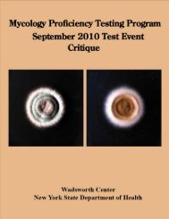
MOLD DESCRIPTIONSM-1 Curvularia sp.Source: EyeScoring:No. LaboratoriesReferee Laboratories with correct ID: 10Laboratories with correct ID: 74Laboratories with incorrect ID: 0Clinical Significance: An infrequent cause of sinusitis, keratitis, endocarditis, mycetoma, and cerebralabscess. Few cases of disseminated infection have been reported in immunocompromised patients.Ecology: Cosmopolitan, pathogen of tropical and subtropical plants.Laboratory Diagnosis:1. Culture – Curvularia grew fast on Sabouraud’s dextrose agar. After 5 days, colonies were initiallywhite, later becoming brownish black, wooly in texture (Figure 1).2. Microscopic morphology – Lactophenol cotton blue or Calcofluor mounts showed brown septatehyphae: conidiophores brown, geniculate with poroconidia (a holoblastic conidium produced througha pore or channel in the cell wall of the conidiophore or conidiogenous cell) slightly curved, brown, 3-4 transverse septations, central cell larger and darker than the other cells (Figure 2).3. Differentiation from other fungi – Curvularia species could be easily differentiated from other dark,muriform (both longitudinal and vertical septa present) fungi by its rapid growth. Microscopically,multicellular, subtly curved conidia with large and dark central cell. In Bipolaris species andDrechslera species, the conidia are distoseptate (multicellular conidia in which the cells are containedwith sacks rather than separated by septa), while in Curvularia species, the conidia are transverselyseptate.4. In vitro susceptibility testing – Most clinical isolates are susceptible to amphotericin B, itraconazole,miconazole and ketoconazole, but resistant to flucytosine and fluconazole.5. Molecular tests – Analysis of genes coding for small subunit rRNA sequences of dematiaceous fungalpathogens provided means of assessing relationships of pathogenic and non-pathogenic forms, andaccurate identification. Electrophoretic karyotyping of Curvularia lunata demonstrated that there are12 chromosomes, size ranging from 1.4 to 4.0 Mb.Comments: All the laboratories reported correct identification.Further Reading:1. Carter, E. and Boudreaux, C. 2004. Fatal cerebral phaeohyphomycosis due to Curvularia lunata in animmunocompetent patient. J Clin Microbiol. 42: 5419-5423.2. Fan, Y.M., Huang, W.M., Li, S.F., Wu, G.F., Li, W., and Chen, R.Y. <strong>2009</strong>. Cutaneousphaeohyphomycosis of foot caused by Curvularia clavata. Mycoses. 52: 544-546.3. Hiromoto, A., Nagano, T., and Nishigori, C. 2008. Cutaneous infection caused by Curvularia speciesin an immunocompetent patient. Br J Dermatol. 158: 1374-1375.9