Journal of - Spinal Research Foundation
Journal of - Spinal Research Foundation
Journal of - Spinal Research Foundation

You also want an ePaper? Increase the reach of your titles
YUMPU automatically turns print PDFs into web optimized ePapers that Google loves.
SPINAL RESEARCH FOUNDATIONTHE JOURNAL OF THE SPINAL RESEARCH FOUNDATIONA multidisciplinary journal for patients and spine specialistsBrian R. Subach, MD, FACSEditor-in-ChiefMarcus M. Martin, PhD and Anne G. Copay, PhDManaging EditorsLee Bryan Claassen, CAE, Laura A. Bologna, and Carrie B. CalifanoAssociate EditorsSPINAL RESEARCH FOUNDATION (SRF)BOARD OF DIRECTORSGuy E. BeattyChairmanMichael H. HowlandSecretaryAndrew T. GreeneTreasurerRaymond F. PugsleyNational Race LiaisonThomas C. Schuler, MD, PhDPresidentBrian D. NaultMemberWilliam H. Evers, Jr., PhDMemberBrian R. Subach, MD, PhDDirector <strong>of</strong> <strong>Research</strong>Paul J. Slosar, Jr., MDMemberKevin M. Burke, Jr.MemberNajeeb M. Thomas, MDMemberTHE JOURNAL OF THE SPINAL RESEARCH FOUNDATIONEDITORIAL BOARDJames P. Burke, MD, PhDAltoona, PAJ. Kenneth Burkus, MDColumbus, GAChristopher H. Comey, MDSpringfield, MAAleksandar Curcin, MD, MBACoos Bay, ORGeorge A. Frey, MDEnglewood, COGerard J. Girasole, MDTrumbull, CTMatthew F. Gornet, MDChesterfield, MORobert J. Hacker, MD &Andrea Halliday, MDSpringfield, ORRegis W. Haid, Jr., MDAtlanta, GALarry T. Khoo, MDLos Angeles, CANoshir A. Langrana, PhDPiscataway, NJMark R. McLaughlin, MD, FACSLanghorne, PADavid P. Rouben, MDLouisville, KYRick C. Sasso, MDIndianapolis, INThomas C. Schuler, MD, FACSReston, VAJames D. Schwender, MDMinneapolis, MNNirav K. Shah, MD, FACSLanghorne, PAPaul J. Slosar, Jr., MDDaly City, CANajeeb M. Thomas, MDMetairie, LAJim A. Youssef, MD &Douglas G. Orndorff, MDDurango, CO<strong>Journal</strong> <strong>of</strong> The <strong>Spinal</strong> <strong>Research</strong> <strong>Foundation</strong> FALL 2012 VOL. 7 No. 2
FALL 2012THE JOURNAL OF THE SPINAL RESEARCH FOUNDATIONVolume 7, Number 2Editor’s NoteBrian R. Subach, MD, FACS . . . . . . . . . . . . . . . . . . . . . . . . . . . . . . . . . . . . . . . . . . . . . . . . . . . 1President’s NoteThomas C. Schuler, MD, FACS ............................................... 2Issue OverviewMarcus M. Martin, PhD and Anne G. Copay, PhD. .................................4We’ve Got your Back Race for <strong>Spinal</strong> HealthReston, VirginiaLaura A. Bologna .............................................................5Spine TalesMixed Martial Arts Fighter - Bill ScottMark R. McLaughlin, MD, FACS ...................................................7Division I Collegiate Swimmer- Emily FergusonEmily Ferguson, Thao Nguyen, PA-C, MPAS, Thomas C. Schuler, MD, FACS .................9Spines in MotionTable <strong>of</strong> ContentsAsk the ExpertJames P. Burke, MD, PhD ......................................................11Biomechanics <strong>of</strong> the SpineRichard A. Banton, DPT, OCS, CMPT, ATC ......................................12Sporting Activities and the Lumbar Spine-Excerpts from The 7 Minute Back Pain SolutionGerard J. Girasole, MD and Cara Hartman, CPT ..................................21<strong>Spinal</strong> Biomechanics <strong>of</strong> the Golf Swing: Chiropractic PerspectivePeter M. Daddio, DC, CCSP ............................................... 26Force Transfer in the SpineDouglas G. Orndorff, MD, Morgan A. Scott, Katie A. Patty, MS ....................30Spine Biomechanics by AgeAakash Agarwal, Vikas Kaul, MS, Anand K. Agarwal, MD, Vijay K. Goel, PhD ........36Spines in Motion Glossary .....................................................47Spine Glossary ................................................................49Regional <strong>Research</strong> Partners. .........................................................55Readership Survey ...................................................................58FALL 2012 VOL. 7 No. 2<strong>Journal</strong> <strong>of</strong> The <strong>Spinal</strong> <strong>Research</strong> <strong>Foundation</strong>
SPINAL RESEARCH FOUNDATIONFrom the EditorBrian R. Subach, MD, FACSWelcome to the fall 2012 issue <strong>of</strong> the <strong>Journal</strong> <strong>of</strong>the <strong>Spinal</strong> <strong>Research</strong> <strong>Foundation</strong>. This journalis specifically dedicated to the science <strong>of</strong> biomechanics.For those <strong>of</strong> you who are unfamiliar with the term,biomechanics is the study <strong>of</strong> movement <strong>of</strong> the humanbody. I prefer to divide the concept further into twoareas: structure and function. Obviously the spine providesstructural support for the torso, head, arms, andlegs, but the intrinsic motion <strong>of</strong> the components <strong>of</strong> thespine (discs, joints, ligaments, etc.) allow for its function.Whether one considers the flexibility <strong>of</strong> an Olympicgymnast, the raw strength <strong>of</strong> a power lifter, or thetorque generated by a scratch golfer, the spine is trulythe basis for all such activities.Our society is certainly an active one. For example,our children play soccer, swim, wrestle, play tennis,and skate. Young adults row, ride, and run, whileas adults, we struggle with the conflict between an activemind and a less than cooperative aging body. Thedegenerative or arthritic process, which affects us all,is directly opposed to the concept <strong>of</strong> normal motion,meaning that aging weakens the structure <strong>of</strong> the spineand clearly slows its function.For this issue, we have brought together a number<strong>of</strong> experts in the field <strong>of</strong> biomechanics that presentboth review articles and their original research in thisarea. Our “Spine Tales” are stories <strong>of</strong> patients whoseimpaired spinal biomechanics have made it impossiblefor them to perform in their respective competitive orrecreational sports. Having overcome their spinal diseasesthrough expert diagnoses and interventions, thesespinal champions are both back at a level <strong>of</strong> functioningwhich would have seemed unobtainable only a fewyears ago.<strong>Spinal</strong> biomechanics, which seems to be a dauntinglycomplex topic to researchers, coaches, and physiciansalike, is truly the study <strong>of</strong> movement. Whetherin a young athlete or an elderly woman, spinal diseaseclearly affects us all. Hopefully, this issue will giveyou some insight into the structure and function <strong>of</strong> thespine, as well as the diseases leading to its impairmentand the interventions available.1 <strong>Journal</strong> <strong>of</strong> The <strong>Spinal</strong> <strong>Research</strong> <strong>Foundation</strong> FALL 2012 VOL. 7 No. 2
FALL 2012From the PresidentThomas C. Schuler, MD, FACSThe Fiscal Crisis is on a Collision Coursewith America’s Health CareOur growing national debt is the single greatestthreat to our national security and our way <strong>of</strong> life.The debt continues to grow because <strong>of</strong> the ongoingannual deficit spending produced by our elected <strong>of</strong>ficials.In reviewing the 2012 data from the Office <strong>of</strong>Management and Budget, it reveals that Washingtonwill spend 3.8 trillion dollars in 2012. Taxes produce2.5 trillion dollars and deficit spending, projected to be1.3 trillion dollars, funds the remainder.known as entitlements, and third, interest. Discretionaryspending under appropriated programs is brokeninto two subcategories: security (the military, FBI,CIA, etc.) and non-security (education, energy, etc.).Mandatory programs, or entitlements, consist mainly<strong>of</strong> Social Security, Medicare, Medicaid, and pensions.The third category is interest on the debt.Approximately 1.3 trillion dollars in 2012 will bespent on the discretionary programs. Two and a halfOFFICE OF BUDGET AND MANAGEMENT PROPOSED 2012 BUDGET2012 OUTLAYS (In Billions <strong>of</strong> Dollars)Appropriated (“discretionary”) programsSecurity .............................................................................................................................. 868Non-security ...................................................................................................................... 450Subtotal Appropriated Programs ................................................................................. 1,319Mandatory ProgramsSocial Security ................................................................................................................... 773Medicare ............................................................................................................................ 478Medicaid ............................................................................................................................ 255Troubled Asset Relief Program (TARP) ............................................................................ 35Other mandatory programs ................................................................................................ 711Subtotal mandatory programs ..................................................................................... 2,252Interest ............................................................................................................................... 225TOTAL OUTLAYS ................................................................................................... 3,796To understand why we continue to run a deficit,we need to define where the government spends itsmoney. The government spends its money in threecategories: first, the appropriated programs (discretionary),second, the mandatory programs, which aretrillion is spent on the mandatory programs, or entitlements,and interest. Approximately 1.5 trillion <strong>of</strong> thatamount is for Social Security, Medicare, and Medicaid.Even if 100% <strong>of</strong> the discretionary programs wereeliminated, Washington does not have enough moneyFALL 2012 VOL. 7 No. 2<strong>Journal</strong> <strong>of</strong> The <strong>Spinal</strong> <strong>Research</strong> <strong>Foundation</strong> 2
FALL 2012Issue OverviewMarcus M. Martin, PhD and Anne G. Copay, PhDEven in our sedentary society, life demands movement.Those who have suffered from back painknow how restricted life can be when they are not ableto move freely. Movement <strong>of</strong> the spine, which mayappear simple and easy, relies on the intricate collaborationand coordination <strong>of</strong> multiple elements. This issue<strong>of</strong> the <strong>Journal</strong> <strong>of</strong> the <strong>Spinal</strong> <strong>Research</strong> <strong>Foundation</strong>explores the complexity <strong>of</strong> spinal movement from theperspectives <strong>of</strong> physical therapists, chiropractors, spinalsurgeons, and, most importantly, through the eyes<strong>of</strong> those afflicted with spinal disease. We have includedthe stories <strong>of</strong> two athletes, a Division I collegiate swimmerand a 1 st Degree Jiu-Jitsu Black Belt. Both werestruck by spinal disease and unable to participate intheir athletic endeavors. After proper treatment and recovery,they are successfully competing again.Dr. Banton, a highly skilled physical therapistat the Virginia Therapy & Fitness Center, is the author<strong>of</strong> the main review for this issue <strong>of</strong> the <strong>Journal</strong><strong>of</strong> the <strong>Spinal</strong> <strong>Research</strong> <strong>Foundation</strong>. He provides acomprehensive introduction to spine biomechanicsand draws a picture <strong>of</strong> the spine as a dynamic unitcomposed <strong>of</strong> multiple joints, each contributing to itsalignment and function. Dr. Daddio from PurcellvilleFamily Doctors provides an expert chiropractor’sview on the biomechanics <strong>of</strong> the golf swing, themechanisms <strong>of</strong> golf injuries, and their avoidance. Heexplores how the manipulation <strong>of</strong> spinal segmentscan accomplish the goal <strong>of</strong> improving the structuralintegrity and stability <strong>of</strong> the spine, facilitating properbiomechanics.Dr. Orndorff, a distinguished orthopedic surgeonwith Spine Colorado, describes the types <strong>of</strong> forces appliedto the spine and the structures affected by thoseforces. A healthy spine should be able to withstand avariety <strong>of</strong> forces but may be tested beyond its limits ifproper technique and proper posture are ignored. Dr.Girasole, who practices at The Orthopaedic & SportsMedicine Center, provides excerpts from his book, The7-Minute Back Pain Solution, and gives us the spinesurgeon’s perspective and techniques to maintain ahealthy spine. Dr. Goel, a world-renowned scientist atthe University <strong>of</strong> Toledo, and his skilled research teamprovide an in-depth review <strong>of</strong> the effect <strong>of</strong> aging onthe biomechanics <strong>of</strong> the spine.Overall, this issue <strong>of</strong> the journal will help ourreaders understand the complexities and motions <strong>of</strong>the spine. Our readers will gain not only knowledge,but also renewed respect for their spines.Marcus M. Martin, PhDDr. Martin’s research interests includeneuroimmunology, virology, and immunology.He is engaged in collaborativeresearch through the <strong>Spinal</strong> <strong>Research</strong><strong>Foundation</strong> with the Medical University<strong>of</strong> South Carolina Children’s Hospital,geared toward the development <strong>of</strong> neuroprotectiveand neuroregenerative compoundsfor the treatment <strong>of</strong> nerve pathology.Dr. Martin’s current research collaborations includeresearch initiatives to apply stem cell therapy for tissue preservation,the development <strong>of</strong> regenerative therapies for intervertebraldiscs, and the development <strong>of</strong> novel methods <strong>of</strong>enhancing bone fusion.Anne G. Copay, PhDDr. Copay has conducted research andauthored several articles in the areas <strong>of</strong>organizational structure, work site healthpromotion, the effect <strong>of</strong> physical activityon energy expenditure and body weight,and the outcomes <strong>of</strong> spine fusion surgeries.Dr. Copay has ongoing research projectsconcerning the effectiveness <strong>of</strong> newspine technologies and the long-term outcomes<strong>of</strong> surgical and non-surgical treatments.FALL 2012 VOL. 7 No. 2<strong>Journal</strong> <strong>of</strong> The <strong>Spinal</strong> <strong>Research</strong> <strong>Foundation</strong> 4
SPINAL RESEARCH FOUNDATION“We’ve Got Your Back” Race for <strong>Spinal</strong> HealthReston, VirginiaLaura A. Bologna, <strong>Spinal</strong> <strong>Research</strong> <strong>Foundation</strong> National Program CoordinatorThe <strong>Spinal</strong> <strong>Research</strong> <strong>Foundation</strong>’s fifth annual“We’ve Got Your Back” Race for <strong>Spinal</strong> Healthwas hosted by the Virginia Spine Institute on May12th, in Reston, Virginia. It attracted more than 800participants, more than 100 volunteers, and raisedover $100,000 in support <strong>of</strong> the foundation’s missionto improve spinal health care through research, education,and patient advocacy.“We want to empower spinal patients with knowledgeand hope, and today celebrated that,” said ThomasC. Schuler, MD, president <strong>of</strong> SRF. “We have had peoplewith major spinal issues who were worried thattheir lives were over, that they couldn’t do things theylove, or carry out their careers. But through modernspinal care– both operative and non-operative– we’vegotten people back to unbelievable levels <strong>of</strong> activity,even returning people to careers in the military and theNational Football League.”Washington Redskins players Rocky McIntosh,Reed Doughty, and Josh Wilson– who have all undergonespinal treatment and returned to the NFL– gavepowerful testaments to the value <strong>of</strong> effective spinaltreatment and are considered <strong>Spinal</strong> Champions.<strong>Spinal</strong> Champions were celebrated on race day withcommemorative race shirts and a special tent where theycould share their stories <strong>of</strong> success. A <strong>Spinal</strong> Championis defined as someone who has suffered from back orneck pain, has overcome it through either non-operativeor surgical treatments, and regained his or her life.Dr. Schuler noted that more than nine out <strong>of</strong> tenpeople will suffer from severe neck and/or low backpain during their lifetimes. Approximately ten percent<strong>of</strong> these people develop chronic pain, which meansthat at any given time, 35 million people in the UnitedStates are directly affected by disabling pain and manymore indirectly. Techniques to cure, manage, and preventthese conditions need to be developed, implemented,and proven.The event also included a <strong>Spinal</strong> Health Fair thatfocused on nutrition, exercise, and prevention <strong>of</strong> spinalproblems. The <strong>Spinal</strong> Health Fair provided educational5 <strong>Journal</strong> <strong>of</strong> The <strong>Spinal</strong> <strong>Research</strong> <strong>Foundation</strong> FALL 2012 VOL. 7 No. 2
FALL 2012materials about the spine, a calcium campaign includinga photo booth with milk mustaches, a massage station,and kids’ activities. The race festivities also includedlive entertainment, refreshments, and free giveaways.“SRF is identifying the best treatments for spinalproblems through a national network <strong>of</strong> research centers,”Schuler said. “This network is expanding to all50 states. We are challenging all <strong>of</strong> our Regional <strong>Research</strong>Partners to host 5K events in their cities to raiseawareness <strong>of</strong> spinal treatment success, help individualsestablish goals to improve their health, and to raisefunds for further research.”The <strong>Spinal</strong> <strong>Research</strong> <strong>Foundation</strong> is proud to havethe only run/walk event in the country designed to celebratethe accomplishments <strong>of</strong> <strong>Spinal</strong> Champions asthey continue to research new techniques to improvespinal health care for future generations. To learn moreabout the <strong>Spinal</strong> <strong>Research</strong> <strong>Foundation</strong>’s “We’ve GotYour Back” Race for <strong>Spinal</strong> Health and other host sites,visit www.spinerf.org/race.FALL 2012 VOL. 7 No. 2<strong>Journal</strong> <strong>of</strong> The <strong>Spinal</strong> <strong>Research</strong> <strong>Foundation</strong> 6
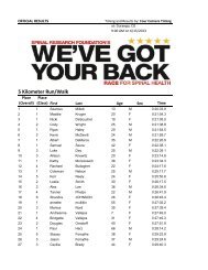
SPINAL RESEARCH FOUNDATIONSpine TaleBill Scott, Mixed Martial Arts FighterMark R. McLaughlin, MD, FACSspine. The results would expectantly remove pressurefrom the spinal nerves in order to relieve pain. <strong>Spinal</strong>disks, which consist <strong>of</strong> cartilage, were removed frombetween the vertebrae; bone tissue taken from Scott’ship was inserted to replace them. The implanted bonetissue, in conjunction with the body’s natural bonegrowthprocesses, created the fusion <strong>of</strong> the vertebrae.Dr. McLaughlin inserted titanium rods and screwsat the site <strong>of</strong> the surgery. The screws, inserted in threeconsecutive vertebrae, served as anchor points for theconnecting rods. The rods and screws kept the lumbarregion stable and prevented motion that hinders theproper fusion <strong>of</strong> the vertebrae.Bill Scott, a Mixed Martial Arts Fighter and 1 st Degree BrazilianJiu-Jitsu Black Belt.When mixed martial arts fighter Bill Scott felt severepain in his lower back and numbness in hisentire right leg in 2003, he thought it was the after-effect<strong>of</strong> a head-on car crash two years earlier. But a prominentneurosurgeon who examined Scott found a seriousspinal problem that was unrelated to the accident.During the consultation with Mark McLaughlin,MD, medical director <strong>of</strong> Princeton Brain & SpineCare, an MRI revealed that several <strong>of</strong> Scott’s lumbarvertebrae had collapsed, shifted to the right side <strong>of</strong> hisbody, and fused together. Although Scott had receiveda surgery to fuse the 3rd and 4th lumbar vertebraemany years ago, he was unaware <strong>of</strong> any problems untilthe fused bones began pressing on his spinal nervesand causing pain. When Dr. McLaughlin told Scottthat a second spinal fusion procedure was necessary,he responded with a question:“I asked him, ‘When will I be able to fight again?’,”said Scott, 47. “He said I shouldn’t, but as far as I wasconcerned, there wasn’t any question that I would, itwas just a matter <strong>of</strong> when.”The primary goal <strong>of</strong> the five-hour surgery, performedby Dr. McLaughlin through a small incision inthe low back, was to realign and fuse the 4th and 5thlumbar vertebrae to eliminate abnormal motion in theLateral view CAT/Myelogram<strong>of</strong> previous spinal fusion surgeryat L3-L4. The arrow indicatesspondylolysthesis at L4-L5.Lateral view CAT/Myelogram <strong>of</strong>Dr. McLaughlin’s fusion extensionsurgery to fuse L4-L5.After the surgery, Dr. McLaughin explained toScott that it would take three months for the vertebraeto fuse properly and wearing a back brace the entiretime was necessary for the best outcome.Scott followed his instructions to the letter and theprocedure was successful. However, Dr. McLaughlinwas skeptical about his patient’s chances <strong>of</strong> returningto his sport.“After the healing was completed, the area where weperformed the surgery was solid as cement, and it still7 <strong>Journal</strong> <strong>of</strong> The <strong>Spinal</strong> <strong>Research</strong> <strong>Foundation</strong> FALL 2012 VOL. 7 No. 2
FALL 2012Bill Scott during a pr<strong>of</strong>essionalMixed Martial Arts fight. Photocourtesy <strong>of</strong> Keith D. Mills.FALL 2012 VOL. 7 No. 2is,” says Dr. McLaughlin.“I had some concernsabout the levels <strong>of</strong> thespine above and belowthe surgery. In 2008 weinserted more screws togive more stability to thelower vertebra, but allthese years later, everythingis holding up verywell.”At the end <strong>of</strong> therecovery period, Dr.McLaughlin reviewed anX-ray <strong>of</strong> Scott’s spine andgave him the OK to startexercising again. But hedidn’t approve a return to fighting. “Dr. McLaughlin isa former champion wrestler himself,” says Scott, “andas much as I respect his medical opinion, I had otherplans.”Scott, a 1 st Degree Brazilian Jiu-Jitsu Black Beltand a former All-American wrestler at MiddlesexCollege, didn’t follow a formal rehab program. Afterspinal fusion, patients are advised to stay active withbrief, gentle exercise and stretching which promotesblood flow and healing. But when Scott returned to hismartial arts training center (Bill Scott BJJ- BrazilianJiu-Jitsu Shore Academy) in his hometown <strong>of</strong> PointPleasant, NJ, he picked up right where he left <strong>of</strong>f.“For my rehab, I just returned to my usual trainingfor Brazilian Jiu-Jitsu and instructing martial arts students.I wanted to get right back in the swing <strong>of</strong> things,and I went at full speed right away. I believe that aperson’s mind-set is extremely important in this type<strong>of</strong> situation. I’ve seen people who have had this type<strong>of</strong> injury become depressed and feel sorry for themselves,and I could have easily gone that route. But Ididn’t allow myself to think that way. I worked hard,and I was determined not to let the situation damagemy life. I wanted to get back to being active and doingwhat I love to do.”These days, in addition to a rigorous schedule <strong>of</strong>teaching martial arts, including self-defense training forlocal and state police <strong>of</strong>ficers in New Jersey, Scott competesin and has won several mixed martial arts bouts.He is currently training for the 2012 Pan Jiu-Jitsu No GiChampionship which takes place this fall.“I’m a fighter; it’s what I do for a living, and it’s alsomy attitude,” says Scott. “When you participate in mysport, you’re going to have injuries now and then. Whenone happens, I get fixed up, move on, and keep fighting.”Working with the unstoppable Scott taught Dr.McLaughlin a great lesson: “I learned that you have tolisten to your patient very closely, not only to understandhow they feel physically, but also where they areemotionally. When I did Bill’s surgery and told himhe really shouldn’t fight anymore, that was like tellinghim not to breathe. Instructing and competing in martialarts are in his DNA, and it was absolutely impossiblefor him to giveit up. When workingwith an elite athletelike Bill, a spine surgeon’sjob is to providethe stability he needsto compete again thebest he can. Scott returnedto fighting andwon matches, which isremarkable consideringthe major surgeryhe had. His energy,enthusiasm, and positiveattitude helpedhim pull through and Bill Scott, <strong>Spinal</strong> Champion!inspire me.”Mark R. McLaughlin,MD, FACSDr. McLaughlin practices neurologicalsurgery with a focus on spine disordersand specific cranial conditions atPrinceton Brain and Spine Care in Princeton,NJ. He served as the President <strong>of</strong>the Young Neurosurgeons’ Committee,a national section <strong>of</strong> the American Association <strong>of</strong> NeurologicalSurgeons. He is the Scientific Program Chairman <strong>of</strong> the AANS/CNS Joint Spine Section, and also an editor <strong>of</strong> Spineuniverse.com, a website dedicated to patient and physician education<strong>of</strong> spinal disorders. He has published more than 65 articles onneurosurgery and spine surgery, and has authored two textbooksabout spine surgery. He has been an invited speaker, presenter,and course director at numerous scientific meetings, andteaches complex spine surgery nationally and internationally. Dr.McLaughlin was recently elected Member-at-Large <strong>of</strong> the JointSpine Section <strong>of</strong> the Congress <strong>of</strong> Neurosurgeons.<strong>Journal</strong> <strong>of</strong> The <strong>Spinal</strong> <strong>Research</strong> <strong>Foundation</strong> 8
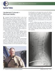
SPINAL RESEARCH FOUNDATIONSpine TaleEmily Ferguson, Division I Collegiate SwimmerEmily Ferguson, Thao Nguyen, PA-C, MPAS, and Thomas C. Schuler, MD, FACSAs a collegiate swimmer at Virginia Tech, my physicalhealth was crucial to my success in the pool aswell as in the classroom. During the fall <strong>of</strong> my junioryear, I began noticing low back pain as a result <strong>of</strong> prolongedsitting through classes. I had to constantly readjustmy posture while sitting because <strong>of</strong> the discomfortI was experiencing. As my back pain became increasinglyunbearable, my performance at practices was alsoaffected. I had a demanding weekly training schedule<strong>of</strong> nine swimming practices, two dryland workouts, andthree lifting sessions; after two months <strong>of</strong> pain, I knew Ihad to get my back evaluated.The first step I took in November <strong>of</strong> 2011 was tovisit the doctors within our athletic department. Myteam physician ordered some x-rays and sent me to aspine specialist in Blacksburg, VA. I had an MRI <strong>of</strong> mylumbar spine completed just before winter break. Theresults came after I had already traveled back home forChristmas; I had two herniated discs. The doctors suggestedI head back to Blacksburg to receive an injectionbefore our swimming team’s annual training trip. Decemberis a crucial training period for swimmers and Ihoped to receive some relief from the pain to participatein the team trip. However, the injections were ineffective,and I was unable to work out during the entire trip.After I got back home, there was about a week beforespring semester classes began. In the meantime,my neighborhood friend had referred my parents to Dr.Schuler. I immediately made an appointment at theVirginia Spine Institute before heading back to school.Once I stepped into VSI I knew I was going to be takencare <strong>of</strong> and in good hands.“Emily came into our <strong>of</strong>fice as a 20 year-old collegiateswimmer from Virginia Tech who had been sufferingfrom back pain for months. Especially for our youngathletes, we like to approach spine care with non-operativemeasures first and foremost,” noted Thao Nguyen,PA-C at the Virginia Spine Institute after her first visitwith Emily in January, 2012. “Our primary objectivewith Emily was to treat her symptoms so she could returnto competitive swimming. It is standard to haveathletes suspend participation in their sports to reducesymptoms; so unfortunately, Emily was advised to discontinueswimming andfocus on physical therapyand the foundation<strong>of</strong> spinal health– corestabilization, strengthening,and aerobic conditioning.Dr. Schuler andI discussed with Emilyand her family that we alwaysexhaust all non-operativetreatment beforesurgery is considered.After rounds <strong>of</strong> physicaltherapy and medications,Emily was not having thesymptom relief we allhoped for; we added injectionsnext.”After these interventions,I returned toschool, but I was unableto redshirt, which meantI could not suspend mystatus as a collegiate athletein order to elongatemy competitive eligibility.I adjusted my goalsto be able to compete se-Figure 1. The pre-op MRI with twodisc herniations.Figure 2. An x-ray after the anteriorlumbar fusion surgery at L4-L5.9 <strong>Journal</strong> <strong>of</strong> The <strong>Spinal</strong> <strong>Research</strong> <strong>Foundation</strong> FALL 2012 VOL. 7 No. 2
FALL 2012Spines in Motion: The Biomechanics <strong>of</strong> the Spinenior year. I came homein May and was frustratedthat I was stillin pain, so I scheduledanother appointmentwith Dr. Schuler.“Emily did not respondto the therapyand injections, so sheunderwent a procedurecalled a discography”Dr. Schuler recalls.“This test helpsus receive informationon whether her discsare the cause <strong>of</strong> her pain and helps us determine if she isa candidate for surgery. Our decision making reviewedEmily’s ongoing history, a physical examination, diagnosticimaging, her response to previous treatments,and the results <strong>of</strong> the discography. We recommended ananterior lumbar fusion surgery, which is a minimally invasivetechnique that would optimize her recovery withthe goal to return her to swimming.”After discussing surgery with my parents and doingsome personal research on the surgical procedure, Idecided to give it a go.“Emily did extremely well after surgery and hada great support group– her family. She started physicaltherapy the day after surgery by walking the halls<strong>of</strong> the hospital, and she went home the next day afterthat,” Thao commented.After surgery I was able to swim competitively forall <strong>of</strong> my senior year at Virginia Tech. Prolonged sittingno longer caused pain.It has been a little more than one year after surgeryand I am able to run, bike, and do any other activitiespain-free. My advice to anyone suffering from back orneck pain is to seek treatment immediately. Your neckand back health are crucial for your entire lifetime.VSI is uniquely exceptional because <strong>of</strong> its comprehensivecare. The clinic, the pain management center,and the Virginia Therapy and Fitness Center are allunder one ro<strong>of</strong>. Overall, I had an amazing experienceFALL 2012 VOL. 7 No. 2with VSI! I was so impressed with the quality <strong>of</strong> serviceand care I received as a patient, that I applied for ajob as a medical assistant with VSI. Today, I am a VSIemployee and am able to help people who are goingthrough the same type <strong>of</strong> pain I went through.Dr. Schuler and Thao were inspired by Emily’sstory. “Emily went on to compete in the swimming pre-Olympic trials after her lumbar fusion surgery, makingher a primary example <strong>of</strong> a true spinal champion!We are proud to share her exceptional story <strong>of</strong> successthrough determination and devotion to her passion <strong>of</strong>swimming.”Thao Nguyen, PA-C, M.P.A.S.Thao is a highly skilled Physician Assistantat The Virginia Spine Institute(VSI). She received her Master <strong>of</strong> PhysicianAssistant Studies degree fromShenandoah University, VA and a Bachelor<strong>of</strong> Science in the College <strong>of</strong> Humanitiesand Science from Virginia CommonwealthUniversity. She completed her residency in medical andsurgical management <strong>of</strong> spinal disorders and is licensed by theVirginia Board <strong>of</strong> Medicine.Thomas C. Schuler, M.D.,F.A.C.S.Dr. Schuler is a distinguished spinesurgeon and founder and CEO <strong>of</strong> theVirginia Spine Institute (VSI). His knowledge,drive, and innovative techniqueshave revolutionized spinal healthcarein the Washington D.C. metropolitanarea. Dr. Schuler is double board certified in spine surgery andorthopaedic surgery. He is nationally recognized as a leader inthe treatment <strong>of</strong> cervical and lumbar spine disorders and wasnamed among the 100 Best Spine Surgeons and Specialistsin America. Additionally, U.S.News & World Report has recentlynamed him among the top 1% <strong>of</strong> physicians in his specialty nationwide.As the spine consultant to the Washington Redskinssince 1993, he returns elite athletes to the playing field safelyand quickly using non-operative and operative techniques. Dr.Schuler also serves as the President <strong>of</strong> the <strong>Spinal</strong> <strong>Research</strong><strong>Foundation</strong> (SRF).<strong>Journal</strong> <strong>of</strong> The <strong>Spinal</strong> <strong>Research</strong> <strong>Foundation</strong> 10
SPINAL RESEARCH FOUNDATIONAsk the ExpertJames P. Burke, MD, PhDWhat are the benefits <strong>of</strong> anactive lifestyle for the spine?An active lifestyle is <strong>of</strong> great importance to the health <strong>of</strong>the spine. The following are specific benefits: 1) Posturalmuscle strength. The spine is supported by muscles,tendons, and ligaments. Exercise that strengthens thepostural muscles contributes to the maintenance <strong>of</strong> goodspine posture. 2) Bone strength. The spine is vulnerableto osteoporosis and vertebral fractures. Weight-bearingexercise helps maintain or increase bone mineral densityand prevent osteoporosis. 3) Body weight. Excess bodyweight, particularly increased abdominal girth, placesextra stress on the spine. As everyone knows, exercisehelps maintain a healthy body weight. 4) Health <strong>of</strong> intervertebraldiscs. The intervertebral discs do not have theirown blood supply; they receive nutrients through diffusionfrom the vertebral bodies. Movement will help“flush” the discs and increase diffusion.What is the best activity/sportfor the spine?Activity in general is beneficial to the spine. Some particularexercises are designed to strengthen the musclesthat support the spine. For instance, plank exercises(exercises based on a push-up position) strengthen yourcore muscles, including the transverse abdominis, rectusabdominis, and the erector spinae, all <strong>of</strong> which supportyour lumbar spine. Stability and balancing exercises,such as balancing on one foot or on unsteady surfaceslike a BOSU ball, balance board, or gymnastics mat,also develop proprioception, core stability, and strength.Can some activities behurtful to the spine?People who suffer from back pain or degenerative discdisease may need to avoid high impact activities, suchas running. The pounding created every time a runner’sfeet hit the ground may cause repeated trauma to the discspace. Aquatic exercises are a great alternative to reducestress on the spine while still benefiting from physicalactivity. Axial bearing exercise is another type <strong>of</strong> exercisethat puts the spine at risk. Axial bearing exercisesare movements where the weight compresses your spinalcolumn from above. Military presses and squats, in whichthe weight is held over head or supported by the shoulders,are examples <strong>of</strong> axial bearing exercises. Performingthose exercises with the spine incorrectly aligned maycause injury. Finally, start-and-stop sports with rapidchanges in direction are very demanding on the spine.Such sports are basketball, football, and racquet sports.If I have spine fusion, will I beable to participate in strenuoussports/activities?After spinal fusion surgery, it is critical to follow a rehabilitationprogram centered on stretching, strengthening,and aerobic conditioning. At the early stage, exercisehelps fuse the vertebrae, but may hurt the fusion ifit is too strenuous. After fusion is achieved, you will beable to carefully resume your habitual activities. However,the more vigorous the activities, the longer it maytake before you can resume them.James P. Burke, M.D., Ph.D.Dr. Burke is a neurosurgeon with AlleghenyBrain and Spine Surgeons inAltoona, Pennsylvania. He is certifiedby the American Board <strong>of</strong> NeurologicalSurgeons. His pr<strong>of</strong>essional associationsinclude American Academy <strong>of</strong>Neurological Surgeons, Congress <strong>of</strong>Neurological Surgeons, North American Spine Society, and theAlpha Omega Alpha National Honor Medical Society. Dr. Burke’sspecializations include brain tumors, concussions, trauma, degenerativespine, spinal stenosis, spinal tumors, and other spinaldeformities.11 <strong>Journal</strong> <strong>of</strong> The <strong>Spinal</strong> <strong>Research</strong> <strong>Foundation</strong> FALL 2012 VOL. 7 No. 2
FALL 2012Biomechanics <strong>of</strong> The SpineRichard A. Banton, DPT, OCS, CMPT, ATCMovement in the human body occurs at jointsurfaces; movement occurs with bones; movement<strong>of</strong> muscles moves the bones; coordinated movements<strong>of</strong> limbs create strong purposeful movementsin a pain-free person. Notice that the list begins withmovement at a joint. It is at this anatomical level thatthe central nervous system interprets and coordinatesa neuro-musculoskeletal response into a functionalmovement. Therefore, logic would tell us that to improvefunction, one must ensure that articular motionis functionally optimal.Flexion/Extension“If there is a single word that encapsulatesall that physical therapy stands for,it is movement.” 1In physical therapy school, it is tradition to studyanatomy and biomechanics in a regional manner. Forexample, the lumbar spine is studied separate from thesacrum which is, in turn, studied separate from the hip.While it is important to understand regionally specificanatomy, successful outcomes in physical therapy areonly achieved by understanding how the regions <strong>of</strong>the body work together. This article summarizes a fewbiomechanical principles <strong>of</strong> the spine as viewed fromthe perspective <strong>of</strong> manual physical therapists.Movements and Motions <strong>of</strong> the SpineRotational movements are movements <strong>of</strong> the vertebraaround an axis. All rotations produce a changein the orientation <strong>of</strong> the vertebra. 2Translational movements are movements <strong>of</strong> thewhole vertebra by the same amount in a given direction.There is no change in the orientation <strong>of</strong> the vertebra.Translation is the “gliding” <strong>of</strong> the vertebra; it rarely occursby itself, but <strong>of</strong>ten accompanies other movements.The movements <strong>of</strong> each spinal segment are limitedby anatomical structures such as ligaments, intervertebraldiscs, and facets. Specifically, anatomical structuresFALL 2012 VOL. 7 No. 2Axial RotationLateral BendingFigure 1. Flexion/Extension (bending), axial rotation(torsion), andlateral bending (side bending). Picture courtesy <strong>of</strong> Medtronic, Inc.<strong>Journal</strong> <strong>of</strong> The <strong>Spinal</strong> <strong>Research</strong> <strong>Foundation</strong> 12
SPINAL RESEARCH FOUNDATIONR. Banton/The <strong>Journal</strong> <strong>of</strong> the <strong>Spinal</strong> <strong>Research</strong> <strong>Foundation</strong> 7(2012) 12–20cause the coupling <strong>of</strong> motions <strong>of</strong> the spine, that is, movementsoccur simultaneously. Flexion, extension, translation,axial rotation, and lateral bending are physiologicallycoupled. The exact pattern <strong>of</strong> coupling depends onthe regional variations <strong>of</strong> anatomical structures.In the cervical and upper thoracic spine, side bendingis coupled with axial rotation in the same direction.In the lumbar spine, lateral bending is coupledwith axial rotation in the opposite direction. In theRegional Coupling PatternsC2–T1T1–T4C0–C1–C2C2–C5C5–T1T1–T4Upper cervicalMiddle cervicalLower cervicalUpper thoracicmiddle and lower thoracic spine, the coupling patternis inconsistent. 1 However, the pattern <strong>of</strong> couplingwill change depending on which movement is initiatedfirst. In the lumbar spine, lateral bending willbe coupled with axial rotation in the same direction iflateral bending is the first movement. Conversely, ifaxial rotation is the first movement, it will be coupledwith lateral bending in the opposite direction. Theterms latexion and rotexion have been applied to thesecoupling patterns (Figure 3 and 4).There has been relatively more interest in latexionand rotexion recently, largely because abnormalcoupling patterns have been linked to instabilities.Changes in coupling patterns have also been observedadjacent to spinal fusions. Finally, these particularcoupling patterns may have relevance in the basic biomechanics<strong>of</strong> the different regions <strong>of</strong> the spine and understandingthem may lead to new discoveries in theevaluation and treatment <strong>of</strong> scoliosis.L1–S1T1–T4 T4–T8T8–L1 T8–L1L1–L4L4–L5L5–S1Middle thoracicLower thoracicUpper lumbarLower lumbarLumbosacralThe Cervical SpineBecause <strong>of</strong> kinematic and clinical uniqueness, thecervical spine is divided up into the occipital-atlantoaxialcomplex (C0-C1-C2), the middle cervical spine(C2-C5), and the lower cervical spine (C5-T1). 3The occipital atlanto-axial region is so unique andcomplex that controversy exists regarding the exactbiomechanics <strong>of</strong> the region. About 60% <strong>of</strong> the entirecervical spine axial rotation occurs from C0-C2 andabout 40% occurs from below. 1 The uniqueness <strong>of</strong>this region is primarily related to the articular surfaces<strong>of</strong> the first two cervical vertebrae. They are unlike anyother vertebrae in the human body because they areboth convex articulating surfaces. This unique geometricshape facilitates significant motion <strong>of</strong> the humanhead. 1 While other vertebral segments acquiretheir stability from vertebral discs, the C1-C2 complexachieves stability through dense ligamentous structures(i.e., alar ligaments and transverse ligaments.)Figure 2. Regional coupling patterns <strong>of</strong> lateral bending and axialrotation. Image modified and adapted from White, A.A. and M.M.Panjabi (1990). Clinical Biomechanics <strong>of</strong> the Spine. Philadelphia,PA, Lippincott.The Occipital-Atlanto-Axial Complex(Upper Cervical Spine)Most controversy regarding movement <strong>of</strong> thespine exists in the upper cervical spine. The uniqueconvex on convex orientation <strong>of</strong> the C1-C2 complex13 <strong>Journal</strong> <strong>of</strong> The <strong>Spinal</strong> <strong>Research</strong> <strong>Foundation</strong> FALL 2012 VOL. 7 No. 2
FALL 2012Spines in Motion: The Biomechanics <strong>of</strong> the Spinehas created debate among researchers for decades.The accepted coupling pattern <strong>of</strong> this region is thatrotation and sidebend <strong>of</strong> the head occurs in oppositedirections. For example, when the head rotates right,C1 sidebends left. This movement pattern is madepossible because <strong>of</strong> the shape <strong>of</strong> the articular condyles<strong>of</strong> C1 and C2. They are both convex in nature;however, the incline <strong>of</strong> the posterior condyle <strong>of</strong> C2is twice as steep as the anterior surface <strong>of</strong> the condyle.Therefore, when the head rotates to the right,the right C1 condyle has farther to glide down theposterior surface <strong>of</strong> C2 than the left C1 condyle hasto glide on the anterior surface <strong>of</strong> the left C2. Onecan easily understand how controversy exists whenyou consider the complex anatomy <strong>of</strong> each segment.Figure 5 demonstrates the anatomical uniqueness <strong>of</strong>the C1-C2 complex.Figure 3. Rotexion: rotation to the right coupled with lateral bendingto the right. Image courtesy <strong>of</strong> the Virginia Therapy and FitnessCenter.Figure 5. Posterosuperior view <strong>of</strong> the upper cervical vertebrae.Image courtesy <strong>of</strong> Medtronic, Inc.Figure 4. Latexion: lateral bending to the right coupled with rotationto the left. Image courtesy <strong>of</strong> the Virginia Therapy and FitnessCenter.FALL 2012 VOL. 7 No. 2The importance <strong>of</strong> coupling in the human spineis that it allows for increased mobility without sacrificingstability. When treating movement patternrestrictions <strong>of</strong> the spine, it is critical for the manualtherapist to understand the unique coupling patterns<strong>of</strong> each region so that maximum mobility and stabilitycan be achieved.The C5-C6 interspace is generally considered tohave the largest range <strong>of</strong> motion in the cervical spine,<strong>Journal</strong> <strong>of</strong> The <strong>Spinal</strong> <strong>Research</strong> <strong>Foundation</strong> 14
SPINAL RESEARCH FOUNDATIONR. Banton/The <strong>Journal</strong> <strong>of</strong> the <strong>Spinal</strong> <strong>Research</strong> <strong>Foundation</strong> 7(2012) 12–20hence the potential reason for the high incidence <strong>of</strong>cervical spondylosis (arthritis) at this segment. 1 Asdiscussed earlier, the cervical spine tries to achievemaximum mobility without sacrificing stability; theuncinate processes are the structures conferring stabilitywith mobility in the cervical spine (Figure 6).The uncinate processes become fully developed atage 18 but do not begin to develop until ages 6 to 9years old. Considering that they help guide flexionand extension <strong>of</strong> the neck, limit lateral bending, andprevent posterior translation, one has to questionthe reasoning behind allowing adolescents to playtackle football or any collision sport before theirnecks are fully developed. Injury to the middle cervicalregion during adolescent years could lead tolifelong complications. 1Figure 6. The vertebral bodies <strong>of</strong> the subaxial cervical spine haveupward projections on the lateral margins called uncinate processes.These processes articulate with the level above to form theuncovertebral joints. The zygapophyseal joints are also known asthe facet joints that are comprised <strong>of</strong> the superior articular process<strong>of</strong> one vertebral body and the inferior articular process <strong>of</strong> theadjacent vertebra. Image courtesy <strong>of</strong> Medtronic.Movement <strong>of</strong> the cervical spine involves a combination<strong>of</strong> uncovertebral and zygapophyseal motion(Figure 6). During neck flexion, the zygapophysealand uncovertebral joints glide in a combined superior,lateral, and anterior direction. Reciprocally, duringneck extension, the joints glide in a combined inferior,medial, and posterior direction.The Thoracic SpineWhile hypermobility <strong>of</strong> the cervical spine hasbeen associated with whiplash injuries, hypomoblity<strong>of</strong> the thoracic spine has been associated with abnormalmechanical influences on both the cervical andlumbar spine. 1The unique feature <strong>of</strong> the thoracic spine is the coordinatedmovement <strong>of</strong> the ribcage with the vertebralsegments. The addition <strong>of</strong> the ribcage increases the requiredcompressive load necessary to cause buckling<strong>of</strong> the spine. 4 There is considerable motion <strong>of</strong> the spineand sternum independent <strong>of</strong> one another, allowing formotion <strong>of</strong> the spine without movement <strong>of</strong> the ribcage.During flexion <strong>of</strong> a thoracic spinal segment, there isanterior translation. This anterior translation facilitatesanterior rotation <strong>of</strong> the adjacent rib. A similar motionoccurs in the middle thoracic spine (T4-T7), but because<strong>of</strong> the anatomical shape <strong>of</strong> the transverse processesand the heads <strong>of</strong> the ribs, there is anterior roll <strong>of</strong>the ribs associated with a superior glide. 2 These minorvariations in biomechanics <strong>of</strong> the thoracic spine andribcage are critical for manual therapists to understandand analyze if their goals are to be successful in treatingand restoring mobility to the thoracic spine and ribs.The pattern <strong>of</strong> coupling in the thoracic spine issimilar to the cervical spine. Lateral bending is coupledwith axial rotation in the opposite direction when lateralbending occurs as the first movement (latexion). Likewise,thoracic rotation is coupled with lateral sidebendingin the same direction when rotation occurs as thefirst movement (rotexion). In the thoracic spine, theribs are oriented so that they approximate when the spinesidebends. However, as further sidebending occurs, theipsilateral ribs glides anteriorly and inferiorly along theplane <strong>of</strong> the costotransverse joint, while the contralateralrib moves superiorly and posteriorly creating rotation inthe opposite direction <strong>of</strong> the sidebend (Figure 7). Conversely,rotation in the thoracic spine is accompanied bysidebending in the same direction, not because <strong>of</strong> facetorientation, but because <strong>of</strong> the ligamentous attachment<strong>of</strong> the ribs to the thoracic vertebrae (Figure 8).Understanding the biomechanics <strong>of</strong> the thoracicspine and ribcage is important when addressing breathingand respiratory issues, chronic pain, facilitated15 <strong>Journal</strong> <strong>of</strong> The <strong>Spinal</strong> <strong>Research</strong> <strong>Foundation</strong> FALL 2012 VOL. 7 No. 2
FALL 2012Spines in Motion: The Biomechanics <strong>of</strong> the SpineDuring respiration, the ribs move upwards, sideways(‘bucket handle”) and forward (“pump handle”).3 This is important to understand when treatingrib hypomobilities because the therapist must not onlyimprove the ability <strong>of</strong> the rib to elevate and depress,but must also make sure the ribs can move mediallyand laterally.The Lumbar SpineThe facet orientation <strong>of</strong> the lumbar spine facilitatesmore flexion and extension than rotation (Figure 9).Figure 7. Bilateral coastal rotation in opposing directions drivesthe superior vertebra into left rotation. 4 Image courtesy <strong>of</strong> Lee, D.(1993). “Biomechanics <strong>of</strong> the thorax: A clinical model <strong>of</strong> in viv<strong>of</strong>unction.” <strong>Journal</strong> <strong>of</strong> Manual and Manipulate Therapy 1(1): 13-21Figure 8. As the superior thoracic vertebra rotates to the right, ittranslates to the left. The right rib rotates posteriorly and the leftrib rotates anteriorly as a consequence <strong>of</strong> the vertebral rotation. 4Image courtesy <strong>of</strong> Lee, D. (1993). “Biomechanics <strong>of</strong> the thorax: Aclinical model <strong>of</strong> in vivo function.” <strong>Journal</strong> <strong>of</strong> Manual and ManipulateTherapy 1(1): 13-21.segments (hypersensitive spine segments), and spinalinstability. A restriction (or other dysfunction) in thespine is associated with illness <strong>of</strong> the organ(s) relatedto the vertebral segment (viscerosomatic reflex). Previousstudies have shown that an increase in somaticdysfunction in the thoracic spine may be linked toviscerosomatic reflex phenomena. For example, Bealdemonstrated that somatic dysfunction <strong>of</strong> T1-T5 waslinked to cardiovascular disease. 5 Similarly, T6 is associatedwith the stomach.FALL 2012 VOL. 7 No. 2Figure 9. Facet joint orientation in the cervical, thoracic, andlumbar spine. Image courtesy <strong>of</strong> Medtronic, Inc.<strong>Journal</strong> <strong>of</strong> The <strong>Spinal</strong> <strong>Research</strong> <strong>Foundation</strong> 16
SPINAL RESEARCH FOUNDATIONR. Banton/The <strong>Journal</strong> <strong>of</strong> the <strong>Spinal</strong> <strong>Research</strong> <strong>Foundation</strong> 7(2012) 12–20In the lumbar spine, flexion and extension motions increasein range from the top to the bottom with exception<strong>of</strong> the lumbosacral joint (L5-S1). The lumbosacraljoint <strong>of</strong>fers more flexion/extension motion than anyother lumbar segments. 1 With regards to lateral bendingin the lumbar spine, each lumbar segment presents withapproximately the same amount <strong>of</strong> movement. Likewise,axial rotation in the lumbar spine is very limitedand nearly equal among each segment.The most important aspect <strong>of</strong> lumbar biomechanicsis the translation that occurs with flexion and extension. 1The measure <strong>of</strong> translation in the lumbar spine is thedetermining factor in the diagnosis <strong>of</strong> spinal instability.Although much research is required in this region to determinemore accurate measures <strong>of</strong> true spinal instability,current literature suggests that 2 mm <strong>of</strong> translationis normal for the lumbar spine. 1 Translation beyond 4mm should be evaluated for clinical instability.The unique coupling patterns associated with thelumbar spine, along with minimal mobility in the transverseplane, may directly or indirectly contribute to thehigher incidence <strong>of</strong> clinical instability at the L4-L5 segment.Panjabi and colleagues have confirmed previouslumbar kinematic investigations that showed the upperlumbar segments L1-L2, L2-L3, and L3-L4 share a couplingpattern different from that <strong>of</strong> L4-L5 and L5-S1. 1In the upper lumbar spine, sidebend and rotation occursin opposite directions, while in the lower lumbar segments,sidebend and rotation occur in the same direction.In addition, Panjabi and associates discovered aninteresting effect <strong>of</strong> posture on coupling patterns. In extension,the coupling motion was a flexion movement;in flexion, the coupling movement was an extensionmovement. 1 In other words, the lumbar spine alwaysshows a tendency to straighten from either flexion orextension. The clinical significance <strong>of</strong> this finding is notyet known, but it does provide reason to further investigatethe biomechanics and kinetics <strong>of</strong> the lumbar spine.The Sacroiliac JointAlthough mobility <strong>of</strong> the sacroiliac joint has beendebated since the early 17th century, it is now acceptedamong all medical pr<strong>of</strong>essionals that there is movementavailable in this joint. 6 Motion at the sacroiliac joint occursduring movement <strong>of</strong> the trunk and lower extremities.Flexion <strong>of</strong> the sacrum is called nutation while extension<strong>of</strong> the sacrum is termed counternutation. Whenthe sacrum nutates, the sacral promontory moves anteriorinto the pelvis. When the sacrum counternutates,the sacral promontory moves backward (Figure 10).Figure 10a. When the sacrum nutates, its articular surface glidesinferoposteriorly relative to the innominate.Figure 10b. When the sacrum counternutates, its articular surfaceglides anterosuperiorly relative to the innominate. Images courtesy<strong>of</strong> Lee, D. (1999). The Pelvic Girdle. Edinburgh, UK, ChurchillLivingstone.17 <strong>Journal</strong> <strong>of</strong> The <strong>Spinal</strong> <strong>Research</strong> <strong>Foundation</strong> FALL 2012 VOL. 7 No. 2
FALL 2012Spines in Motion: The Biomechanics <strong>of</strong> the SpineThe sacroiliac joint is shaped like an “L” that hasfallen backwards on its long arm. When the sacrumnutates , the sacrum glides inferiorly down the shortarm <strong>of</strong> the “L” and posteriorly down the long arm <strong>of</strong>the “L” resulting in a relative anterior rotation <strong>of</strong> thepelvis. Conversely, counternutation involves the sacrumgliding anteriorly along the long arm and superiorlyalong the short arm.During leg flexion, it is expected that the sacrumwill nutate on the side <strong>of</strong> the flexed leg. Conversely,during leg extension, the sacrum counternutates on theside <strong>of</strong> the extended leg. Physical therapists <strong>of</strong>ten askpatients to flex and extend their legs during evaluationso that they can assess the mobility <strong>of</strong> the sacroiliacjoint. They assess the osteokinematic <strong>of</strong> the bone or, inother words, how the bone moves in relation to anotherbone. When the sacrum nutates relative to the pelvis, atranslation motion, or arthrokinematic occurs. Arthrokinematicrefers to motion within the joint regardless <strong>of</strong>the motion <strong>of</strong> the bones.When these movements do not occur naturally, thesacroiliac joint is diagnosed as restricted or jammed.Conversely, if these translatory motions are deemed tobe excessive in nature, the sacroiliac joint is diagnosedas hypermobile. Successful results in physical therapyare achieved if the therapist and physician can concuron the state <strong>of</strong> the sacroiliac joint and appropriate treatment.Locked or jammed sacroiliac joints respond wellto appropriate manipulation and mobilization, whilehypermobile sacroiliac joints respond well to prolotherapy,belting, and stabilization. 6FALL 2012 VOL. 7 No. 2DISC PRESSURE (MPa)-0.0-0.1-0.2-0.390 100 110 120BACK REST INCLINE (DEGREES)Figure 11. Decrease in the intradiscal pressure with greater backrest.Image modified and adapted from White, A.A. and M.M.Panjabi (1990). Clinical Biomechanics <strong>of</strong> the Spine. Philadelphia,PA, Lippincott.Biomechanics and SittingBiomechanics during sitting is <strong>of</strong> particular interestto ergonomics and the millions <strong>of</strong> people who performtheir occupation while sitting. <strong>Research</strong> currentlysuggests that lumbar support has the greatest influenceon lumbar lordosis and the inclination <strong>of</strong> the backresthad the most influence in reducing pressure within thelumbar disc.3 As the inclination <strong>of</strong> the lumbar supportincreases, more weight is distributed on the backrestand less muscle activation is required from the erectorspinae muscles <strong>of</strong> the spine (Figure 11). When theerector spinae muscles are resting there is considerablyless load placed upon the vertebral discs as opposedto when they are contracted.In addition to using a lumbar support, it is also recommendthat using an arm rest to support the trunkcan further decrease the amount <strong>of</strong> load placed uponthe vertebral discs during sitting. 3120ºanglelumbar supportarm reststhighsupportFigure 12. Example <strong>of</strong> good seat design. Image modified andadapted from White, A.A. and M.M. Panjabi (1990). Clinical Biomechanics<strong>of</strong> the Spine. Philadelphia, PA, Lippincott.<strong>Journal</strong> <strong>of</strong> The <strong>Spinal</strong> <strong>Research</strong> <strong>Foundation</strong> 18
SPINAL RESEARCH FOUNDATIONR. Banton/The <strong>Journal</strong> <strong>of</strong> the <strong>Spinal</strong> <strong>Research</strong> <strong>Foundation</strong> 7(2012) 12–20Biomechanics and LiftingIntradiscal pressures vary with position and activities7 (Figure 13).NOYESUPPER BODYWEIGHTMUSCLEFORCESUPPER BODYWEIGHTMUSCLEFORCESOBJECTWEIGHTDISC LOADOBJECTWEIGHT DISCLOADFigure 14. Disc pressure is a combined result <strong>of</strong> the object weight,the upper body weight, the back muscles forces, and their respectivelever arms to the disc centers. On the left, the object is fartheraway from the disc centers and creates greater muscle forces anddisc pressures than on the right. Image modified and adapted fromWhite, A.A. and M.M. Panjabi (1990). Clinical Biomechanics <strong>of</strong> theSpine. Philadelphia, PA, Lippincott.Figure 13. (A&B) Comparison <strong>of</strong> disc pressures in different positionsand during various activities. Image modified and adapted fromWhite, A.A. and M.M. Panjabi (1990). Clinical Biomechanics <strong>of</strong> theSpine. Philadelphia, PA, Lippincott.It has been demonstrated that intradiscal pressuresincrease when heavy weights are lifted. The heavier theweight, the larger the increase in intradiscal pressures. 3Proper lifting techniques reduce the disc load (Figure 14).Interestingly, a protruding abdomen acts as aweight carried further away from the body (Figure 15).To avoid injury to a vertebral disc during lifting,the intradiscal pressure must be countered. Normalbiomechanics and normal discs are necessary toachieve counter pressure. In a normal disc, the annularFigure 15. The weight here is the adipose tissue rather than an externalobject. The further the abdomen protrudes, the longer the leverarm to the disc centers and the greater the disc pressures. Imagemodified and adapted from White, A.A. and M.M. Panjabi (1990).Clinical Biomechanics <strong>of</strong> the Spine. Philadelphia, PA, Lippincott.19 <strong>Journal</strong> <strong>of</strong> The <strong>Spinal</strong> <strong>Research</strong> <strong>Foundation</strong> FALL 2012 VOL. 7 No. 2
FALL 2012Spines in Motion: The Biomechanics <strong>of</strong> the Spinefibers will be tensed by increased intra-discal pressurefrom the trunk flexing forward. “Normal” being thekey word here, as the annular fiber orientation in anormal disc is 60 degrees from vertical as comparedto a degenerative disc, whose annular fibers becomemore horizontal. For that reason, degenerated discs areunable to resist shear forces and therefore are morelikely to be injured during lifting.The lumbar spine achieves stability and balanceduring lifting because as the spine flexes forward,the accompanying counternutation <strong>of</strong> the sacrum increasestension in the thoraco-lumbar fascia. Forwardflexion <strong>of</strong> the lumbar spine also triggers contraction <strong>of</strong>the pelvic floor and transversus abdominus muscles,which biomechanically tightens the thoraco-lumbarfascia. This combined action on the posterior ligamentoussystem acts as an anti-shearing force on the lumbarspine. If the erector spinae muscles are contractedin a flexed lumbar spine, the effect is an increasedcompression on the zygapophyseal joints. This wouldfacilitate transference <strong>of</strong> load through the corticalbone <strong>of</strong> the neural arches, decreasing compression onthe lumbar vertebrae, and thereby countering the intradiscalpressure.In summary, normal biomechanical flexion is theposition <strong>of</strong> power for the lumbar spine. In the absence<strong>of</strong> adequate flexion <strong>of</strong> the lumbar vertebra or in the absence<strong>of</strong> adequate ligamentous and muscular support,lifting could be hazardous to the spine.ConclusionOne <strong>of</strong> the primary reasons for studying spinebiomechanics is to accurately identify and analyzechanges that occur with pathology. For example,Panjabi and co-workers found an increase in lumbarspine translation movement in the presence <strong>of</strong> lumbardisc degeneration. 3 Increased translation in thelumbar spine has been linked to lower back pain. 3Conversely, research has observed that up to 43% <strong>of</strong>people with low back pain have decreased or absentmotion at L4-L5 and L5-S1 levels. 3 Some hypothesessuggest an increase in lumbar motion, while otherspropose a decrease in lumbar motion as a cause <strong>of</strong> lowback pain. The answer is likely both. Additional researchis required to learn more regarding the effectsFALL 2012 VOL. 7 No. 2<strong>of</strong> faulty biomechanics on the spine. Recent researchhas added clarity to the biomechanical model <strong>of</strong> thespine, allowing manual therapists to evaluate andtreat with techniques that are more specific, therebyimproving outcomes. 5 Further research will always benecessary to establish reliability and validity <strong>of</strong> treatmenttechniques and their effects on the biomechanics<strong>of</strong> the spine.References1. Pettman E. Manipulative Thrust Techniques. An Evidence-Based Approach. Abbotsford, Canada: Apherna Publishing;2006.2. Grieve GP. Common Vertebral Joint Problems. Edinburg:Churchill Livingstone; 1988.3. White AA, Panjabi MM. Clinical Biomechanics <strong>of</strong> the Spine.Philadelphia, PA: Lippincot; 1990.4. Flynn WT. The Thoracic Spine and Rib Cage: MusculoskeletalEvaluation and Treatment. Boston, MA: Butterworth-Heinemann; 1996.5. Beal MC. Palpatory testing for somatic dysfunction inpatients with cardiovascular disease. J Am Osteopath Assoc.1983;82:822-831.6. Lee D. The Pelvic Girdle. Edinburgh, UK: ChurchillLivingstone; 1999.7. Nachemson A. The load on lumbar disks in different positions<strong>of</strong> the body. Clin Orthop Relat Res. Mar-Apr 1966;45:107-122.Richard A Banton,DPT, OCS, CMPT, ATCRichard is a certified manual physicaltherapist (CMPT) <strong>of</strong> the North AmericanInstitute <strong>of</strong> Manual Therapy (NAIOMT).He has served as co-clinic director forVirginia Therapy and Fitness Centersince its inception in 2004. He has beenpracticing physical therapy since 1998, working with a variety <strong>of</strong>orthopedic, neurologic, and pediatric conditions. His degrees insports medicine (ATC) and physical therapy (DPT) have allowedhim to work with athletes from the high school and collegiatelevels to pr<strong>of</strong>essionals– Olympic athletes, the Washington Redskins,NASCAR and the LPGA.<strong>Journal</strong> <strong>of</strong> The <strong>Spinal</strong> <strong>Research</strong> <strong>Foundation</strong> 20
SPINAL RESEARCH FOUNDATIONSporting Activities and the Lumbar SpineExcerpts and Summaries from The 7-Minute Back Pain Solutionby Gerard J. Girasole, MD and Cara Hartman, CPTSports are great fun and wonderful activities thatkeep our cardiovascular system functioning welland our muscles and bones strong and supple. But dueto the many kinds <strong>of</strong> motions needed to play sports,athletes and those who work out have a greater risk<strong>of</strong> sustaining a lower back injury. With sports such asskiing, basketball, football, dance, ice-skating, soccer,running, golf and tennis, the spine endures a significantamount <strong>of</strong> stress and absorption <strong>of</strong> pressure,twisting, turning and even bodily impact during play.An estimated 5 to 10 percent <strong>of</strong> athletic injuriesinvolve the lower lumbar spine area, and most are simplestrains. They’re usually caused by a specific eventor trauma, like overreaching for a backhand volley onthe tennis court or getting tackled on the football field,though some are due to repetitive minor injuries thatresult in micro-trauma.The reason why lower back injuries are so prevalentin sports-related injuries is directly related to theintricate anatomy <strong>of</strong> the lumbar spine. Five vertebraecompose the lumbar segment <strong>of</strong> the spine; each <strong>of</strong> thefive levels consists <strong>of</strong> a functional component known asthe motion segment. The combination <strong>of</strong> two vertebralbodies, an intervertebral disc between the adjacent vertebralbody, and two facet joints form the motion segment.The facet joints are classified as synovial jointsand allow for mobility <strong>of</strong> the lower back. However, althoughthese joints provide articulation <strong>of</strong> the vertebraeand flexibility <strong>of</strong> the lumbar spine, the joints are susceptibleto injury in instances <strong>of</strong> hyperextension or usingincorrect form during other physical activities.The most common form <strong>of</strong> lower back pain iswhen a motion segment is injured, in turn activatingthe supporting paraspinal muscles to protect thespine. These muscles then become inflamed due to theincrease in stress, and the result is debilitating pain.However, the inflammation in the paraspinal musclesis not identical to inflammation found in joints. Jointinflammation due to a trauma is an initial reaction thatsignals a cascade <strong>of</strong> biological responses to increaseblood cells, immune cells, and other cells to rush to thesite <strong>of</strong> injury and begin the healing process.Figure 1. The motion segment (outlined by black box) <strong>of</strong> thelumbar spine. Image provided by Medtronic, Inc.Lower back pain or muscle strain happens whenyour muscle fibers are abnormally stretched or torn.Lumbar strain also happens when the ligaments, thetough bands <strong>of</strong> tissues that hold the bones together, aretorn from their attachment. (Differentiating a strainfrom a sprain can be difficult as these injuries showsimilar symptoms.)To reiterate, inflammation in a muscle is not thesame as inflammation in a joint. If you have lowerback pain or strain from an injury, there is a slightdisruption <strong>of</strong> the muscle fibers, which triggers an inflammatoryresponse in the muscle, inducing evenmore muscle soreness. Much <strong>of</strong> this soreness is due toa buildup <strong>of</strong> by-products that are formed from the initialinjury, predominately lactic acid, which is highlycaustic. These by-products prevent the muscles fromworking properly and act as noxious stimuli. The byproductsirritate the muscles and impede the normalflow <strong>of</strong> nicely oxygenated red blood cells to the area,which are needed to clear out the substances thathave built up.The uniqueness <strong>of</strong> spinal anatomy andthe increased stress that sporting activities or exerciseplace on the spine increases vulnerability to injury.The vulnerability is specific to the type <strong>of</strong> exerciseoccurring-either open chain or closed chain exercise.21 <strong>Journal</strong> <strong>of</strong> The <strong>Spinal</strong> <strong>Research</strong> <strong>Foundation</strong> FALL 2012 VOL. 7 No. 2FacetjointVertebralbodyIntervertebraldisc
FALL 2012Spines in Motion: The Biomechanics <strong>of</strong> the SpineDuring an open chain exercise, such as running ortennis, your feet constantly leave the ground, and thenwhen they make contact with the ground again, forceis delivered to the lower lumbar spine, where the motionsegments are. During closed chain exercises, suchas riding a stationary bike or using a StairMaster, astairstepper, or an elliptical machine, your feet neverleave the ground and there is no direct force deliveredto the lower lumbar spine.Due to the pounding nature <strong>of</strong> open-chain activity,there can be repetitive trauma to the disc spaces.In those who have weak muscles (especially in theircore) or any form <strong>of</strong> degenerative disc problem, thechances increase that they will develop chronic lowerback pain if they engage in open-chain exercise.Running and Your Lower BackRunning is a very common and enjoyable sport formillions <strong>of</strong> people. Not only can it be stress relievingto go for a contemplative jog or a longer run beforeor after a tough day at work, but running provides anexcellent cardiovascular workout, which is good forheart and lung health as well as weight loss. But becauserunning is an open-chain exercise, it is very demandingon your lower lumbar spine. Your core mustbe strong for you to be able to balance yourself for anextended period <strong>of</strong> time when you run. Any muscleimbalances you have place stress on your spine whenyou run. In addition, running or jogging is an extensionactivity, meaning that your spine and pelvis aretilted backward, which puts significant pressure onthe entire spine. Most runners or joggers run in thisFALL 2012 VOL. 7 No. 2extended position, which puts repetitive compressiveloads onto their spine’s motion segments every timetheir feet leave and then hit the ground. Run like thisfor several miles at a time, multiple times a week, andeventually your back might start screaming in protest.To avoid this, you want to run using perfect form.This means you should seek out your ideal neutral spineposition, one in which your muscles all counterbalanceeach other so that you maintain a perfect alignment. Ifyou hyperextend (your spine moves backward) or flex(your spine moves forward) due to fatigued muscles,this will cause even more stress to be placed on the lumbarspine, and not dispersed throughout your core muscles.Tired muscles also provide less support, as well asplacing more pressure on the spine, which can also leadto damage to not only the discs themselves but also tothe facet joints. When that happens, you are at increasedrisk for developing low back pain.Racquet Sports and Your Lower BackTennis is a popular sport for all ages. The specificrepetitive movements when serving and when hittingtennis balls are what make it so much fun, but this alsomeans that tennis is notorious for causing lower backpain in recreational players as well as pr<strong>of</strong>essionals.More specifically, a tennis player goes throughcountless trunk rotations and twisting motions whileperforming forehand and backhand shots. While doingthe normal routine <strong>of</strong> a forehand or a backhandshot, there is a change from extension to flexion, <strong>of</strong>tenwhile running and turning at the same time, and thiscreates a constant load on your spine’s discs and facetjointcomplexes. Not only that, but these shots aredone with rapid start-and-stop motions, making tennisan open chain exercise that places significant stress onthe lumbar spine, especially as all these motions areabsorbed by your lower back and pelvis.The back muscles must endure repeated sudden forwardand lateral movements and the start/stop movements.It is almost impossible to think about BackMindfulness in the less than split second it takes to goafter a ball. Back Mindfulness is a new mind-set forthinking about, using, and strengthening your back.And then there’s the overhead serve. This is donein a static position, but when throwing up the ball and<strong>Journal</strong> <strong>of</strong> The <strong>Spinal</strong> <strong>Research</strong> <strong>Foundation</strong> 22
SPINAL RESEARCH FOUNDATIONG. Girasole and C.Hartman//The <strong>Journal</strong> <strong>of</strong> the <strong>Spinal</strong> <strong>Research</strong> <strong>Foundation</strong> 7(2012) 21–25Figure 2. The spine is hyperextended in the initiation phase <strong>of</strong> thetennis serve.Figure 3. As the tennis player follows through with the serve, theback transitions from extension to flexion.then bringing your racquet down to hit it, it is necessaryto hyperextend your lower back, which compresses thelumbar discs and the joints, which rely on the musclesaround them to stabilize them (Figure 1 and 2.) Peoplewho have weak core muscles will not be able to withstandthis repetitive hyperextension. Eventually, theywill develop lower back pain, and if it is not rectified,chronic lower back problems can result.Golf and Your Lower BackGolf grows more and more popular every day. It’s awonderful sport that anyone, from kids to seniors, canenjoy– but it’s also a sport that compromises the lowerback like no other. It is widely believed that at least 80percent <strong>of</strong> all amateurs play with lower back pain orget injured at some point in their playing days. This isespecially true for older golfers. I’ve found that many<strong>of</strong> my patients are incredibly depressed about having togive up their cherished golf games because their backhurts too much during and after play. It has also beenestimated that a whopping 90 percent <strong>of</strong> pr<strong>of</strong>essionalgolfers suffer from lower back pain, and back pain isone <strong>of</strong> the leading disabilities on the PGA Tour.In nearly all cases, golf-related lower back painstems from improper postural alignment and muscleimbalance, either during play or from everyday life.The reason for all this pain is that the golf swing is avery traumatic motion for the entire body, but especiallythe lower back. There is significant torque involvedin the proper mechanics <strong>of</strong> the swing. In orderto hit a golf ball correctly and accurately, you mustundertake an extremely complicated set <strong>of</strong> motions relyingon many different muscle groups and then pivotthrough the lower back and the hips.A golfer needs to know about his or her “spine angle,”which is the angle formed during a proper swingso you can hit the ball correctly. If you flex too farforward or extend too far back, it is almost impossibleto hit the ball correctly. The only way that you canachieve the correct spine angle and maintain it throughthe rigors <strong>of</strong> a golf swing is to have a strong core. Yourspine and its motion segments rely on the muscles surroundingthem, as well as those in the pelvis, to stabilizethem and disperse the forces and loads that occurduring the intricate golf swing.Owing to the fact that the swing is so complicatedand the spine angle is so important, an experiencedpr<strong>of</strong>essional golfer who has excellent swing mechanicsand is physically fit can still have lower back pain.23 <strong>Journal</strong> <strong>of</strong> The <strong>Spinal</strong> <strong>Research</strong> <strong>Foundation</strong> FALL 2012 VOL. 7 No. 2
FALL 2012Spines in Motion: The Biomechanics <strong>of</strong> the SpineFigure 4. Spine angle is the optimum spine posture throughoutthe entire golf swing. A spine angle <strong>of</strong> 30° should be maintainedfrom beginning to follow-through <strong>of</strong> the swing to produce the mostaccurate results.Not surprisingly, amateur golfers and weekendwarriors are more likely to have poor swing mechanics,lack fitness, and avoid proper warm-ups andstretches, resulting in injury and chronic, nagginglower back pain.The lumbar spine is designed to endure stressesthat come with everyday motions, such as bending,flexing and rotation. When these stresses are magnifiedby certain activities, such as a golf swing or a tennisshot, especially when combined with poor techniqueand poor muscle strength, you overload your spine.StretchingWhat is the one thing the most competent and highperformingpr<strong>of</strong>essional athletes– from ballet dancersto football players to golfers– have in common? Theystretch every day.Stretching is an activity that must be done properlyand consistently in order to be beneficial. It shouldnever be considered a full workout, but rather a supplementto your regular fitness routine. Daily stretchingis the best protection against intervention; instead<strong>of</strong> implementing a stretching routine as a reaction toacute pain, you should develop a consistent and regularstretching schedule to prevent lower back injury. Thenfuture bouts <strong>of</strong> pain can be minimized and current paincan be alleviated. Here’s what stretching does:• It maintains, improves, and increases flexibility.Muscle flexibility allows your joints tomove through their normal ranges <strong>of</strong> motion.A tight muscle can prevent your normal range<strong>of</strong> motion– which in turn can lead to an injuryand pain.• It lengthens the muscles and tendons, aidingin the prevention <strong>of</strong> injuries. By increasing thelength and flexibility <strong>of</strong> your muscles throughthese stretches, less force is placed on thespine, and this, believe it or not, reduces theincidence <strong>of</strong> lower back pain.• It aids in the repair <strong>of</strong> muscles and tendons,preventing soreness after exercise or sports.• It increases the blood flow to the muscles,bringing them the nourishment they need whilehelping to remove waste and by-products. Thebetter your blood flow to your muscles, thebetter your chances <strong>of</strong> a normal recovery frommuscle and joint injury.• It may slow the degeneration <strong>of</strong> muscles andjoints. It <strong>of</strong>ten triggers the release <strong>of</strong> endorphins,those feel-good neurotransmitters inyour brain that are a wonderful stress relieverand your body’s very own pain relief system.• It helps you get a good night’s sleep, as stretchingbefore bed is not only relaxing but lengthensyour muscles and helps with stiffness thenext morning. This is especially important forthose who find that spending many hours lyingin bed makes their back pain worse. And, <strong>of</strong>course, a good night’s rest is so important toyour overall health and well-being.• It improves your postural alignment. Tightmuscles contribute to poor posture, whilestretching makes muscles more flexible andless tight. With better posture when standingFALL 2012 VOL. 7 No. 2<strong>Journal</strong> <strong>of</strong> The <strong>Spinal</strong> <strong>Research</strong> <strong>Foundation</strong> 24
SPINAL RESEARCH FOUNDATIONG. Girasole and C.Hartman//The <strong>Journal</strong> <strong>of</strong> the <strong>Spinal</strong> <strong>Research</strong> <strong>Foundation</strong> 7(2012) 21–25Gerard J. Girasole, MDDr. Girasole is a board certified orthopaedicsurgeon at The Orthopaedic& Sports Medicine Center. He has extensiveexperience in the treatment <strong>of</strong>lumbar disc disease, neck pain, scoliosis,herniated discs, and spinal stenosisusing both operative and non-operativetechniques. Dr. Girasole is on the forefront <strong>of</strong> minimally invasivesurgical techniques for patients with low back pain and he is veryactive in the training <strong>of</strong> other surgeons in the various techniques.He has recently completed a FDA trial study for the treatment<strong>of</strong> patients with back pain with a Total Disc Replacement procedure.He is very active in the Academic Society for the Spine andteaches yearly at several accredited spinal courses.and sitting, you automatically reduce the pressureon your discs– causing you to hurt a lotless or not at all– and you stand taller andlook leaner.• It helps with balance and coordination, andthus has a positive impact on how you performyour regular daily activities.Pr<strong>of</strong>essional athletes are not the only people whosuffer from lower back pain. As you can imagine, it isalso very common in the weekend warrior or sporadicexerciser and in regular exercisers, doing their best tostay healthy. Of course, it is always great to work outwhenever possible, but you need to apply Back Mindfulnessto any physical activity, including sports.Cara Hartman, CPTCara Hartman is a back-pain survivorwho lived through years <strong>of</strong> sufferinguntil she transformed her life by becominga personal trainer certified with theNational Exercise Trainers Association(NETA). She also has a sports injuryspecialist certification with the NationalExercise & Sports Trainers Association (NESTA). Cara’s specialtiesare core and flexibility training as well as back pain prevention,and she developed the exercises in the book and fine-tunedthem with Dr. Girasole’s expert advice. She carefully instructsher clients with back pain during exercise how to heal their backsand strengthen their core muscles while providing prevention tipsfor daily life.25 <strong>Journal</strong> <strong>of</strong> The <strong>Spinal</strong> <strong>Research</strong> <strong>Foundation</strong> FALL 2012 VOL. 7 No. 2
FALL 2012<strong>Spinal</strong> Biomechanics <strong>of</strong> the Golf Swing:Chiropractic PerspectivePeter M. Daddio, DC, CCSPThe biomechanics <strong>of</strong> the lumbar spine during thegolf swing and its relationship to low back pain hasbecome a major concern among both pr<strong>of</strong>essional andamateur golfers. Golfers desire to hit the ball furtherand straighter while preventing low back pain. Theseconcerns have led to an increase in the number <strong>of</strong> studiesrelated to the biomechanics <strong>of</strong> the lumbar spine andits relationship to the improvement <strong>of</strong> the golf swing.Sports medicine pr<strong>of</strong>essionals have to deal with this issuein their <strong>of</strong>fices more <strong>of</strong>ten due to the increase in thenumber <strong>of</strong> amateur golfers. In addition, most golfersdo not engage in crucial <strong>of</strong>f-season exercise and preparationfor the beginning <strong>of</strong> the season. This contributesto the high incidence <strong>of</strong> injuries early in the season. Agood <strong>of</strong>f-season workout is extremely important.As a chiropractor, I do not only examine the actions<strong>of</strong> the muscular, neurological, and ligamentous structures<strong>of</strong> the lumbar spine, but I also evaluate the relationship<strong>of</strong> the lower extremities to the biomechanicalfunction <strong>of</strong> the lumbar spine. When we look at whathas to occur in the lumbar spine to initiate a proper golfswing, we see that the multifidus muscles are extremelyimportant. Numerous studies show that the stability <strong>of</strong>the spinal segments during motion is essential to minimizethe torque onthe joints. The deepmuscles <strong>of</strong> the spine,particularly the trans-Figure 1. Image courtesy <strong>of</strong> Sean Horan.verse abdominusand the multifidi, aremore effective andanatomically suitedfor specific spinestabilization and areactivated first. Subsequently,the superficialmuscles <strong>of</strong> the spine facilitate proper rotation <strong>of</strong>each individual spinal segment. 1,2 Ligamentous stabilityensures balance surrounding the joint. The neurologicalinnervations <strong>of</strong> these s<strong>of</strong>t tissues must be efficientto allow the necessary actions to take place. There mustnot be any interference with the nerve function.MultifidusDiaphragmTransversusabdominisMuscles <strong>of</strong>pelvic floorFigure 2. Inner core muscles including the multifidi andtransverse abdominus muscles that support the spine. Imagecourtesy <strong>of</strong> emedicinehealth.com.Panjabi has stated that clinical spinal instability isan important cause <strong>of</strong> low back pain. 3 He describesthe stabilizing system <strong>of</strong> the spine as being dividedinto three subsystems: the spinal column, the spinalmuscles, and the neural control unit. There are alsostudies that indicate a strong relationship between theneuromuscular control system and spinal stability.Any decrease in the efficiency <strong>of</strong> the neuromuscularsystem will decrease spinal stability. Therefore it is essentialto evaluate the golfers both at the driving rangeduring their swings and at their <strong>of</strong>fice visits.Pr<strong>of</strong>essional golfers, as opposed to amateur golfers,tend to possess a better combination <strong>of</strong> strengthand flexibility in their torsos, shoulders, and hips. Theyalso tend to have greater single leg balance. 4 Anotheradvantage <strong>of</strong> pr<strong>of</strong>essional golfers is their superior clubhead speed, which is related to the amount <strong>of</strong> spinalrotation and shoulder girdle protraction at the top <strong>of</strong>the backswing. 5 The amount <strong>of</strong> spinal segmental rotationis directly related to the proper function <strong>of</strong> thethree subsystems Panjabi described. 3EvaluationEvaluation <strong>of</strong> proper technique when performingany activity is important in order to produce aFALL 2012 VOL. 7 No. 2<strong>Journal</strong> <strong>of</strong> The <strong>Spinal</strong> <strong>Research</strong> <strong>Foundation</strong> 26
SPINAL RESEARCH FOUNDATIONP. Daddio//The <strong>Journal</strong> <strong>of</strong> the <strong>Spinal</strong> <strong>Research</strong> <strong>Foundation</strong> 7(2012) 26–29neuromuscular advantage. 7 For instance, if duringthe evaluation it is determined that the erector spinaeand the external oblique muscles are firing asthe primary spinal stabilizers instead <strong>of</strong> the stronger,deeper transverse abdominus and multifidi muscles,then the patient may be compensating for low backpain. 6 Consequently, this causes the patient to developoveruse injuries due to poor biomechanics duringthe golf swing.KyphosisLordosisFrontCervicalThoracicLordosisLumbarNormal weightbearinglineFigure 3. Bone spurring (osteophyte formation) in the lumbarspine. Image courtesy <strong>of</strong> Dr. Shane Mangrum.The use <strong>of</strong> lumbar spine radiographs is importantto determine the presence <strong>of</strong> any disc degeneration,joint subluxation (dislocation or displacement betweenarticulating bones), or osteophyte formation(Figure 3). These factors can change the biomechanicalstress on the joints, as well the range <strong>of</strong> motion atthe different levels. Specifically, the decreased motionin the lower levels <strong>of</strong> the lumbar spine can havea much greater negative effect on the stability <strong>of</strong> thespine during the golf swing. It is very common tosee degenerative disc disease present at the L5 levelin many <strong>of</strong> my patients who present with vague lowback pain after playing a round <strong>of</strong> golf.Figure 4. Curvatures <strong>of</strong> the Spine. Image courtesy <strong>of</strong> srs.org.The films will also help to evaluate the lordoticcurve <strong>of</strong> the lumbar spine (Figure 4). An increased lordoticcurve causes stress on the posterior segments,particularly during the backswing, and also reduces therange <strong>of</strong> motion in this area. This reduction limits thebackswing and directly affects the power generated tohit the ball farther. Similarly, a decreased lumbar lordoticcurve will also decrease the segmental range <strong>of</strong>motion in the lower spine. Its effect on the backswingwill be similar, however the approach to improve properbiomechanics in the region will be significantly different.8 Another factor to consider is the sacral base angle.An increased sacral base angle will cause a decreasedrange <strong>of</strong> motion in the L5-S1 level.Sacroiliac joint dysfunction is commonly seen withcases <strong>of</strong> lumbar instability and low back pain. Since thesacroiliac joint must compensate for decreased flexibilityin the lower lumbar segments, it is subjected to over-27 <strong>Journal</strong> <strong>of</strong> The <strong>Spinal</strong> <strong>Research</strong> <strong>Foundation</strong> FALL 2012 VOL. 7 No. 2
FALL 2012Spines in Motion: The Biomechanics <strong>of</strong> the Spineuse injuries. The exact location <strong>of</strong> subluxations must bedetermined prior to treatment because the sacroiliacjoint is comprised <strong>of</strong> two separate joints, an upper anda lower one. Also, there is the potential for neurologicaldysfunction, trigger points, and muscle spasm in thegluteal muscles, particularly the gluteus medius. Thesefactors will create an area that blocks the range <strong>of</strong> motion<strong>of</strong> the lumbar spine on the backswing.A sacroiliac joint subluxation can also affect the hipon the side <strong>of</strong> the subluxation. As previously stated, thehips are very important in creating the torque duringthe backswing, which is necessary to produce the overallstrength <strong>of</strong> the swing. As a habit, I always check theiliotibial band on the side <strong>of</strong> the hip fixation to rule outtrigger points, which may inhibit the flexibility neededthroughout the swing. There is always the potential fortrigger points (commonly known as muscle knots) todevelop, especially with chronic structural instability,which would consequently change the postural biomechanicsduring the follow through and acceleration <strong>of</strong>the swing.The next step <strong>of</strong> evaluation is <strong>of</strong> the lower extremitiesand their effects on the postural biomechanics <strong>of</strong>the lumbosacral region. The lower extremities play animportant role in the outcome <strong>of</strong> the golf swing, particularlythe mechanics <strong>of</strong> the foot and ankle joint. First, itis extremely important to rule out any structural defectsin the foot and ankle that may interfere with the normalsegmental movements <strong>of</strong> the spine. For instance, a verycommon abnormal finding is the presence <strong>of</strong> pronation(Figure 6). This translates into muscular imbalances andspinal joint dysfunction. 8 If the pronation is ignored,the stability <strong>of</strong> the spinal segments is compromised.Since there will always be some biomechanical stressput on the spine and sacroiliac joints during the swing,it is very important to have balance and stability in yourfoundation, particularly on your backswing. The transfer<strong>of</strong> force from the back foot on the backswing, tothe front foot on the downswing and acceleration, willdetermine the distance <strong>of</strong> the ball.The importance <strong>of</strong> posture and proper spinal biomechanicsduring the golf swing is essential to preventinginjuries in the low back and maximizing the distance andaccuracy <strong>of</strong> golf shots. The lumbar spine must be in aFALL 2012 VOL. 7 No. 2SacrumFigure 5. The two sacroiliac joints are seen at the articulation <strong>of</strong>the sacrum and the ilium. Image courtesy <strong>of</strong> spine-health.com.stable position during the stance phase; the lower spinalsegments to approximately L3-L4 are locked in flexionand the upper lumbar vertebrae are in extension. Thetransitional level where there is a slight shift from flexionto extension becomes a stress point for the lumbarspine. It is necessary for there to be normal neuromuscularfunction in this phase. When evaluating a patient’sstance phase, I have found that if there is interferenceEversion(Frontal Plane)Sacroiliac jointsSymphysispubisPronationAbduction(Horizontal)Iliac boneCoccyxPronation is made up <strong>of</strong> movement in all 3 planes <strong>of</strong> motion(frontal, sagittal and horizontal)Dorsiflexion(Sagital)Figure 6. Pronation <strong>of</strong> the right foot. Image courtesy <strong>of</strong> docpods.com.<strong>Journal</strong> <strong>of</strong> The <strong>Spinal</strong> <strong>Research</strong> <strong>Foundation</strong> 28
SPINAL RESEARCH FOUNDATIONP. Daddio//The <strong>Journal</strong> <strong>of</strong> the <strong>Spinal</strong> <strong>Research</strong> <strong>Foundation</strong> 7(2012) 26–29Figure 7. Frontal and sagittal plane views <strong>of</strong> a swing sequence for an elite male golfer. Image courtesy <strong>of</strong> Sean Horan at golfmedicine.com.with the neuromuscular innervations, the deep muscles,particularly the multifidi, the erector spinae, and externaloblique muscles are activated too soon.Clinical Approach<strong>Spinal</strong> manipulation is a hands-on therapeutic approachnecessary to treat the lumbar instability presentwith improper biomechanics. It has been shown innumerous studies that the high velocity, low amplitudethrust <strong>of</strong> spinal manipulation actually increases thefunction <strong>of</strong> the multifidus muscle by activating the ligamentstretch receptors. In turn, the multifidus musclecontracts and decreases the load stress on the joint. 9By alleviating the tension on the spinal segments, themanipulation can improve the structural integrity andstability <strong>of</strong> the joints to allow for proper biomechanics.The manipulation treatment also includes the use<strong>of</strong> a technique called Cox flexion/distraction. This techniquerequires a flexion/ distraction table that allows thepractitioner to flex the lumbar spine and apply manualtraction to the spinous processes <strong>of</strong> each individual segment<strong>of</strong> the lumbar spine. Both the erector spinae andthe deep muscles <strong>of</strong> the spine can be affected by thedynamic motion traction. Along with the manipulation,the use <strong>of</strong> electrical muscle stimulation is very effectivein reducing spasms and muscular imbalances.Once the proper spinal positioning is achieved, spinalstrengthening exercises are recommended to provideadditional stability. If necessary, orthotics may berecommended to stabilize any pronation or other footissues and to maintain spinal stability.The knowledge <strong>of</strong> sports biomechanics can definitelyimprove performance and reduce the risk <strong>of</strong> injuries.Proper training methods prior to beginning theactivity should include both strengthening as well asstretching programs.References1. Lee D. “The Pelvic Girdle” 3rd Edition, ChurchHill Livingstone,Edinburgh; 20042. Panjabi and White. “Clinical Biomechanics <strong>of</strong> Spine” LippincottWilliams &Wilkins; 19903. Panjabi, “Clinical <strong>Spinal</strong> Stability and Low Back Pain; <strong>Journal</strong><strong>of</strong> Electromyography and Kinesiology 13 (2003) 371-3794. J Strength Cond Res. 2007 Nov; 21 (4) 1166-715. J Applied Biomechanics 2011, 27 (3); 242-2516. J <strong>of</strong> Biomechanics. Vol. 41 issue 13 P2829-33 Sept. 18, 20087. Dynamic Chiropractic. Vol. 11 issue 06, March 12,19938. Christianson, K. “<strong>Spinal</strong> Biomechanics: What role do the feetplay?” Dynamic Chiropractic. Vol. 25 issue 24 Nov. 19, 20079. Slosberg, M. “Manipulation Improves Recruitment <strong>of</strong> MultifidusMuscles, Reduces Disability. Dynamic Chiropractic. Vol.30 issue 05 Feb. 26, 2012Peter M. Daddio, D.C., CCSPDr. Daddio is a Certified ChiropracticSports Physician. He is a 1985 graduate<strong>of</strong> New York Chiropractic College.He has lectured to numerous groups onthe topic <strong>of</strong> sports medicine. From 1996-1999, he was the Team Chiropractorfor the NFL Washington Redskins. Heserved as Medical director and TeamPhysician for the Italian National Lacrosse Team at the 2006World Lacrosse Championships as well as International Tournamentin Amsterdam. He has been in private practice specializingin spinal sports injuries.29 <strong>Journal</strong> <strong>of</strong> The <strong>Spinal</strong> <strong>Research</strong> <strong>Foundation</strong> FALL 2012 VOL. 7 No. 2
FALL 2012Force Transfer in the SpineDouglas G. Orndorff, MD, Morgan A. Scott, Katie A. Patty, MSIn comparison to the rest <strong>of</strong> the body, the motion andsegments that make up the spine are the most dynamicand poorly understood <strong>of</strong> all. The spine is a mechanicalsystem that supports the torso against loadsand allows motion, within limits. The spine is organizedinto three regions: cervical, thoracic and lumbar.All sections total 33 vertebrae separated by 23 intervertebraldiscs. Each segment <strong>of</strong> the spine (vertebrae,discs, ligaments, and muscles) interacts and articulatesin a controlled manner by mimicking a complex system<strong>of</strong> levers, pivots, and passive restraints. Knowledge<strong>of</strong> biomechanics and how the forces and alignmentaffect the individual components <strong>of</strong> the spine isessential to understanding all aspects <strong>of</strong> clinical analysisand management <strong>of</strong> spinal problems.Figure 1. Compressive forces action on the intervertebral disc.Image provided by Medtronic, Inc.FALL 2012 VOL. 7 No. 2Compressive ForcesForces that act along the axis <strong>of</strong> the spine, or downwardonto the spine or disc, are referred to as compressiveforces. These forces act to flatten the spine andassociated components, such as discs and vertebral bodies.Compressive forces are caused by multiple internaland external forces that create a transfer <strong>of</strong> force. Commonexternal forces include gravity, and external contactforces, such as a weight being lifted. Internal forcesinclude contracting musculature or passive actions <strong>of</strong>ligaments or tissues. An example <strong>of</strong> the spine undera compressive load is a person beginning to jump; theacceleration upward applies the compressive force ontothe spine.The effects <strong>of</strong> a compressive load are sharedamong the anatomical features <strong>of</strong> the spine, includingthe vertebral bodies, the facet joints, spinal ligaments,and the intervertebral discs. An intervertebral discconsists <strong>of</strong> multiple components. The annulus fibrosusis the outer edge <strong>of</strong> the disc and is composed <strong>of</strong> type-1collagen; it completely encircles the more hydratedcenter, the nucleus pulposus. 1 Each section <strong>of</strong> the discreacts differently to varying forces. In association withthe facet joints and vertebral bodies, the discs are subjectedto the all <strong>of</strong> the compressive loads. 2 The intervertebraldiscs act as shock absorbers. As the compressiveforce is applied to the vertebral body, the hydrateddisc obtains enough fluid pressure to resist the forceand pushes the surrounding structures in all directionsaway from the nucleus center. The disc will slightlycompress, decreasing in size during the physiologicalaction. This action prevents the vertebral bodies fromcoming in close contact with each other and maintainsthe integrity <strong>of</strong> the associated nerves and spinalstructure. However, in a degenerative disc, the nucleuspulposus loses the water content, thus decreasing thehydrostatic pressure inside the disc space (Figure 2).The internal pressure required to resist compression isdecreased, and as a result, the endplates are subjectedto less pressure at the center. This decreased pressurecauses the loads to be distributed more around the periphery(Figure 3).High compressive loads applied to discs causepermanent deformation; however, Virgin (1951)found no herniation <strong>of</strong> the nucleus pulposus. 3 Thiswork and additional studies by Hirsch et al. suggestthat disc herniation is not caused solely by excessive<strong>Journal</strong> <strong>of</strong> The <strong>Spinal</strong> <strong>Research</strong> <strong>Foundation</strong> 30
SPINAL RESEARCH FOUNDATIONDG Orndorff/The <strong>Journal</strong> <strong>of</strong> the <strong>Spinal</strong> <strong>Research</strong> <strong>Foundation</strong> 7(2012) 30–35Normal discDecreased water<strong>of</strong> the nucleuspulposusDistortion <strong>of</strong> fibersLoss<strong>of</strong>discheightTears in the lamellaeFigure 2. Loss <strong>of</strong> water content from the nucleus pulposus when comparing a normal lumbar disc to a degenerativelumbar disc. Image provided by Medtronic, Inc.compressive loading. 4 Mark<strong>of</strong>f et al. conducted astudy analyzing the effects <strong>of</strong> axial loading in associationwith repetitive flexion on the disc herniationmechanism. 1 He found that axial twist and repetitiveflexion increased delamination <strong>of</strong> the annulus (separation<strong>of</strong> the fibers <strong>of</strong> the annulus). Axial twistingalone did not affect the integrity <strong>of</strong> the annulus, implyingthat the tendency for the disc to herniate to theside and towards the back <strong>of</strong> the spine, as seen in theclinical situation, is not inherent in the structure <strong>of</strong> thedisc. The herniation must depend on certain loadingsituations more than the compression. 1Tension ForcesA tension force in the spine acts to elongate thediscs and vertebral body. An example <strong>of</strong> a tensile forceon the spine is when a person hangs freely from a bar,allowing the spine to elongate due to the gravitationalforces. The vertebral discs again transfer many <strong>of</strong>the tensile forces in the spine through the outer portion<strong>of</strong> the disc (the annulus fibrosus) which is basicallya system <strong>of</strong> collagen strings; as the strings arepulled in opposite directions, tensile force is appliedonto the disc. 1 These tensile stresses occur during thephysiologic motions <strong>of</strong> flexion, extension and lateralbending. During bending loads, the intervertebral discexperiences equal forces in both tension and compression.Bending and torsional loads are more damagingto the spine than compressive loads. 1 Due to theunique structure <strong>of</strong> the intervertebral disc, general motionmay increase or decrease the stiffness <strong>of</strong> the disc.Studies on tension and compression on the intervertebraldisc demonstrate that the stiffness <strong>of</strong> the intervertebraldisc decreases during tension and increasesduring compression. 1 The increased stiffness duringcompression is a direct result <strong>of</strong> the fluid pressure <strong>of</strong>the nucleus pulposus. Clinically, the spine is neverloaded purely in tension; other forces act with tensionand are applied to parts <strong>of</strong> the vertebral disc duringphysical motions.Intradiscal Pressure and PostureIntradiscal pressure is the hydrostatic pressuremeasured in the nucleus pulposus <strong>of</strong> a healthy intervertebraldisc. Pressure is applied to the intervertebraldiscs during all daily activities including walking, sittingand lying down. Each position carries with it aFigure 3. Illustration <strong>of</strong> the changes that occur in the vertebral endplate due to degeneration. Image provided by Medtronic, Inc.31 <strong>Journal</strong> <strong>of</strong> The <strong>Spinal</strong> <strong>Research</strong> <strong>Foundation</strong> FALL 2012 VOL. 7 No. 2
FALL 2012Spines in Motion: The Biomechanics <strong>of</strong> the SpineFigure 4. Intradiscal pressure measurements at various positions.different level <strong>of</strong> force exerted onto the spine and associateddiscs.Several studies measure the intradiscal pressure <strong>of</strong>a person during varying tasks. In the original studies,a pressure transducer (or gauge that reads the internalpressure <strong>of</strong> the disc) was placed into the L3-L4 disc.The results indicated that 60% <strong>of</strong> an individual’s bodyweight affects the intradiscal pressure at L3-L4. 5 Thispressure increased to 200% <strong>of</strong> the individual’s bodyweight during sitting, standing with 20 degrees <strong>of</strong>flexion, or forward bending. The pressure increased to300% <strong>of</strong> the individual’s body weight when the personwas asked to hold a 20 kg weight while remaining ina flexed position.Another study was conducted to expand on thepreviously measured pressures associated with variouspositions or postures. 6 The intradiscal pressurewas measured for every position outlined in Figure4. The lowest measured pressure occurred when lyingdown supine. Using relaxed standing as the base<strong>of</strong> comparison (intradiscal pressure = 0.50 megapascal),straightening the back while standing increasedthe intradiscal pressure by 10%. Similarly, sittingwith the back consciously straightened (as is typicallyrecommended) increased the intradiscal pressureby 10%. The authors postulated that the difference inpressure readings was a result <strong>of</strong> increased muscle activity.An unexpected result <strong>of</strong> this study was the 10%decrease in pressure measured while sitting relaxedwithout backrest. The intradiscal pressure further decreasedby 46% when slouching and resting againstthe chair backrest. It is assumed that the individual isrelaxed in this position and that this relaxation causesthe forces created by muscle activity and gravity to betransferred from the spine to the backrest <strong>of</strong> the chair.Another interesting finding outlined in this study wasthe effect <strong>of</strong> prolonged lying down on intradiscal pressure.After the lying down for seven hours, the intradiscalpressure increased by 140% (from 0.10 to 0.24megapascal). Presumably, rehydration <strong>of</strong> the disc duringrelaxation or limited pressure is responsible for theincreased hydrostatic pressure <strong>of</strong> the disc.Beyond posture, the intervertebral discs can begreatly affected by what enters the body, specificallynicotine. The majority <strong>of</strong> the testing done in regards tothe effects <strong>of</strong> smoking on the intervertebral discs andFALL 2012 VOL. 7 No. 2<strong>Journal</strong> <strong>of</strong> The <strong>Spinal</strong> <strong>Research</strong> <strong>Foundation</strong> 32
SPINAL RESEARCH FOUNDATIONDG Orndorff/The <strong>Journal</strong> <strong>of</strong> the <strong>Spinal</strong> <strong>Research</strong> <strong>Foundation</strong> 7(2012) 30–35low back pain has been via animal studies. Utilizingrabbits as a model for studying degeneration, it wasidentified that nicotine in the blood resulted in necrosis(tissue death) and increased the stiffness <strong>of</strong> the nucleuspulposus. Similarly, a decrease in vascularity and narrowing<strong>of</strong> the vessels was seen in the annulus fibrosus.Necrosis in the disc decreases the ability to utilize oxygen,leading to an inability to synthesize collagen withinthe disc, ultimately causing degeneration. 9In addition to animal studies, there are many survey-basedstudies from general health organizationsconnecting the use <strong>of</strong> tobacco to increased low backpain. The United States collected self-report surveysfrom 502 adolescents; results indicated that smokersexperienced low back pain more <strong>of</strong>ten than nonsmokers.10 Surveys completed by 73,507 Canadiansrevealed the prevalence <strong>of</strong> chronic low back pain in23.3% <strong>of</strong> daily smokers compared to 15.7% in nonsmokers.11Kyphotic spine<strong>of</strong> a 12 week old fetusFigure 5. Fetus with C-shaped kyphotic spine.Image provided by Medtronic, Inc.Sagittal AlignmentThe sagittal view <strong>of</strong> the spine is a vertical planethat separates the body into a left and right side. Thisview shows the side <strong>of</strong> the spine and outlines thethree distinct curves <strong>of</strong> the spinal column– the cervical,thoracic, and lumbar curves. The cervical andlumbar sections curve toward the front <strong>of</strong> the body(lordosis). The thoracic section <strong>of</strong> the spine curvestoward the back <strong>of</strong> the body (kyphosis). These curvaturescreate the traditional S shape <strong>of</strong> the spine. Infantsare born with a C-shaped curve; the shape <strong>of</strong> theadult spine develops progressively (Figure 5). As theinfant learns to lift his or her head, sit, then crawl, thecervical lordosis forms as a results <strong>of</strong> the gravitationalrequirements <strong>of</strong> sustaining the neck posture. The secondportion <strong>of</strong> the curvature, the lumbar lordosis, iscreated as the child begins to walk. The thoracic sectionmaintains the kyphotic curvature that was presentat birth. These three distinct curves in the spineare important to transmit the forces applied duringall activities, acting as a shock absorber. The curvesact as a spring and support more weight than if thespine were straight. 12 The physics <strong>of</strong> the spine shapeincreases resistance to axial compression (a compressiveforce applied from the head toward the feet).The increased number <strong>of</strong> curves in the spine actuallyincreases the resistance to force one hundred fold incomparison to a straight spine. The curvature <strong>of</strong> thespine varies greatly with posture.Sagittal alignment is an important portion <strong>of</strong>the biomechanics <strong>of</strong> the spine. If there is exaggeratedlordosis or kyphosis in sections <strong>of</strong> the spine, itcould increase the sheer forces seen within the vertebraljoints or other portions <strong>of</strong> the spine. This increasedin forces can result in increased wear on thevertebral bodies and degradation <strong>of</strong> the intervertebraldiscs. Sagittal imbalance is a common concern followingsurgical procedures that require a fusion <strong>of</strong>one or multiple levels <strong>of</strong> the spine. A fusion can alterthe mechanics <strong>of</strong> the lumbar spine it two ways. First,it can alter the natural sagittal alignment, causing thespine to become straighter and increasing the pressureapplied to discs and the spinal column. This is referredto as flat back syndrome. Second, it does not allowthe fused vertebrae to be mobile, causing the resultantforces to be transferred to the next vertebra or the adjacentsegment. The extra forces on the mobile segmentscan increase degeneration and wear.33 <strong>Journal</strong> <strong>of</strong> The <strong>Spinal</strong> <strong>Research</strong> <strong>Foundation</strong> FALL 2012 VOL. 7 No. 2
SPINAL RESEARCH FOUNDATIONDG Orndorff/The <strong>Journal</strong> <strong>of</strong> the <strong>Spinal</strong> <strong>Research</strong> <strong>Foundation</strong> 7(2012) 30–35herniated discs to muscle strain. If improper postureor lifting is repeated continuously, this will fatigue orweaken the spinal structure increasing the chance <strong>of</strong>injury. The same continual exposure to force on thespine will cause general wear and tear on the spinalcolumn as we age. This decreases the space betweenthe vertebral bodies and allows the forces on the spineto affect the nerves and spinal column, ultimately resultingin increased pain and decreased function. Asmentioned, musculature is essential for spinal stabilityto provide the constant forces that allows forstiff upright motion <strong>of</strong> the spine. Increasing musclestrength in both the back and abdominal core willhelp maintain the natural alignment <strong>of</strong> the spine andspinal stability.References1. Markolf KL, and Morris JM. The Structural Component <strong>of</strong> theIntervertebral Disc. J. Bone and Joint Surg.1974; 56-A:675-687.2. Bernhard M, White AA, and Panjabi MM. (2006). BiomechanicalConsiderations <strong>of</strong> <strong>Spinal</strong> Stability. The Spine (132-156).Philadelphia: Saunders.3. Virgin W. Experimental investigations into physical properties<strong>of</strong> intervertebral disc. J Bone Joint Surg Br. 1951; 33:607.4. Hirsch G, Nachemson A. A new observation on the mechanicalbehavior <strong>of</strong> lumbar discs. Acta Orthop Scan. 1954; 23; 254.5. Nachemson A. Morris JM. In vivo measurements <strong>of</strong> intradiscalpressure. J Bone Joint Surg AM.1964; 46:1077.6. Wilke HJ, Neff P, Caimi, M and Hoogland T. New in Vivo Measurements<strong>of</strong> Pressures in the Intervertebral Disc in Daily Life.Spine.1999; 24(8): 755-762.7. Dolan P, Adams MA, Hutton WC. Commonly adopted posturesand their effect on the lumbar spine. Spine 1988;13:197–201.8. Adams MA, Hutton WC. The Effect <strong>of</strong> Posture on the LumbarSpine. J Bone Joint Surg.1985;67-B(4):625-629.9. Iwahashi M, Matsuzaki H, Tokuhashi Y, et al. Mechanism <strong>of</strong>intervertebral disc degeneration caused by nicotine in rabbitsto explicate intervertebral disc disorders caused by smoking.Spine.2002;27(13):1396-401.11. Alkherayf F, Agbi C. Cigarette smoking and chronic low backpain in the adult population. Clin Invest Med.2009;32(5):E360-E367.12. Smith TJ, Fernie GR. Functional Biomechanics <strong>of</strong> the Spine.Spine.1991;16(10):1197-203.13. Jaumard NV, Welch WC, Winkelstein BA. <strong>Spinal</strong> Facet JointBiomechanics and Mechanotransduction in Normal, Injuryand degenerative conditions. J Biomech. Eng. 2011;133:1-15.14. Gardner-Morse M, Stokes IA, Laible JP. Role <strong>of</strong> Musclesin Lumbar Spine Stability in Maximum Extension Efforts. JOrtho. <strong>Research</strong>.1995; 13:802-808.15. Panjabi MM. Clinical spinal instability and low back pain. J.Electromyography and Kinesiology. 2003;13:371-379.16. White AA, Johnson RM, Panjabi MM. Biomechanical analysis<strong>of</strong> clinical stability in the cervical spine. JClin Orthp RelatRes. 1975;(109):85-96.Douglas G. Orndorff, MDDr. Douglas Orndorff is a boardcertifiedorthopedic surgeon with SpineColorado. His special interests includemotion preservation surgery, cervicaland lumbar degenerative and traumaconditions, spinal deformity and thetreatment <strong>of</strong> spinal tumors. Dr. Orndorffcompleted his undergraduate studies atthe University <strong>of</strong> Denver. He earned his medical degree at theUniversity <strong>of</strong> Colorado School Of Medicine. Dr. Orndorff completedhis internship in General Surgery and his residency in OrthopaedicSurgery at the University <strong>of</strong> Virginia. He completed afellowship in spine surgery at the University <strong>of</strong> Wisconsin.Dr. Orndorff’s pr<strong>of</strong>essional objectives are to practice comprehensiveorthopaedic and spinal surgery within the Four Cornerscommunity and to be actively involved in academic research andeducation.Dr. Orndorff is a member <strong>of</strong> the North American Spine Society,the AO/ASIF International Spine Society and a Member <strong>of</strong> theAmerican Academy <strong>of</strong> Orthopaedic Surgery.Dr. Orndorff is a passionate golfer and an avid outdoorsmanwho enjoys skiing, mountain biking, cycling and fly-fishing. Heand his wife have three children.10. Feldman DE, Rossignol M, Shrier I, et al. Smoking. A riskfactor for development <strong>of</strong> low back pain in adolescents.Spine.1999;24(23):2492-6.35 <strong>Journal</strong> <strong>of</strong> The <strong>Spinal</strong> <strong>Research</strong> <strong>Foundation</strong> FALL 2012 VOL. 7 No. 2
FALL 2012Spine Biomechanics and AgeAakash Agarwal, Vikas Kaul, MS, Anand K. Agarwal, MD, Vijay K. Goel, PhDFrom a biomechanical perspective, several skeletalcomponents working in harmony allow usthe day-to-day human experiences <strong>of</strong> walking, running,and, in some exceptional cases, hitting a perfectgolf swing. 1 Our spine happens to be one <strong>of</strong> the criticalcomponents. The individual vertebrae, interconnectingligaments, shock-absorbing discs, and facetjoints– all play a role in the performance <strong>of</strong> theseactivities. However, as we age, each <strong>of</strong> these componentsstarts to deteriorate, which is evidenced byits poor biomechanical characteristics. Ultimately,degenerative cascades result in diseases like osteoporosis,spinal stenosis, spondylolysis, and spondylolisthesis.The objective <strong>of</strong> this review is to provide: a)a very brief introduction to biomechanically-relevantanatomy, b) a concise synthesis <strong>of</strong> the accumulatedbiomechanical evidence demonstrating degenerationcorrelated with age, and c) appropriate surgicalinterventions in some age-related spinal diseases. 2A discussion about muscular degeneration has beenavoided in the interest <strong>of</strong> brevity, the overall context,and the focus <strong>of</strong> the review.AnatomyNormal Adult AnatomyThe vertebral column (spine) comprises the neckand the back. It forms a key part <strong>of</strong> the axial skeletalsystem: it protects the spinal cord, supports the weight<strong>of</strong> the trunk, provides posture, and <strong>of</strong>fers a partiallystiff and flexible axis for the body. 3The vertebral column in an adult consists <strong>of</strong> 33vertebrae divided into five regions: 7 cervical (C1-C7), 12 thoracic (T1-T12), 5 lumbar (L1-L5), 5 sacral(S1-S5), and 4 coccygeal. The first 24 vertebrae aremovable while the rest fuse by late adulthood. Intervertebraldiscs (IVD) between vertebrae contributeto the flexibility <strong>of</strong> the column (Figure 2). However,there is no IVD between the occiput and C1, or betweenC1 and C2. The vertebral column has four curvatures:cervical, thoracic, lumbar, and sacral. Thethoracic and sacral curvatures are concave anteriorlyFigure 1: A. Transverse view <strong>of</strong> a vertebra showing different anatomicalfeatures; B. A vertebra showing spinal stenosis causedby hypertrophied bone around the spinal canal. Modifiedand adapted from Primum non nocere. (2010). <strong>Spinal</strong> Stenosis.Retrieved July 27, 2012, from: http://gardenrain.wordpress.com/2009/03/19(kyphosis), while the cervical and lumbar curvaturesare concave posteriorly (lordosis). 4, 5A vertebra consists <strong>of</strong> a vertebral body, a vertebral(neural) arch protecting the spinal cord, andseveral processes as sites for muscle attachment (Figure1). The bony tissue in a vertebra is composed <strong>of</strong>cancellous and cortical bone. Cancellous bone is ahighly porous structure consisting <strong>of</strong> a network <strong>of</strong>rod-and-plate shaped trabeculae surrounding an interconnectedpore space that is filled with bone marrow.The main distinguishing factor between corticaland cancellous bone is their porosity: 5-20% and 40-95% for cortical and cancellous bone, respectively.The vertebral body is the anterior part <strong>of</strong> a vertebrathat supports body weight and transmits ground reactionforces. A hyaline cartilage covers the cranial andcaudal ends <strong>of</strong> the vertebral body. It also providesprotection to the vertebral bodies and helps in diffusion<strong>of</strong> nutrients and waste fluids between the IVDand the vertebral body. 6, 7The superior and inferior processes <strong>of</strong> adjacentvertebrae– known as facets or zygapophyseal joints–articulate in a way to constrain motion. Depending onthe position and orientation, these joints also bear part<strong>of</strong> the body weight and provide resistance to motionacross the adjacent vertebrae, like slippage. 5FALL 2012 VOL. 7 No. 2<strong>Journal</strong> <strong>of</strong> The <strong>Spinal</strong> <strong>Research</strong> <strong>Foundation</strong> 36
SPINAL RESEARCH FOUNDATIONA. Agarwal, V. Kaul, A. Agarwal, V. Goel/The <strong>Journal</strong> <strong>of</strong> the <strong>Spinal</strong> <strong>Research</strong> <strong>Foundation</strong> 7(2012) 36–46Figure 2: Bi-level spinal segment showing normal bone (topvertebra), osteoporotic bone (middle vertebra) and a vertebralcompression fracture due to osteoporosis (bottom vertebra). Thetwo adjoining vertebrae along with the ligaments and the intervertebraldisc (consisting <strong>of</strong> gel-like nucleus pulposus surroundedby concentric layers <strong>of</strong> annulus fibrous) form a functional spinalunit– FSU. Modified and adapted from Clinica Neuros. (2010).Vertebroplasty. Retrieved July 27, 2012, from: http://neuros.net/en/vertebroplasty.phpIVD is somewhat cylindrical in shape and is madeup <strong>of</strong> three structures: annulus fibrosus (AF), nucleuspulposus (NP), and cartilaginous end plates (CEPs). NPis the central hydrated region <strong>of</strong> the IVD and developshydrostatic pressure due to the compressive stresses exertedon it. 7 NP is surrounded by laminated AF circumferentiallyand by CEPs at the cranial and caudal ends.AF and CEPs absorb the hydrostatic pressure developedby the NP and prevent it from protruding out to the adjoiningareas like the spinal canal. IVD allows bendingand rotation <strong>of</strong> the spine and helps in the transmission<strong>of</strong> loads all the while dissipating some <strong>of</strong> the energy.The ligaments that stabilize the spine are passivestructures: anterior longitudinal ligament (ALL), posteriorlongitudinal ligament (PLL), ligamenta flava(LF), ligamentum nuchae (LN), interspinous ligament(ISL), intertransverse ligament (IL), and capsular ligament(CL). Apart from these, there are additional ligamentspresent in the upper cervical region. 8A functional spinal unit (FSU) consists <strong>of</strong> twoadjacent vertebrae, the intervertebral disc and all adjoiningligaments between them (Figure 2). It is thesmallest physiological motion segment <strong>of</strong> the spine toexhibit the key anatomical and biomechanical characteristicssimilar to those <strong>of</strong> the entire spine. 9Changes in Anatomy and Disease Manifestationwith AgeWith age, most <strong>of</strong> the spinal structures show degenerativechanges and some <strong>of</strong> these are described below:Trabecular Architecture and Cortical ThicknessIn a vertebral body, trabecular bone tends to becomemore rod-like and its thickness decreases withage. Also, there is an increase in the anisotropy (theresistance to forces is no longer in all directions) <strong>of</strong> thetrabecular structure. This increased anisotropy allowsincreased axial load carrying capacity. However, thereis a down side: increased vulnerability to fractures dueto <strong>of</strong>f-axis impacts. 10Cortical thickness also changes with age and osteoporosis.In an osteoporotic spine, upper thoracicvertebral bodies have 15% thinner ventral corticalbone when compared with control specimens. Lowerthoracic and lumbar vertebral bodies also show a 30%decrease in the dorsal cortical thickness compared tocontrol. Even in a normal spine with no signs <strong>of</strong> osteoporosis,a significant decrease in the cortical thicknessbelow the T8 vertebral body has been found. 11Osteoporosis and Vertebral Compression Fractures (VCF)Osteoporosis is a condition <strong>of</strong> skeletal fragility inwhich bone strength gets reduced to the extent thatfractures occur with minimal force, <strong>of</strong>ten during routineactivities. Primary osteoporosis is classified intotwo major types: one related to menopausal estrogenloss (type-I) and the other to aging (type-II). Type Iosteoporosis signifies a loss <strong>of</strong> trabecular bone aftermenopause in women, while type II osteoporosis suggestsa loss <strong>of</strong> cortical and trabecular bone in both menand women as a result <strong>of</strong> age-related bone loss. One<strong>of</strong> the major predictors <strong>of</strong> osteoporosis is bone mineraldensity (BMD). On average, BMD reaches its peak at24-25 years <strong>of</strong> age and decreases thereafter. 12-1437 <strong>Journal</strong> <strong>of</strong> The <strong>Spinal</strong> <strong>Research</strong> <strong>Foundation</strong> FALL 2012 VOL. 7 No. 2
FALL 2012Spines in Motion: The Biomechanics <strong>of</strong> the SpineA VCF is the collapse <strong>of</strong> a vertebra due to traumaor a weakened vertebra in a subject with osteoporosis(Figure 2). VCFs are associated with pain and progressivevertebral body collapse resulting in spinal kyphosis.The initial acute pain can be incapacitating andmay become chronic in some cases. Chronic pain isa result <strong>of</strong> incomplete vertebral healing accompaniedby progressive vertebral body collapse, pseudarthrosisat the involved region, or altered spinal kinematics.Prolonged inactivity after a VCF can lead to furtherbone loss, muscle attrition, and an increased risk <strong>of</strong>additional fracture. 12Disc DegenerationIVDs undergo wear and tear with age, which is exacerbatedby lower nutrition supply and waste exchange(Figure 3). 15, 16 Intervertebral disc degeneration (IDD)begins as early as 20 years <strong>of</strong> age, and autopsies suggestthat 97% <strong>of</strong> individuals over 50 years <strong>of</strong> age have discdegeneration. 17-20 Manifestation <strong>of</strong> degeneration beginswith decrease in the proteoglycan content <strong>of</strong> the NP andhence its water content, which results in reduced intradiscalpressure and height. Radial tears, fissures, protrusions,osteophyte formations, facet joint arthritis, andSchmorl’s nodes (IVD protrusions into the vertebralbody) may form along with pain, worsening the degree<strong>of</strong> degeneration. 15, 21 Disc degeneration is classified interms <strong>of</strong> grades ranging from grades I to V: Grade I refersto disc with homogeneous and distinct NP and AF,with normal disc height. On the other extreme, grade Vrefers to inhomogeneous and indistinct NP and AF, withcollapsed disc space. 22Annular Tear and Disc HerniationAnnular tear mostly accompanies IDD, but itcould also occur early in the cascade due to strenuousactivities (Figure 3). This condition leads to extrusion<strong>of</strong> disc nucleus into the spinal canal, leading to herniation.Confined herniation occurs from the protrusion<strong>of</strong> nucleus pulposus, with outermost layer <strong>of</strong> AF remainingintact. 23, 24Figure 3: A diagram showing concentric and radial annular tearswith nucleus pulposus extruding into spinal canal causing discherniation. Modified and adapted from Lynn Kerew ChiropracticCorporation. (2012). Annular Fissures: Definition and Treatment.Retrieved July 27, 2012, from: http://lynnkerewchiropractic.com/blog/annular-fissures-definition-and-treatment<strong>Spinal</strong> Stenosis, Osteoarthritis, and SpondylolisthesisFacet joints undergo degeneration due to overuseor high loading conditions. Typically, facet degenerationstarts with degeneration <strong>of</strong> cartilage, leadingto synovial space inflammation and joint space narrowing,ultimately resulting in osteophyte formation.This may lead to stenosis or spondylolisthesis. 25-27 Ithas been suggested that disc degeneration precedesfacet degeneration. 26, 28, 29Hyaline cartilage traverses the synovial joint surface<strong>of</strong> a facet joint and is critical for its proper function.Its loss is a common finding with age. Radiographically,this can be seen as reduction in joint spaceon magnetic resonance imaging (MRI). In most cases,this loss is accompanied by a variable amount <strong>of</strong> inflammatorydestruction <strong>of</strong> the cartilage and synoviallined joint capsule, known as inflammatory pannus.Depending on the severity and duration <strong>of</strong> the disease,the cartilage may be completely lost and the barebones may start to rub against each other, leading toeburnation, i.e. new bone formation. It is achieved byincreasing its surface area through osteophyte formation(Figure 4). This is called osteoarthritis. 30Spondylolisthesis is the slippage <strong>of</strong> one vertebrarelative to the adjacent vertebra, and it <strong>of</strong>ten resultsfrom asymmetric degeneration <strong>of</strong> either the IVD orthe facet joints. The imbalance in stresses, due toasymmetric degeneration <strong>of</strong> these joints, can lead toFALL 2012 VOL. 7 No. 2<strong>Journal</strong> <strong>of</strong> The <strong>Spinal</strong> <strong>Research</strong> <strong>Foundation</strong> 38
SPINAL RESEARCH FOUNDATIONA. Agarwal, V. Kaul, A. Agarwal, V. Goel/The <strong>Journal</strong> <strong>of</strong> the <strong>Spinal</strong> <strong>Research</strong> <strong>Foundation</strong> 7(2012) 36–46Figure 4: A diagram showing theeburnation <strong>of</strong> facet surfaces leadingto osteoarthritis. Osteoarthritis<strong>of</strong> facets are normally is seen witha narrowed intervertebral disc atthat level. Modified and adaptedfrom Skye Physiotherapy & Pilates.(2010). Lumbar Facet Joint Arthritis.Retrieved July 27, 2012, from: http://www.skyephysiopilates.com/article.php?aid=50an asymmetric deformitywhich furtherexacerbates the asymmetricloading. Thisbecomes a closedloop cycle that promotesprogression <strong>of</strong>instability. The de formitymay occur alongany <strong>of</strong> the three axes:vertical axis (axial rotation),lateral translation,or anteriortranslation (Figure5). The degenerativechanges that occurduring spondylolisthesiscan also lead toFigure 5: A diagram showing thespondylolisthesis <strong>of</strong> L5 vertebra. Itoccurs after the fracture <strong>of</strong> parsinterarticularis, when vertebra is lessresisted to forward slippage. Modifiedand adapted from STANFORD Hospitaland Clinics. (2012). Spondylolysisand Spondylolisthesis. Retrieved July27, 2012, from: http://stanfordhospital.org/clinicsmedServices/COE/orthopaedics/spineCenter/patientEducation/spondylolysis.html.spinal stenosis. 31, 32<strong>Spinal</strong> stenosis is an abnormal narrowing <strong>of</strong> thespinal canal that could occur in any region <strong>of</strong> the vertebralcolumn. IDD leads to altered local and segmentalmechanics, generating compensatory changes likehypertrophy <strong>of</strong> bone,facets, and ligaments.Along with degenerativechanges occurringwithin thenormal aging spine,the surrounding musculaturealso undergoatrophy with age.Therefore the spinalcolumn stability dependson the hypertrophiedbone, facetjoints, and ligaments.These hypertrophiedstructures result inthe narrowing <strong>of</strong> thespinal canal or the foramen,which ubiquitouslyoccurs in agingspine (Figure 1). 33Biomechanical changes with ageCreep Characteristics <strong>of</strong> IVDWith the application <strong>of</strong> a sudden load, a non-degenerateddisc creeps (i.e. deforms) slowly, as comparedto a degenerated disc. This implies that the viscoelasticproperty (i.e., resistance to deformation andreturn to original shape) <strong>of</strong> an IVD attenuates with ageas the disc degenerates. This will result in less shockabsorption and uneven stress distribution (Figure 6). 34Figure 6: Temporal change in the displacement <strong>of</strong> an intervertebraldisc under a constant load (i.e., creep) with differentstages <strong>of</strong> degeneration. As degeneration worsens, two effects areobserved: a) the final displacement increases and b) the rate <strong>of</strong>deformation increases significantly, in particular, immediately afterthe load is applied. 34 Modified and adapted from Kazarian, L.E.Creep characteristics <strong>of</strong> the human spinal column. Orthop ClinNorth Am 6, 3-18 (1975).Kinematics <strong>of</strong> Functional <strong>Spinal</strong> Unit (FSU)The instantaneous axis <strong>of</strong> rotation (IAR) <strong>of</strong> a normalFSU is confined to a small area in space but adegenerated FSU has its IAR spread out signifyinginstability (Figure 7). 35 The range <strong>of</strong> motion (ROM)increases in the initial stages <strong>of</strong> IDD, but decreasesin the advanced stages. A spontaneous fusion betweenthe vertebrae may occur if left untreated. 36-37 In vivostudies suggest that flexion-extension, lateral bending,and axial rotation decrease on an average by 23.3%,39 <strong>Journal</strong> <strong>of</strong> The <strong>Spinal</strong> <strong>Research</strong> <strong>Foundation</strong> FALL 2012 VOL. 7 No. 2
FALL 2012Spines in Motion: The Biomechanics <strong>of</strong> the SpineFigure 7: In a normal FSU, theinstantaneous center <strong>of</strong> rotation(COR) stays within a narrow regionin the posterior aspect <strong>of</strong> the FSU(shown in the yellow circle; F:Flexion; E: Extension). In the case<strong>of</strong> a degenerated disc, the CORmay vary over a wide area, evenoutside the FSU. 41 Modified andadapted from Fujiwara, A. et al.The effect <strong>of</strong> disc degenerationand facet joint osteoarthritis onthe segmental flexibility <strong>of</strong> thelumbar spine. Spine (Phila Pa 1976)25, 3036-3044 (2000).30.6% and 25.1%, respectivelyfor 50+ years<strong>of</strong> age compared to 20-29 years <strong>of</strong> age (Figure8). 38 The IAR and ROMchanges due to the degeneration<strong>of</strong> all <strong>of</strong>these following structures:ligament, facetjoints, and IVD. Thesechanges in the IAR andthe ROM could lead tohypomobility, hypermobility,immobility, orparadoxical motion withage. Paradoxical motionimplies that motion occursin a direction oppositeto the spinal bend. 39For example, spondylolisthesisis related withAF fibers with less tension, and hence most <strong>of</strong> the loadtransfers as compression through AF (Figure 10). 19, 43-45Compressive Strength <strong>of</strong> Vertebral BodyAs a direct result <strong>of</strong> decrease in BMD with age,the maximum compressive strength at the central region<strong>of</strong> the vertebral body decreases. 14 The decrease inFigure 9: Comparison <strong>of</strong> the stress pr<strong>of</strong>iles between a normal anda degenerated disc. In a normal disc, a plateau in the stress pr<strong>of</strong>ileis observed whereas, in the degenerated disc, spikes are seen inthe annular regions and diminished stress pr<strong>of</strong>ile is observed in thenucleus pulposus. 43 Modified and adapted from McMillan, D.W.,McNally, D.S., Garbutt, G. & Adams, M.A. Stress distributions insideintervertebral discs: the validity <strong>of</strong> experimental “stress pr<strong>of</strong>ilometry’.Proc Inst Mech Eng H 210, 81-87 (1996).Figure 8: Variation in the intersegmental mobility with disc degenerationfor men and women in three anatomical planes (Withpermission from Fujiwara A, Lim TH, An HS, et al. The effect <strong>of</strong> discdegeneration and facet joint osteoarthritis on the segmental flexibility<strong>of</strong> the lumbar spine. Spine 1994;19(12):1371–80). 42hypermobility in younger patients and hypomobilityin older patients. 36, 40Stresses and Intradiscal Pressure Across IVDThe dehydration <strong>of</strong> the nucleus over time leads toan increase <strong>of</strong> stresses within AF and loss <strong>of</strong> disc height(Figure 9). The decrease in hydration also results inreduced intradiscal pressure, which in turn leaves theFALL 2012 VOL. 7 No. 2Figure 10: Decrease <strong>of</strong> intradiscal pressure with increasing grade<strong>of</strong> disc degeneration. The pressure measurements were taken inthe prone body position. Horizontal and vertical refers to the alignment<strong>of</strong> the pressure-sensitive membrane <strong>of</strong> the pressure sensor. 45Modified and adapted from Sato, K., Kikuchi, S. & Yonezawa, T. Invivo intradiscal pressure measurement in healthy individuals andin patients with ongoing back problems. Spine (Phila Pa 1976) 24,2468-2474 (1999).<strong>Journal</strong> <strong>of</strong> The <strong>Spinal</strong> <strong>Research</strong> <strong>Foundation</strong> 40
SPINAL RESEARCH FOUNDATIONA. Agarwal, V. Kaul, A. Agarwal, V. Goel/The <strong>Journal</strong> <strong>of</strong> the <strong>Spinal</strong> <strong>Research</strong> <strong>Foundation</strong> 7(2012) 36–46compressive strength along with the thinning <strong>of</strong> corticalbone may lead to VCF causing anterior wedgingin the vertebral body. This moves the centre <strong>of</strong> gravityforward, resulting in a flexed posture which is hard tocompensate by the muscles and ligaments alone. 46Load SharingIDD changes the structure and hence alters theload distribution resulting in excess loading <strong>of</strong> AF andthe facet joints (Figure 11). The compressive load onthe neural arch increases with age as a result <strong>of</strong> IDD.These FSUs with higher loads on the neural archesshow bone loss within the vertebral body and hypertrophyin the facet joints and pars interarticularis. 47, 48Figure 11: Effects <strong>of</strong> lumbar disc degeneration on compressiveload sharing. In a normal disc, the neural arch resists only 8% <strong>of</strong>the applied compressive force, and the remainder is distributedbetween the anterior and posterior aspects <strong>of</strong> the vertebral body.Disc degeneration forces the neural arch to resist 40% <strong>of</strong> theapplied compressive force, whereas the anterior vertebral bodyresists only 19%. 47Surgical InterventionsVertebroplasty and KyphoplastyVertebroplasty is a percutaneous technique totreat and stabilize a vertebral body fracture by injectingpolymethymethacrylate (PMMA) in the vertebralbody, under imaging guidance. 49 It eases pain in approximately80% <strong>of</strong> patients. The main disadvantageassociated with this technique is high percentage <strong>of</strong>extravertebral cement leakage, which could cause neurologicaldamage. Also, with this technique, it is notpossible to restore vertebral body height, which meansthat spinal deformity cannot be fully corrected. 50-52Kyphoplasty is a percutaneous technique where byinflatable balloon tamps are inserted into the vertebralbody under fluoroscopic guidance. The balloons arethen inflated and the end plates <strong>of</strong> the vertebral bodyare restored as close as possible to the original height.After this, the restored height is stabilized usually usingPMMA. 53 The added benefits <strong>of</strong> kyphoplasty oververtebroplasty are restoration <strong>of</strong> vertebral body height,reduced kyphotic deformity, and less likelihood <strong>of</strong> cementleakage. 54, 55As mentioned previously, in a VCF the center <strong>of</strong>gravity <strong>of</strong> the body moves anterior, and thus a decrease<strong>of</strong> moment arm is compensated by increase <strong>of</strong> forcesin the erector spinae. Even with injection <strong>of</strong> PMMA,this compensatory mechanism is not overtaken andit is stipulated that adjacent level VCFs (secondaryVCFs) may be due to these higher forces. Therefore,restoration <strong>of</strong> height and angle is important to restorethe natural biomechanics <strong>of</strong> the FSU and to mitigatesecondary VCFs. 56<strong>Spinal</strong> Fusion and Dynamic StabilizationFusion, involving autografts, allografts, or manmadegrafts, stabilizes the involved segments <strong>of</strong> thespine by supporting the anterior or posterior column.The fusion process may be augmented by using interbodycages with or without a rigid or dynamic fixationdevice. Overtime, the bone will grow around the porouscage forming a bone bridge that connects the vertebraeabove and below. It is the most common procedurefor disc degeneration, spondylolisthesis (withor without spinal stenosis), and low back pain. It isestimated that 200,000 spinal fusions were performedin 2002 in the US alone. 57 This procedure immobilizesthe FSU, putting more stress on adjacent FSUs, andhence accelerates the degeneration <strong>of</strong> adjacent discs.Stress must be applied to bones in order to promotefusion. Stress shielding is a major biomechanicalproblem which could lead to pseudarthrosis(non-union). This stress shielding could be due to thepresence <strong>of</strong> intact facet joints at that level and pediclescrewand rod system (Figure 12). On the contrary, ifthe spinal instrumentation is avoided, it may lead tomigration <strong>of</strong> the cage (or bone graft). Recently, facetfixation with an interbody cage (or bone graft), or use<strong>of</strong> a semi-rigid pedicle-screw and rod system has been41 <strong>Journal</strong> <strong>of</strong> The <strong>Spinal</strong> <strong>Research</strong> <strong>Foundation</strong> FALL 2012 VOL. 7 No. 2
FALL 2012Spines in Motion: The Biomechanics <strong>of</strong> the Spineshown to increase the chances <strong>of</strong> union by reducingstress shielding. 57-59The philosophy behind dynamic stabilization isthe restoration <strong>of</strong> physiological motion, load distribution,and intradiscal pressure. After lying supine atnight or after a period <strong>of</strong> reduced gravity, the intradiscalpressure decreases (from 2.4MPa to 0.5MPa), andtherefore leads to increase in its height and volume. 60By analogy, mechanical decompression should leadto similar observations. Biomechanical tests conductedin vitro have shown disc pressure reductionand maintenance <strong>of</strong> lordosis using these dynamic stabilizationsystems. 16, 61Figure 12: The figure above shows Von Mises stress (in MPa)contours <strong>of</strong> a validated L4-L5 spinal model with rigid (titanium)posterior instrumentation (left) and intact (right), for extension <strong>of</strong>7.5Nm with 400N <strong>of</strong> follower load. The stress shielding <strong>of</strong> neuralarch is seen in the case <strong>of</strong> posterior instrumentation (left) whileintact (right) shows normal stresses on the neural arch.Pedicle Screw Based Dynamic Stabilization SystemsThis approach can be used for the treatment <strong>of</strong> facetjoint pain, spinal stenosis, instability in general, orstructural deformities- all due to disc degeneration. 62,63 The Graf TM system is quite well known and established,but yet not accepted due to poor outcome andhigh revision rates. Studies on Dynesys TM have shownpromising results like maintenance <strong>of</strong> disc height evenafter 26 months <strong>of</strong> implantation 64 and no progression<strong>of</strong> disc degeneration even after 34 months. 65 However,it was found that it shows less intradiscal pressurechange in lateral bending when compared withintact and posteriorly destabilized units and almost nochange during axial rotation and in neutral position (asshown by rigid fixation too). 66 The data also showedthat this system doesn’t affect the motion <strong>of</strong> adjacentFALL 2012 VOL. 7 No. 2vertebrae. 67 Screw loosening is a potential problemwhich develops with time. 64 Conflicting data is availablein the literature as well.Interspinous Spacers (ISS)Some examples <strong>of</strong> ISS are Wallis (Abbott Spine,France), DIAM (Medtronic, USA), and X-Stop (St.Francis, USA). Typically, these are used for lumbarspinal stenosis as an alternative to laminectomy andspinal fusion, but can be used for other IDD relateddisorders. 68 After decompression, the ISS is insertedbetween the spinal processes using sizing distracter.Problems associated with ISS are fractures <strong>of</strong> the spinousprocesses, subsidence <strong>of</strong> the implant into thebone, and dislocation. Some authors claim it restrictsmotion instead <strong>of</strong> restoring it. 69Disc Arthroplasty and NucleoplastyDisc arthroplasty is a procedure in which the degeneratedIVD is replaced with a disc prosthesis (orartificial disc such as CHARITÉ TM , Depuy Spine, Inc.and ProDisc TM , Synthes Spine, Inc.). Although studiessuggest that they reduced pain up till 24 months fromsurgery, their long term effectiveness is unknown.Also, there is evidence <strong>of</strong> migration, prolapsed, andfailure <strong>of</strong> prosthesis. 70, 71Studies on CHARITÉ TM arthroplasty have shownflexion-extension ROM ranging from 9 to 16 degreesat L4-L5 and 7 to 9 degrees at L5-S1. 72, 73 Similarly forProDisc-L ® , flexion-extension ROM obtained rangedfrom 6 to 7.7 degrees for functional spinal units betweenL3-S1. 74 As these ROMs are within physiologicalrange for an intact spinal segment, these studiessuggest that accelerated adjacent segment degenerationis mitigated.Disc prostheses have different designs, leading todifferences in their biomechanics after implantation.Single articulating prostheses have a static center <strong>of</strong>rotation, and therefore the motion <strong>of</strong> the FSU is constrainedto a single IAR. This leads to stress concentrationat the bone-implant interface and in the posteriorelements with associated changes in the quality <strong>of</strong>motion. While dual articulating prostheses allow formobility in the IAR dynamically over the range <strong>of</strong> motion,this motion may not be physiologic, due to lack<strong>Journal</strong> <strong>of</strong> The <strong>Spinal</strong> <strong>Research</strong> <strong>Foundation</strong> 42
SPINAL RESEARCH FOUNDATIONA. Agarwal, V. Kaul, A. Agarwal, V. Goel/The <strong>Journal</strong> <strong>of</strong> the <strong>Spinal</strong> <strong>Research</strong> <strong>Foundation</strong> 7(2012) 36–46<strong>of</strong> inherent stiffness as in native disc. Therefore, theylack flexural stiffness and shock absorption properties.Elastomeric prostheses (non-articulating) reproducethe dynamic position <strong>of</strong> the IAR in the human spine,while maintaining stiffness that may stabilize the motion<strong>of</strong> the IAR to approximate that <strong>of</strong> the native disc.They may provide both the normal motion with restrainand shock absorption. 75, 76Nucleoplasty is the replacement <strong>of</strong> the nucleusportion <strong>of</strong> the disc by a synthetic implant or hydrogel.This procedure is designed for an earlier stage <strong>of</strong>degeneration in which the AF is still intact. Variousmaterials are utilized in these implants, including metalsand ceramics, injectable fluids (NuCore TM , Spinewave), hydrogels, inflatables, and elastic coils. In thismethod, most <strong>of</strong> AF and the endplates remain intact,preserving the tissue’s natural structure and function.This approach is less invasive than a disc arthroplasty,and the major problem <strong>of</strong> implant fixation to the vertebraethrough the endplate does not occur. The time requiredfor this procedure is much shorter compared tothe total disc replacement and may approach the timerequired for a discectomy. 77, 78Discectomy and Annular ClosureDiscectomy is a minimally invasive proceduredesigned to reduce the pain caused by herniated disctissue pressing against nerve roots in the spine. Theaim is to remove pain and mitigate recurrent herniation.This procedure ends up damaging the AF. Suturingthe AF was thought to be insufficient thereforemany commercially available devices (like Xclose TMand INclose TM ) have an anchoring mechanism withsuturing. These could only help contain NP in placebut cannot compensate the damage made to the AF.Another commercially available device (Barricad TM )can fully bridge the damage portion <strong>of</strong> AF and acts asa barrier for NP. Also, preliminary studies show thatit reinforces the entire posterior portion <strong>of</strong> the AF andprevents further herniation. Hence, some authors considerit to be a novel device. 79, 80ConclusionThe cost <strong>of</strong> spine-related surgeries runs in billions<strong>of</strong> dollars every year. 81 With an aging population,these costs are bound to increase even further. In orderfor clinicians to choose the best solution for a givensubject, it is imperative to they possess a basic understanding<strong>of</strong> the biomechanics involved. By the sametoken, engineers must have a deep background in theanatomy involved in surgery. By covering the biomechanicallyrelevant anatomy <strong>of</strong> the spinal column, thechanges in some key biomechanical characteristicswith age, and appropriate surgical interventions, it ishoped that an interested clinician would feel at easewith some <strong>of</strong> the jargon and its clinical relevance.References1. (Borg-Stein, J., Elson, L. & Brand, E. The aging spine insports. Clin Sports Med 31, 473-486 (2012).2. Papadakis, M., Sapkas, G., Papadopoulos, E.C. & Katonis, P.Pathophysiology and biomechanics <strong>of</strong> the aging spine. OpenOrthop J 5, 335-342 (2011).3. Kulshreshtha, A.K. Studies on the anatomy <strong>of</strong> the humanvertebral column. Indian J Med Sci 15, 958-963 (1961).4. Veleanu, C. & Diaconescu, N. Contribution to the clinicalanatomy <strong>of</strong> the vertebral column. Considerations on thestability and the instability at the height <strong>of</strong> the “vertebralunits”. Anat Anz 137, 287-295 (1975).5. Wood, P.M. Applied anatomy and physiology <strong>of</strong> the vertebralcolumn. Physiotherapy 65, 248-249 (1979).6. Yeager, V.L. Anatomy <strong>of</strong> the lumbar vertebral column. SeminNeurol 6, 341-349 (1986).7. Humzah, M.D. & Soames, R.W. Human intervertebral disc:structure and function. Anat Rec 220, 337-356 (1988).8. Putz, R. The detailed functional anatomy <strong>of</strong> the ligaments <strong>of</strong>the vertebral column. Ann Anat 174, 40-47 (1992).9. Charriere, E., Sirey, F. & Zysset, P.K. A finite element model <strong>of</strong>the L5-S1 functional spinal unit: development and comparisonwith biomechanical tests in vitro. Comput Methods BiomechBiomed Engin 6, 249-261 (2003).10. Ding, M., Odgaard, A., Linde, F. & Hvid, I. Age-relatedvariations in the microstructure <strong>of</strong> human tibial cancellousbone. J Orthop Res 20, 615-621 (2002).11. Ritzel, H., Amling, M., Posl, M., Hahn, M. & Delling, G. Thethickness <strong>of</strong> human vertebral cortical bone and its changes inaging and osteoporosis: a histomorphometric analysis <strong>of</strong> thecomplete spinal column from thirty-seven autopsy specimens.J Bone Miner Res 12, 89-95 (1997).43 <strong>Journal</strong> <strong>of</strong> The <strong>Spinal</strong> <strong>Research</strong> <strong>Foundation</strong> FALL 2012 VOL. 7 No. 2
FALL 2012Spines in Motion: The Biomechanics <strong>of</strong> the Spine12. Melton, L.J., 3rd Epidemiology <strong>of</strong> spinal osteoporosis. Spine(Phila Pa 1976) 22, 2S-11S (1997).13. Laurent Benhamou, C. Bone ultrastructure: evolution duringosteoporosis and aging. Osteoporos Int 20, 1085-1087 (2009).14. Ebbesen, E.N., Thomsen, J.S., Beck-Nielsen, H., Nepper-Rasmussen, H.J. & Mosekilde, L. Age- and gender-relateddifferences in vertebral bone mass, density, and strength. JBone Miner Res 14, 1394-1403 (1999).15. Urban, J.P. & Roberts, S. Degeneration <strong>of</strong> the intervertebraldisc. Arthritis Res Ther 5, 120-130 (2003).16. Schnake, K.J., Putzier, M., Haas, N.P. & Kandziora, F.Mechanical concepts for disc regeneration. European Spine<strong>Journal</strong> 15, 354-360 (2006).17. Miller, J.A., Schmatz, C. & Schultz, A.B. Lumbar discdegeneration: correlation with age, sex, and spine level in 600autopsy specimens. Spine (Phila Pa 1976) 13, 173-178 (1988).18. Haefeli, M. et al. The course <strong>of</strong> macroscopic degeneration inthe human lumbar intervertebral disc. Spine (Phila Pa 1976)31, 1522-1531 (2006).19. Adams, M.A. & Roughley, P.J. What is intervertebral discdegeneration, and what causes it? Spine (Phila Pa 1976) 31,2151-2161 (2006).20. Battie, M.C. & Videman, T. Lumbar disc degeneration:epidemiology and genetics. J Bone Joint Surg Am 88 Suppl 2,3-9 (2006).21. Ho, P.S. et al. Progressive and regressive changes in thenucleus pulposus. Part I. The neonate. Radiology 169, 87-91(1988).22. Christian, W.A., Pfirrmann, M. & Alexander Metzdorf, M.Magnetic Resonance Classification <strong>of</strong> Lumbar IntervertebralDisc Degeneration. Spine 26, 1873-1878 (2001).23. Hadjipavlou, A.G., Tzermiadianos, M.N., Bogduk, N. &Zindrick, M.R. The pathophysiology <strong>of</strong> disc degeneration: acritical review. J Bone Joint Surg Br 90, 1261-1270 (2008).24. Aprill, C., Laslett, M. & McDonald, B. Side <strong>of</strong> symptomaticannular tear and site <strong>of</strong> low back pain: is there a correlation?Spine (Phila Pa 1976) 28, 1347-1348; author reply 1348-1350(2003).25. Fujiwara, A. et al. The relationship between facet jointosteoarthritis and disc degeneration <strong>of</strong> the lumbar spine: anMRI study. European Spine <strong>Journal</strong> 8, 396-401 (1999).26. Fujiwara, A. et al. The relationship between disc degeneration,facet joint osteoarthritis, and stability <strong>of</strong> the degenerativelumbar spine. <strong>Journal</strong> <strong>of</strong> <strong>Spinal</strong> Disorders 13, 444-450 (2000).27. Sanchez-Masian, D., Beltran, E., Mascort, J. & Lujan-Feliu-Pascual, A. Intervertebral disc disease: anatomy,pathophysiology and clinical presentation. Clin Vet PequenosAn 32, 7-12 (2012).28. Fujiwara, A. et al. The relationship between facet jointosteoarthritis and disc degeneration <strong>of</strong> the lumbar spine: anMRI study. Eur Spine J 8, 396-401 (1999).29. Eubanks, J.D., Lee, M.J., Cassinelli, E. & Ahn, N.U.Does lumbar facet arthrosis precede disc degeneration? Apostmortem study. Clin Orthop Relat Res 464, 184-189 (2007).30. Yoshimura, N., Dennison, E., Wilman, C., Hashimoto, T. &Cooper, C. Epidemiology <strong>of</strong> chronic disc degeneration andosteoarthritis <strong>of</strong> the lumbar spine in Britain and Japan: acomparative study. J Rheumatol 27, 429-433 (2000).31. Morel, E. et al. [Sagittal balance <strong>of</strong> the spine and degenerativespondylolisthesis]. Rev Chir Orthop Reparatrice Appar Mot91, 615-626 (2005).32. Kosaka, H. et al. Pathomechanism <strong>of</strong> loss <strong>of</strong> elasticity andhypertrophy <strong>of</strong> lumbar ligamentum flavum in elderly patientswith lumbar spinal canal stenosis. Spine 32, 2805-2811 (2007).33. Sairyo, K. et al. Pathomechanism <strong>of</strong> ligamentum flavumhypertrophy: A multidisciplinary investigation based onclinical, biomechanical, histologic, and biologic assessments.Spine 30, 2649-2656 (2005).34. Kazarian, L.E. Creep characteristics <strong>of</strong> the human spinalcolumn. Orthop Clin North Am 6, 3-18 (1975).35. White, A.A., 3rd & Panjabi, M.M. The basic kinematics <strong>of</strong> thehuman spine. A review <strong>of</strong> past and current knowledge. Spine(Phila Pa 1976) 3, 12-20 (1978).36. Tanaka, N. et al. The relationship between disc degenerationand flexibility <strong>of</strong> the lumbar spine. Spine J 1, 47-56 (2001).37. Urban, J.P., Ralphs, J. & Roberts, S. The Nucleus <strong>of</strong> theIntervertebral Disc from Development to Degeneration.AMER. ZOOL. (2000).38. Dvorak, J., Vajda, E.G., Grob, D. & Panjabi, M.M. Normalmotion <strong>of</strong> the lumbar spine as related to age and gender. EurSpine J 4, 18-23 (1995).39. Shaffer, W.O., Spratt, K.F., Weinstein, J., Lehmann, T.R. &Goel, V. The Consistency and Accuracy <strong>of</strong> Roentgenograms forMeasuring Sagittal Translation in the Lumbar Vertebral MotionSegment - an Experimental-Model. Spine 15, 741-750 (1990).40. Takayanagi, K. et al. Using cineradiography for continuousdynamic-motion analysis <strong>of</strong> the lumbar spine. Spine 26, 1858-1865 (2001).41. Rolander, S.D. Motion <strong>of</strong> the lumbar spine with specialreference to the stabilizing effect <strong>of</strong> posterior fusion. Anexperimental study on autopsy specimens. Acta Orthop Scand,Suppl 90:91-144 (1966).FALL 2012 VOL. 7 No. 2<strong>Journal</strong> <strong>of</strong> The <strong>Spinal</strong> <strong>Research</strong> <strong>Foundation</strong> 44
SPINAL RESEARCH FOUNDATIONA. Agarwal, V. Kaul, A. Agarwal, V. Goel/The <strong>Journal</strong> <strong>of</strong> the <strong>Spinal</strong> <strong>Research</strong> <strong>Foundation</strong> 7(2012) 36–4642. Fujiwara, A. et al. The effect <strong>of</strong> disc degeneration and facetjoint osteoarthritis on the segmental flexibility <strong>of</strong> the lumbarspine. Spine (Phila Pa 1976) 25, 3036-3044 (2000).43. Adams, M.A., McNally, D.S. & Dolan, P. ‘Stress’ distributionsinside intervertebral discs. The effects <strong>of</strong> age and degeneration.J Bone Joint Surg Br 78, 965-972 (1996).44. McMillan, D.W., McNally, D.S., Garbutt, G. & Adams, M.A.Stress distributions inside intervertebral discs: the validity <strong>of</strong>experimental “stress pr<strong>of</strong>ilometry’. Proc Inst Mech Eng H 210,81-87 (1996).45. Sato, K., Kikuchi, S. & Yonezawa, T. In vivo intradiscalpressure measurement in healthy individuals and in patientswith ongoing back problems. Spine (Phila Pa 1976) 24, 2468-2474 (1999).46. Agarwal, A., Yeh, J. & Pflugmacher, R. in <strong>Spinal</strong> infectionsand trauma. (ed. S. Rajasekaran) (Jaypee Brothers MedicalPublishers, New Delhi; 2011).47. Pollintine, P., Przybyla, A.S., Dolan, P. & Adams, M.A. Neuralarch load-bearing in old and degenerated spines. J Biomech 37,197-204 (2004).48. Adams, M.A. & Dolan, P. Spine biomechanics. J Biomech 38,1972-1983 (2005).49. Peh, W.C., Gilula, L.A. & Zeller, D. Percutaneousvertebroplasty: a new technique for treatment <strong>of</strong> painfulcompression fractures. Mo Med 98, 97-102 (2001).50. Hide, I.G. & Gangi, A. Percutaneous vertebroplasty: history,technique and current perspectives. Clin Radiol 59, 461-467(2004).51. Heini, P.F., Walchli, B. & Berlemann, U. Percutaneoustranspedicular vertebroplasty with PMMA: operative techniqueand early results. A prospective study for the treatment <strong>of</strong>osteoporotic compression fractures. Eur Spine J 9, 445-450(2000).52. Guglielmi, G., Andreula, C., Muto, M. & Gilula, L.A.Percutaneous vertebroplasty: indications, contraindications,technique, and complications. Acta Radiol 46, 256-268 (2005).53. Martinez-Quinones, J.V., Aso-Escario, J. & Arregui-Calvo,Y.R. [Percutaneous vertebral augmentation: vertebroplasty andkyphoplasty: operative technique]. Neurocirugia (Astur) 16,427-440 (2005).54. Da Fonseca, K. et al. [Surgical technique <strong>of</strong> balloonkyphoplasty]. Unfallchirurg 109, 401-405 (2006).55. Runge, M. & Bonneville, J.F. [Balloon assisted kyphoplasty:new technique for treatment <strong>of</strong> vertebral compressionfractures]. J Radiol 88, 1200-1202 (2007).56. Rohlmann, A., Zander, T. & Bergmann, G. <strong>Spinal</strong> loads afterosteoporotic vertebral fractures treated by vertebroplasty orkyphoplasty. Eur Spine J 15, 1255-1264 (2006).57. Boden, S.D. Overview <strong>of</strong> the biology <strong>of</strong> lumbar spine fusionand principles for selecting a bone graft substitute. Spine(Phila Pa 1976) 27, S26-31 (2002).58. Nagel, D.A., Edwards, W.T. & Schneider, E. Biomechanics <strong>of</strong>spinal fixation and fusion. Spine (Phila Pa 1976) 16, S151-154(1991).59. Ferrara, L.A. & Goel, V.K. The biomechanics <strong>of</strong> spinal fusion.ArgoSpine News & <strong>Journal</strong> 22, 57-61 (2010).60. LeBlanc, A.D., Evans, H.J., Schneider, V.S., Wendt, R.E., 3rd& Hedrick, T.D. Changes in intervertebral disc cross-sectionalarea with bed rest and space flight. Spine (Phila Pa 1976) 19,812-817 (1994).61. Sengupta, D.K. & Mulholland, R.C. Fulcrum assisted s<strong>of</strong>tstabilization system: a new concept in the surgical treatment <strong>of</strong>degenerative low back pain. Spine (Phila Pa 1976) 30, 1019-1029; discussion 1030 (2005).62. Korovessis, P., Papazisis, Z., Koureas, G. & Lambiris,E. Rigid, semirigid versus dynamic instrumentation fordegenerative lumbar spinal stenosis: a correlative radiologicaland clinical analysis <strong>of</strong> short-term results. Spine (Phila Pa1976) 29, 735-742 (2004).63. Brechbuhler, D., Markwalder, T.M. & Braun, M. Surgicalresults after s<strong>of</strong>t system stabilization <strong>of</strong> the lumbar spine indegenerative disc disease--long-term results. Acta Neurochir(Wien) 140, 521-525 (1998).64. Schnake, K.J., Schaeren, S. & Jeanneret, B. Dynamicstabilization in addition to decompression for lumbar spinalstenosis with degenerative spondylolisthesis. Spine (Phila Pa1976) 31, 442-449 (2006).65. Putzier, M., Schneider, S.V., Funk, J.F., Tohtz, S.W. &Perka, C. The surgical treatment <strong>of</strong> the lumbar disc prolapse:nucleotomy with additional transpedicular dynamicstabilization versus nucleotomy alone. Spine (Phila Pa 1976)30, E109-114 (2005).66. Schmoelz, W., Huber, J.F., Nydegger, T., Claes, L. & Wilke,H.J. Influence <strong>of</strong> a dynamic stabilisation system on loadbearing <strong>of</strong> a bridged disc: an in vitro study <strong>of</strong> intradiscalpressure. Eur Spine J 15, 1276-1285 (2006).67. Schmoelz, W. et al. Dynamic stabilization <strong>of</strong> the lumbar spineand its effects on adjacent segments: an in vitro experiment. J<strong>Spinal</strong> Disord Tech 16, 418-423 (2003).68. Senegas, J. Mechanical supplementation by non-rigid fixationin degenerative intervertebral lumbar segments: the Wallissystem. Eur Spine J 11 Suppl 2, S164-169 (2002).69. Lindsey, D.P. et al. The effects <strong>of</strong> an interspinous implant onthe kinematics <strong>of</strong> the instrumented and adjacent levels in thelumbar spine. Spine (Phila Pa 1976) 28, 2192-2197 (2003).45 <strong>Journal</strong> <strong>of</strong> The <strong>Spinal</strong> <strong>Research</strong> <strong>Foundation</strong> FALL 2012 VOL. 7 No. 2
FALL 2012Spines in Motion: The Biomechanics <strong>of</strong> the Spine70. Court, C. et al. The effect <strong>of</strong> static in vivo bending on themurine intervertebral disc. Spine J 1, 239-245 (2001).71. Putzier, M. et al. Charite total disc replacement--clinical andradiographical results after an average follow-up <strong>of</strong> 17 years.Eur Spine J 15, 183-195 (2006).72. McAfee, P.C. et al. A prospective, randomized, multicenterFood and Drug Administration investigational deviceexemption study <strong>of</strong> lumbar total disc replacement withthe CHARITE artificial disc versus lumbar fusion: part II:evaluation <strong>of</strong> radiographic outcomes and correlation <strong>of</strong>surgical technique accuracy with clinical outcomes. Spine 30,1576-1583; discussion E1388-1590 (2005).73. Lemaire, J.P., Carrier, H., Sariali el, H., Skalli, W. & Lavaste,F. Clinical and radiological outcomes with the Charite artificialdisc: a 10-year minimum follow-up. J <strong>Spinal</strong> Disord Tech 18,353-359 (2005).74. Zigler, J. et al. Results <strong>of</strong> the prospective, randomized,multicenter Food and Drug Administration investigationaldevice exemption study <strong>of</strong> the ProDisc-L total discreplacement versus circumferential fusion for the treatment <strong>of</strong>1-level degenerative disc disease. Spine (Phila Pa 1976) 32,1155-1162; discussion 1163 (2007).75. Lee, C.K. & Goel, V.K. Artificial disc prosthesis: designconcepts and criteria. Spine J 4, 209S-218S (2004).76. Geisler, F.H. The CHARITE Artificial Disc: design history,FDA IDE study results, and surgical technique. Clin Neurosurg53, 223-228 (2006).77. Karaman, H. et al. Effectiveness <strong>of</strong> nucleoplasty applied forchronic radicular pain. Med Sci Monit 17, CR461-466 (2011).78. Manchikanti, L., Derby, R., Benyamin, R.M., Helm, S. &Hirsch, J.A. A systematic review <strong>of</strong> mechanical lumbar discdecompression with nucleoplasty. Pain Physician 12, 561-572(2009).79. Shiraishi, T. & Crock, H.V. Re-exploration <strong>of</strong> the lumbar spinefollowing simple discectomy: a review <strong>of</strong> 23 cases. Eur SpineJ 4, 84-87 (1995).80. Chiang, C.J. et al. The effect <strong>of</strong> a new anular repairafter discectomy in intervertebral disc degeneration: anexperimental study using a porcine spine model. Spine (PhilaPa 1976) 36, 761-769 (2011).81. Tso, P., Walker, K., Mahomed, N., Coyte, P.C. & Rampersaud,Y.R. Comparison <strong>of</strong> lifetime incremental cost:utility ratios<strong>of</strong> surgery relative to failed medical management for thetreatment <strong>of</strong> hip, knee and spine osteoarthritis modelled using2-year postsurgical values. Can J Surg 55, 181-190 (2012).Vijay K. Goel, PhDAs a researcher for over 35 years, Dr.Goel is considered a pioneer and globalexpert in the field <strong>of</strong> spine biomechanics.He currently serves as endowedchair and McMaster-Gardner pr<strong>of</strong>essor<strong>of</strong> orthopedic bioengineering in the College<strong>of</strong> Engineering and the College <strong>of</strong>Medicine at Toledo University, as wellas the co-director at the Engineering Center for Orthopaedic<strong>Research</strong> Excellence (E-CORE). He has two major textbooks,over 300 full-length peer-reviewed publications, and over 550conference presentations to his credit. Dr. Goel is the Presidentand CEO <strong>of</strong> GAMMA Spine, LLC as well as the vice president<strong>of</strong> <strong>Research</strong> and Design for Turning Point, LLC. He serves onthe editorial boards for several prestigious scientific journals suchas SPINE, <strong>Journal</strong> <strong>of</strong> <strong>Spinal</strong> Disorders, European Spine <strong>Journal</strong>,and the International <strong>Journal</strong> <strong>of</strong> Spine Surgery. Dr. Goel is alsoan international guest lecturer and visiting pr<strong>of</strong>essor.Aakash Agarwal,PhD CandidateAakash received the B. Tech degreein mechanical engineering from the NationalInstitute <strong>of</strong> Technology, India in2010 and is currently a PhD candidatein Biomedical Engineering at the University<strong>of</strong> Toledo, Ohio. He serves as aresearch assistant at Engineering Center<strong>of</strong> Orthopedic <strong>Research</strong> Excellence (E-Core) at the University<strong>of</strong> Toledo, working on projects relating to bone remodeling, scoliosistreatment efficacy <strong>of</strong> different surgical techniques, spinalimplant testing, and spinal-product design analysis using finiteelement modeling.FALL 2012 VOL. 7 No. 2<strong>Journal</strong> <strong>of</strong> The <strong>Spinal</strong> <strong>Research</strong> <strong>Foundation</strong> 46
SPINAL RESEARCH FOUNDATIONSpines in Motion GlossarySpine Biomechanics: PerspectivesGlobal: The global perspective is all-inclusive; the entire skull,spinal column, and pelvis are in view.Regional: The regional perspective is more narrowly focused; however,the region varies in size, it could include the entire cervicothoracicregion (C1-T12), just the thoracic region (T1-T12), or onlymultiple thoracic segments (T4-T9).Local: The local perspective usually pertains to a single motionsegment-the functioning unit <strong>of</strong> the spine.GLOBALREGIONAL LOCALCompressionLoadPivot PointTensionGlobal Biomechanics: ForcesForce: An action applied to a body which results in a change <strong>of</strong> stateor movement.Compression: A force that acts along the axis <strong>of</strong> the spine to pushmaterial together; the intervertebral discs act as shock absorbers andare flattened by compressive forces, such as gravity.Tension: A force that acts along the axis <strong>of</strong> the spine to pull materialapart, elongating the material; vertebral bodies and intervertebral discsare elongated by tension forces.Shear: A force acting perpendicular to the surface it is acting on; ashear force acting on the spine may result in spondylolisthesis (anteriordisplacement or forward slippage <strong>of</strong> a vertebra in relation to the vertebrabelow).Torque: Rotational forces perpendicular to the axis <strong>of</strong> the spine; anexample would be twisting from side to side; excessive torque canresult in torsional fractures.Creep: The gradual deformation <strong>of</strong> a body due to a constant application<strong>of</strong> a load over a period <strong>of</strong> time; deformed intervertebral discs mayoccur as a result <strong>of</strong> continual loads and stress placed on the spine.Relaxation: A reduction in stress or load overtime, but strain anddeformation remains constant.47 <strong>Journal</strong> <strong>of</strong> The <strong>Spinal</strong> <strong>Research</strong> <strong>Foundation</strong> FALL 2012 VOL. 7 No. 2
FALL 2012Global Biomechanics: MovementFlexion: A bending motion that results in decreasingthe angle <strong>of</strong> a joint; flexion <strong>of</strong> the spine refers to theact <strong>of</strong> bending forward.Extension: A bending motion that results in increasing theangle <strong>of</strong> a joint; extension <strong>of</strong> the spine refers to standingstraight from a forward bend; extension is the opposite <strong>of</strong>flexion.FlexionRotationRotation: Movement that moves around a longitudinalaxis; for example, by turning your head to look to yourright or left, you are rotating your spine.Extention with rotationLocal Biomechanics: The Motion SegmentMotion Segment: The functional component <strong>of</strong> the spine that includestwo adjacent vertebral bodies, the common intervertebral disc,and the facet joints that allow for three degrees <strong>of</strong> freedom (rotation,flexion/extension, lateral bending).Ligaments act asPassive RestraintsVertebral bodies andbony projections actas LeversActuators: The active forces that drive the movement <strong>of</strong> the motion segment;the anterior and posterior muscles exert active control on the levers.Levers: Serve as attachment sites for actuators and restraints;the vertebral body and bony processes are insertionpoints for the muscles and ligaments.Pivots: The points that allow for rotation;the intervertebral discs and facet joints arepivot points that allow the vertebral bodies tohave rotational movement around their axes.Lever(TransverseProcess)Disc and facet jointsact as PivotsPivot(Disc Nucleus)Muscles act asActuatorsLever LeverRestraints: The forces opposing the actuators;provide stability and prevent excessivemotion beyond normal ranges; the ligaments<strong>of</strong> the motion segments provide passiverestraint on the levers.Images courtesy <strong>of</strong> Medtronic, Inc.Actuator(Muscle)Restraint(Ligament)Posterior ViewPivot(Facet)LeverPivotLeverRestraintPosterior ViewFALL 2012 VOL. 7 No. 2<strong>Journal</strong> <strong>of</strong> The <strong>Spinal</strong> <strong>Research</strong> <strong>Foundation</strong> 48
SPINAL RESEARCH FOUNDATIONSpine GlossaryAnatomical PlanesAxial plane: Divides the body into upper and lowerparts, inferior and superior portions; also referred toas the transverse plane.Coronal plane: Divides the body into front and backparts, anterior and posterior portions; also referred toas the frontal plane.Sagittal plane: Divides the body into left and rightparts; is referred to as the midsagittal or medianplane when the body is bisected along the midlineinto equal halves.Anatomical ViewsAnterior: References the front surface <strong>of</strong> the body; located more towards the front portion <strong>of</strong> the body relativeto another structure; for example: the nose is anterior to the ears.Posterior: References the back surface <strong>of</strong> the body; located more towards the back portion <strong>of</strong> the body relativeto another structure; the opposite <strong>of</strong> anterior; for example: the spine is posterior to the abdomen.Distal: Further distance from a point <strong>of</strong> reference; typically applied to an extremity; for example: the wrist isdistal to the elbow.Proximal: Closer distance from a point <strong>of</strong> reference; typically applied to an extremity; the opposite <strong>of</strong> distal; forexample: the knee is proximal to the toes.Inferior: Located below a point <strong>of</strong> reference; directed downward closer to the feet; for example: the knees areinferior to the hips.Superior: Located above a point <strong>of</strong> reference; directed upward closer to the head; opposite <strong>of</strong> inferior; forexample: the head is superior to the shoulders.Lateral: Located away from the midline <strong>of</strong> the body; for example: the shoulder is located lateral to the sternum.Medial: Located closer to the midline <strong>of</strong> the body; opposite <strong>of</strong> lateral; for example: the belly button is medial to the hips.49 <strong>Journal</strong> <strong>of</strong> The <strong>Spinal</strong> <strong>Research</strong> <strong>Foundation</strong> FALL 2012 VOL. 7 No. 2
FALL 2012Spines in Motion: The Biomechanics <strong>of</strong> the Spine<strong>Spinal</strong> Anatomy<strong>Spinal</strong> column: The vertebral column extending from the base<strong>of</strong> the skull to the tailbone; consists <strong>of</strong> 33 bones including sevencervical vertebrae, twelve thoracic vertebrae, five lumbar vertebrae,five fused sacrum vertebrae.Atlas (C1)Axis (C2)C7T1RegionsCervical: Relating to the neck or cervix; the first seven vertebrae; C1-C7.Thoracic: Relating to the thorax; the twelve vertebrae located inferiorlyto the cervical region and superiorly to the lumbar region; serves asattachment points for ribs; T1-T12.Lumbar: Relating to section <strong>of</strong> the spine between the ribs and thepelvis; the five vertebrae superior to the sacrum; L1-L5.T12L1Sacrum: An inverted triangle-shaped bone composed <strong>of</strong> five fusedvertebrae without intervertebral discs; articulates with the lumbarspine at the lumbosacral joint; articulates with the hip bone at thesacroiliac joint; superior to the coccyx; S1-S5.Coccyx: The small triangle-shaped bone composed <strong>of</strong> four t<strong>of</strong>ive fused rudimentary vertebrae; adjacent to the sacrum; knownas the tailbone.Ilium: Large upper part <strong>of</strong> the pelvis that serves as an articulationpoint for the sacrum; iliac bone frequently serves as a source forautogenous bone grafts.Sacrum(S1-S5)CoccyxCurvatureKyphosis: <strong>Spinal</strong> curvature <strong>of</strong> the thoracic and sacral regions; from a sagittal view, there is anteriorconcavity and posterior convexity; normal thoracic kyphosis is 20° to 40°; there is a wide range <strong>of</strong>sacral kyphosis.Lordosis: <strong>Spinal</strong> curvature <strong>of</strong> the cervical and lumbar regions; from a sagittal view, there is anteriorconvexity and posterior concavity; normal cervical lordosis is 20° to 40°; normal lumbar lordosis is30° to 50°.FALL 2012 VOL. 7 No. 2<strong>Journal</strong> <strong>of</strong> The <strong>Spinal</strong> <strong>Research</strong> <strong>Foundation</strong> 50
SPINAL RESEARCH FOUNDATIONVertebral AnatomyArticular process: There are four articular processestwosuperior and two inferior- that extend posteriorlyfrom each vertebra at the junction between the pedicleand the lamina.Facet joints: A synovial joint formed by the superiorarticular process and the inferior process <strong>of</strong> adjacentvertebrae; also known as the zygapophyseal joint.Lamina: Each vertebra has two laminae which areflattened bony plates extending medially from thepedicles to the spinous process.Motion Segment: The functional component <strong>of</strong> thespine that includes two adjacent vertebral bodies, thecommon intervertebral disc, and the facet joints thatallow for three degrees <strong>of</strong> freedom-rotation, flexion/extension, lateral bending.Pedicle: Each vertebra has two pedicles, which arebony processes connecting the dorsal side <strong>of</strong> thevertebral body with the laminae.Spinous process: Bony projection extendingposteriorly from the midline <strong>of</strong> the vertebra;serves as an insertion point for ligaments.Transverse process: The left and right processesextending laterally from each pedicleon the vertebra.Vertebral body: The main weight-bearingsegment <strong>of</strong> the vertebra located anterior to thespinal cord; its cavity is composed <strong>of</strong> cancellousbone encircled by a protective cortical rim; the flattop and bottom surfaces are the attachment sites <strong>of</strong>the intervertebral discs.BodyTransverseprocessSuperiorarticular processVertebral foramen: The opening formed by the anterior andposterior vertebral arches encasing the spinal cord and nerve roots.The vertebral foramen forms the spinal canal by stacking thevertebrae from the first cervical vertebra to the last lumbar vertebra.LaminaCorticalrimSuperiorarticularprocessFacetjointInferiorarticularprocessMotionsegmentCancellousbonePedicleVertebral foramenSpinous process51 <strong>Journal</strong> <strong>of</strong> The <strong>Spinal</strong> <strong>Research</strong> <strong>Foundation</strong> FALL 2012 VOL. 7 No. 2
FALL 2012Spines in Motion: The Biomechanics <strong>of</strong> the SpineIntervertebral Disc AnatomyIntervertebral Disc: Fibrocartilagenous structure located betweenadjacent vertebrae; serves as a cartilaginous ligament by holdingthe vertebrae together; also has elastic properties to allow somemovement at each vertebral level; there are a total <strong>of</strong> 23 discs.Annulus fibrosus: The outer protective layer <strong>of</strong> the intervertebraldiscs that surrounds the nucleus pulposus and is composed <strong>of</strong> rings <strong>of</strong>collagen fibers.Nucleus pulposus: The inner gel-like portion <strong>of</strong> the interver tebraldiscs that holds most <strong>of</strong> the water content <strong>of</strong> the discs.AnnulusfibrosusNucleuspulposusNervous Tissue<strong>Spinal</strong> cord: Part <strong>of</strong> the central nervous system; bundle <strong>of</strong>grey and white nervous tissue that extends from the medullaoblongata at the base <strong>of</strong> the brain, runs through the spinalcanal, and terminates at the conus medullaris at the firstlumbar vertebra. The spinal cord functions to relay sensoryand motor information to and from the brain, and also actsindependently <strong>of</strong> the brain to regulate certain reflexes.Cauda equina: Bundle <strong>of</strong> spinal nerve roots in the spinalcanal that extend below the conus medullaris.Cerebrospinal fluid: Clear, colorless fluid produced in thebrain and spinal cord; this fluid circulates throughout thecentral nervous system to deliver nutrients, remove waste,and give protection to the brain and spinal cord by acting asa shock absorber.NerveWhitematterGraymatterArachnoidAnatomyPia mater: The delicate, innermost layer <strong>of</strong> the meninges-Duraprotective membranes covering <strong>of</strong> the brain and spinal cord.materSubarachnoid space: The cerebrospinal fluid filled spacebetween the pia mater and the arachnoid mater.Arachnoid mater: The middle <strong>of</strong> the three meningeallayers; named for its spider web-like filaments that extendthrough the subarachnoid space to the pia mater.Subdural space: The small space located between the arachnoid mater and the dura mater.Dura mater: The tough, outermost later <strong>of</strong> the coverings <strong>of</strong> the central nervous system.Epidural space: The outermost layer <strong>of</strong> the spinal canal, located outside the dura mater.<strong>Spinal</strong>cordSubarchnoid spacePiamater*To learn more about your spinal health, please visit www.spinerf.org. Images courtesy <strong>of</strong> Medtronic, Inc.FALL 2012 VOL. 7 No. 2<strong>Journal</strong> <strong>of</strong> The <strong>Spinal</strong> <strong>Research</strong> <strong>Foundation</strong> 52
SPINAL RESEARCH FOUNDATIONNeck and Back Pain Affects MillionsThe <strong>Spinal</strong> <strong>Research</strong> <strong>Foundation</strong> is anon-pr<strong>of</strong>it organization dedicated to improvingspinal health care through research,education, and patient advocacy. Located inReston, Virginia, the <strong>Foundation</strong> collaborateswith spinal research partners acrossthe country to prove the success <strong>of</strong> traditionalapproaches, as well as develop newtechniques and technologies. These resultsare shared with both the medical pr<strong>of</strong>essionand the general public to improve the overallquality and understanding <strong>of</strong> optimal spinalhealth care.The <strong>Spinal</strong> <strong>Research</strong> <strong>Foundation</strong> has made remarkable progress inscientific research associated with neck and back pain. The <strong>Foundation</strong>collects data relative to patients’ treatments and outcomes and hasembarked on projects designed to better understand the biochemistry<strong>of</strong> neuropathic pain and develop new drug and surgical regimens toaddress it. The <strong>Foundation</strong> continues to expand its research efforts,partnering with other research institutions to further the advancement<strong>of</strong> spine related research. The <strong>Spinal</strong> <strong>Research</strong> <strong>Foundation</strong>has been involved in numerous studies:• The use <strong>of</strong> novel perioperative drug therapy to improvesurgical outcomes.• The evaluation <strong>of</strong> medical devices for relief <strong>of</strong> back pain.More than 85% <strong>of</strong> the population will sufferfrom severe neck and/or low back painduring their lifetime. Eight percent <strong>of</strong> thesepeople develop chronic pain, which meansthat at any given time, around 25 million peoplein the United States are directly affectedby this condition and many more indirectly.Techniques to cure, manage, and preventthis limiting and disabling condition need tobe developed. Educating the public, healthcare providers, and insurance providers isthe first step in advancing spinal health care.You can help!The <strong>Spinal</strong> <strong>Research</strong> <strong>Foundation</strong> isAmerica’s leading non-pr<strong>of</strong>it healthorganization dedicated to spinal health.Friends like you have made it possiblefor us to make huge strides andgroundbreaking research discoveries.Join us in our mission to improve spinalhealth care. Support cutting edgeresearch by making a donation to the<strong>Spinal</strong> <strong>Research</strong> <strong>Foundation</strong>.• The evaluation <strong>of</strong> analgesic drug regimens.• The development <strong>of</strong> non-operative techniques to resolvedisabling neck and back pain.• Investigating the use <strong>of</strong> BMP (Bone MorphogeneticProtein) in minimally invasive spinal surgery to minimizepost-operative pain and dysfunction.• The development <strong>of</strong> cervical and lumbar discreplacement technologies.• The development <strong>of</strong> disc regeneration technologythrough the use <strong>of</strong> stem cells derived from bone marrow.• The investigation <strong>of</strong> lactic acid polymers to preventfibroblast in-growth in surgical wounds.• A nation-wide multi-center prospective spinetreatment outcomes study.Support Cutting-edge <strong>Research</strong>• Visit www.SpineRF.org to make a secure online donation.• Call (703)766-5404 to make a donation over the phone.• The <strong>Spinal</strong> <strong>Research</strong> <strong>Foundation</strong> is a non-pr<strong>of</strong>it 501(c)(3)organization. Donations are tax deductible.Stay Informed• Visit our website <strong>of</strong>ten to keep up-to-date on the <strong>Foundation</strong>’sactivities and research breakthroughs.www.SpineRF.org
Visit WWW.SPINERF.ORGStraight Talkabout <strong>Spinal</strong> HealthIf you are concerned aboutspinal health, and want moreinformation than what is availableon a commercial medical website,the <strong>Spinal</strong> <strong>Research</strong> <strong>Foundation</strong>can help.Our free resources and activeonline community are available24/7 to support you and informyou about spinal health, spinalconditions, treatment options,exercise and wellness, and hopefor living pain free.LEARN How to Take Control <strong>of</strong> Your <strong>Spinal</strong> HealthAsk an expert a spinal health question and read about thelatest spinal health researchCONNECT with Others in Interactive ForumsBecome part <strong>of</strong> the SPINERF.ORG community and meetpeople who are learning to manage their spinal healthchallenges and regaining their livesCELEBRATE Your <strong>Spinal</strong> Health Success StoryInspire and empower others with hope by sharing yourexperience and how you have overcome a spinal disorderLEARN • CONNECT • CELEBRATE<strong>Spinal</strong> <strong>Research</strong> <strong>Foundation</strong>WWW.SPINERF.ORG
SPINAL RESEARCH FOUNDATION<strong>Spinal</strong> <strong>Research</strong> <strong>Foundation</strong> <strong>Research</strong> PartnersThe <strong>Spinal</strong> <strong>Research</strong> <strong>Foundation</strong> has named 24 <strong>Research</strong> Partners across the countrythat share one core mission: improving spinal health care through research, education,and patient advocacy. These centers <strong>of</strong>fer the best quality spinal health care whilefocusing on research programs designed to advance spinal treatments and techniques.★★★Allegheny Brain and Spine SurgeonJames P. Burke, MD, PhD201 Howard Ave, Building E-1Altoona, PA 16601814-946-9150centralpabrainandspinesurgeons.comAtlanta Brain and Spine CareRegis W. Haid, Jr., MD2001 Peachtree Rd, NE, Ste 575Atlanta, GA 30309404-350-0106atlantabrainandspine.comColorado ComprehensiveSpine InstituteGeorge A. Frey, MD3277 South Lincoln StEnglewood, CO 80113303-762-0808coloradospineinstitute.comThe Hughston ClinicJ. Kenneth Burkus, MD6262 Veterans ParkwayColumbus, GA 31909706-324-6661hughston.comIndiana Spine GroupRick C. Sasso, MD13225 N. Meridian StCarmel, IN 46032317-228-7000indianaspinegroup.comInova <strong>Research</strong> CenterZobair M. Younossi, MD, MPH3300 Gallows RdFalls Church, VA 22042703-776-2580Menlo Medical ClinicAllan Mishra, MD1300 Crane StMenlo Park, CA 94025650-498-6500MUSC Darby Children’s<strong>Research</strong> InstituteInderjit Singh, PhD59 Bee St, MSC 201Charleston, SC 294251-800-424-MUSCNew England NeurosurgicalAssociates, LLCChristopher H. Comey, MD300 Carew St, Ste OneSpringfield, MA 01104413-781-221155 <strong>Journal</strong> <strong>of</strong> The <strong>Spinal</strong> <strong>Research</strong> <strong>Foundation</strong> FALL 2012 VOL. 7 No. 2
FALL 2012Oregon Neurosurgery SpecialistsRobert J. Hacker, MDAndrea Halliday, MD3355 RiverBend Dr, Ste 400Springfield, OR 97477541-686-8353oregonneurosurgery.comThe Orthopaedic and SportsMedicine CenterGerard J. Girasole, MD888 White Plains RdTrumbull, CT 06611203-268-2882osmcenter.comThe Orthopedic Center <strong>of</strong> St. LouisMatthew F. Gornet, MD14825 N. Outer Forty Rd, Ste 200Chesterfield, MO 63017314-336-2555toc-stl.comPrinceton Brain and Spine CareMark R. McLaughlin, MD, FACS1203 Langhorne-Newtown Rd, Ste 138Langhorne, PA 19047215-741-3141princetonbrainandspine.comRiver City Orthopaedic SurgeonsDavid P. Rouben, MD9300 Stonestreet Rd, Ste 200Louisville, KY 40272502-935-8061rivercityortho.comRutgers UniversityDepartment <strong>of</strong> Biomedical EngineeringNoshir A. Langrana, PhD, PE599 Taylor RdPiscataway, NJ 08854732-445-4500South Coast Orthopaedic AssociatesAleksandar Curcin, MD, MBA2699 N. 17th StCoos Bay, OR 97420541-266-3600scoastortho.comSouthern Brain and SpineNajeeb M. Thomas, MD4228 Houma Blvd, Ste 510Metairie, LA 70006504-889-7200sbsdocs.netSpine ColoradoJim A. Youssef, MDDouglas G. Orndorff, MD1 Mercado St, Ste 100Durango, CO 81301970-375-3697spinecolorado.comThe Spine Clinic <strong>of</strong> Los AngelesLarry T. Khoo, MD1245 Wilshire Blvd, Ste 717Los Angeles, CA 90017213-481-8500spineclinicla.comSpineCare Medical GroupPaul J. Slosar, Jr., MDSan Francisco Spine Institute1850 Sullivan AveDaly City, CA 94015650-985-7500spinecare.comTwin Cities Spine CenterJames D. Schwender, MD913 East 26th St, Ste 600Minneapolis, MN 55404612-775-6200tcspine.comUniversity <strong>of</strong> Minnesota MedicalCenter, FairviewDavid W. Polly, Jr., MD2450 Riverside Ave, SouthMinneapolis, MN 55454612-672-7575The Virginia Spine InstituteThomas C. Schuler, MD, FACS, PresidentBrian R. Subach. MD, FACSDirector <strong>of</strong> <strong>Research</strong>1831 Wiehle AveReston, VA 20190703-709-1114spinemd.comVirginia Therapy & Fitness CenterRichard A. Banton, PT, DPT, ATCE. Larry Grine, PT, MSPT, ATC, CSCS1831 Wiehle AveReston, VA 20190703-709-1116vtfc.comFALL 2012 VOL. 7 No. 2<strong>Journal</strong> <strong>of</strong> The <strong>Spinal</strong> <strong>Research</strong> <strong>Foundation</strong> 56
Donate Your Used Books, CDs, and DVDs to“Spine Tales”and Support the<strong>Spinal</strong> <strong>Research</strong> <strong>Foundation</strong>Do you have old books, movies, or CDs taking up valuablespace and collecting dust in your home or <strong>of</strong>fice?Would you like to help improve the lives <strong>of</strong> millions <strong>of</strong>people suffering from neck or back pain, perhaps evensomeone you know or love?“Spine Tales” can help!We will collect your donated items and list them forsale on our bookstore at Amazon.com. Proceeds willbenefit the <strong>Spinal</strong> <strong>Research</strong> <strong>Foundation</strong>. Its life-changingresearch, education, and patient advocacy programs arededicated to improving spinal health care and returningpatients to their families, their careers, and their lives.Please bring your gently used books, CDs,and DVDs to the location where you seethis poster displayed, or contact The <strong>Spinal</strong><strong>Research</strong> <strong>Foundation</strong> at (703) 766-5404,www.spineRF.org.Let “Spine Tales” help you fight clutter AND spinaldisorders, all in one generous act!
<strong>Journal</strong> <strong>of</strong> the <strong>Spinal</strong> <strong>Research</strong> <strong>Foundation</strong>Readership SurveyWe need your input!Please complete this survey and mail to:The <strong>Spinal</strong> <strong>Research</strong> <strong>Foundation</strong>1831 Wiehle Avenue, 2nd FloorReston, VA 20190Or complete online at www.SpineRF.orgI would prefer to receive this publicationq Less <strong>of</strong>tenq More <strong>of</strong>tenq As is (twice a year)Of the last four issues <strong>of</strong> the <strong>Journal</strong> (Success <strong>of</strong> <strong>Spinal</strong> HealthCare, Evolution <strong>of</strong> Spine Health Care, Obesity and Disease,Spines <strong>of</strong> Service), how many have you read?q Zero q One q Two q Three q FourWhich articles from this issue did you read?(Check all that apply)q Editor’s Noteq President’s Noteq Issue Overviewq We’ve Got Your Back: Reston, VAq Spine Tale: Scottq Spine Tale: Fergusonq Ask the Expertq Biomechanics <strong>of</strong> the Spineq Sporting Activities and the Lumbar Spineq <strong>Spinal</strong> Biomechanics <strong>of</strong> the Golf Swingq Force Transfer in the Spineq <strong>Spinal</strong> Biomechanics by AgeOn a scale <strong>of</strong> 1 (not interested) to 5 (very interested), what is yourinterest level on the following topics:1 2 3 4 5(not(veryinterested)interested)Spine Conditions q q q q qTreatment Options q q q q qPatient Stories q q q q qAsk the Expert q q q q q<strong>Research</strong> Updates q q q q qOn a scale <strong>of</strong> 1 (very bad) to 5 (excellent), how would you rate thefollowing aspects <strong>of</strong> the <strong>Journal</strong>:1 2 3 4 5(very bad)(excellent)Text Size q q q q qImages q q q q qOverall Content q q q q qLength q q q q qDepth <strong>of</strong> Explanation q q q q qLevel <strong>of</strong> Difficulty q q q q qOn a scale <strong>of</strong> 1 (not interested) to 5 (very interested), how wouldyou rate your overall interest with the <strong>Journal</strong>?1 2 3 4 5q q q q qHave you visited the <strong>Spinal</strong> <strong>Research</strong> <strong>Foundation</strong>’s Website(www.spinerf.org)?q Yesq NoWhich <strong>of</strong> the following best describes you?q I suffer or have suffered from a spine conditionq I know someone who has suffered from a spine conditionq I am a medical pr<strong>of</strong>essionalq None <strong>of</strong> the AboveWhat is your age group?q Under 30 q 50 to 59q 30 to 39 q 60 to 69q 40 to 49 q Over 70What is your gender?q Maleq FemaleDo you have any additional comments or concerns?What other features/topics are you interested in seeing infuture issues?
Fold HerePostage isRequiredHelp supportspinal researchwith yourstamp!<strong>Spinal</strong> <strong>Research</strong> <strong>Foundation</strong>1831 Wiehle Ave. Suite 100Reston, Virginia 20190Fold Here
“It has been my pleasure and passion to care for patients with spinal problemsthroughout my career. Finding better ways to diagnose and treat these problemshas been the driving force behind my clinical care and my research. Todaythe greatest challenge is figuring out how to define the value <strong>of</strong> this care andhelping patients who are in need maintain access to care that can help them.”SPINALHERODavid W. Polly, Jr., MDPr<strong>of</strong>essor, Chief <strong>of</strong> Spine ServiceDepartment <strong>of</strong> Orthopaedic SurgeryUniversity <strong>of</strong> MinnesotaThe <strong>Spinal</strong> <strong>Research</strong> <strong>Foundation</strong> recognizes our outstandingclinicians and researchers in the field <strong>of</strong> spinal health care andresearch and pr<strong>of</strong>iles them as <strong>Spinal</strong> Heroes. These dedicatedspine care pr<strong>of</strong>essionals embrace excellence in both researchand education, contributing significantly to improvements inthe diagnosis and treatment <strong>of</strong> spinal disorders. We recognizeDavid W. Polly, Jr., MD as a <strong>Spinal</strong> Hero.FALL 2012 VOL. 7 No. 2<strong>Journal</strong> <strong>of</strong> The <strong>Spinal</strong> <strong>Research</strong> <strong>Foundation</strong>
Thank You!The Board <strong>of</strong> Directors <strong>of</strong> The <strong>Spinal</strong> <strong>Research</strong> <strong>Foundation</strong>is grateful for the continued investment <strong>of</strong> our donors andextends its appreciation to all who have contributed.<strong>Research</strong>Through the generous support <strong>of</strong> our donors, The <strong>Spinal</strong><strong>Research</strong> <strong>Foundation</strong> has been able to significantly expandthe scope <strong>of</strong> our scientific research and educationalprograms. These gifts have been utilized to embark onprojects geared toward understanding the mechanisms <strong>of</strong>spinal diseases and developing new treatments for theseconditions. This work would not be possible without thesupport <strong>of</strong> our donors.EducationPatient AdvocacyTo make a donation in order to improve the quality <strong>of</strong> spinalhealth care in America visit:www.SpineRF.orgor contact us at:The <strong>Spinal</strong> <strong>Research</strong> <strong>Foundation</strong>1831 Wiehle Ave, Ste 100Reston, VA 20190Phone: 703-766-5405Fax: 703-709-1397Volunteerism