Increased Glutathione and Glutathione Peroxidase in Lungs of ...
Increased Glutathione and Glutathione Peroxidase in Lungs of ...
Increased Glutathione and Glutathione Peroxidase in Lungs of ...

Create successful ePaper yourself
Turn your PDF publications into a flip-book with our unique Google optimized e-Paper software.
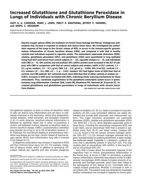
<strong>Increased</strong> <strong>Glutathione</strong> <strong>and</strong> <strong>Glutathione</strong> <strong>Peroxidase</strong> <strong>in</strong><strong>Lungs</strong> <strong>of</strong> Individuals with Chronic Beryllium DiseaseSUZY A. A. COMHAIR, MARC J. LEWIS, PERCY R. BHATHENA, JEFFREY P. HAMMEL,<strong>and</strong> SERPIL C. ERZURUMDepartments <strong>of</strong> Pulmonary <strong>and</strong> Critical Care Medic<strong>in</strong>e, Cancer Biology, <strong>and</strong> Biostatistics <strong>and</strong> Epidemiology, Lerner Research Institute,Clevel<strong>and</strong> Cl<strong>in</strong>ic Foundation, Clevel<strong>and</strong>, OhioReactive oxygen species (ROS) are mediators <strong>of</strong> chronic tissue damage <strong>and</strong> fibrosis. Endogenous antioxidantsmay <strong>in</strong>crease <strong>in</strong> response to oxidants <strong>and</strong> reduce tissue <strong>in</strong>jury. We <strong>in</strong>vestigated the antioxidantresponse <strong>of</strong> the lungs to the chronic release <strong>of</strong> ROS, as occurs <strong>in</strong> the immune-specific granulomatous<strong>in</strong>flammation <strong>of</strong> chronic beryllium disease (CBD), <strong>and</strong> compared it with that <strong>in</strong> healthycontrols <strong>and</strong> <strong>in</strong>dividuals exposed to cigarette smoke. The antioxidants superoxide dismutase (SOD),catalase, glutathione peroxidase (GPx), <strong>and</strong> glutathione (GSH) were quantitated <strong>in</strong> lung epitheliall<strong>in</strong><strong>in</strong>g fluid (ELF) <strong>and</strong> serum from control subjects (n 10), cigarette smokers (n 8), <strong>and</strong> <strong>in</strong>dividualswith CBD (n 9). GPx activity <strong>and</strong> extracellular GPx (eGPx) prote<strong>in</strong> were <strong>in</strong>creased <strong>in</strong> the ELF <strong>of</strong> subjectswith CBD <strong>in</strong> comparison with that <strong>of</strong> control subjects <strong>and</strong> smokers (eGPx <strong>in</strong> ELF: controls, 1.3 0.2 g/ml, smokers, 1.9 0.3 g/ml, CBD, 3.8 0.8 g/ml; p 0.002; GPx U/ml ELF, controls 1.4 0.3, smokers 1.8 0.4, CBD, 4.5 1, p 0.02). Smokers’ ELF had higher levels <strong>of</strong> GSH than that <strong>of</strong>controls, but CBD patients’ ELF conta<strong>in</strong>ed much more GSH than that <strong>of</strong> either controls or smokers (p 0.001). Increases <strong>in</strong> GSH were correlated with eGPx, <strong>in</strong>dicat<strong>in</strong>g similar <strong>in</strong>duc<strong>in</strong>g mechanisms for theseantioxidants. Thus, coord<strong>in</strong>ate augmentation <strong>of</strong> the glutathione antioxidant system occurs <strong>in</strong> granulomatouslung <strong>in</strong>flammation. Comhair SAA, Lewis MJ, Bhathena PR, Hammel JP, Erzurum SC. <strong>Increased</strong>glutathione <strong>and</strong> glutathione peroxidase <strong>in</strong> lungs <strong>of</strong> <strong>in</strong>dividuals with chronic berylliumdisease. AM J RESPIR CRIT CARE MED 1999;159:1824–1829.Occupational exposure to dusts or fumes <strong>of</strong> beryllium metalor salts can lead to lung <strong>in</strong>flammation <strong>and</strong> cell <strong>in</strong>jury <strong>in</strong> <strong>in</strong>dividualsemployed <strong>in</strong> the electronics, dental alloy preparation,nuclear weapons, metal extraction, <strong>and</strong> aerospace <strong>in</strong>dustries(1–6). In some <strong>in</strong>stances, beryllium-<strong>in</strong>duced <strong>in</strong>flammation isfollowed by epithelial repair, but <strong>in</strong> other circumstances achronic cell-mediated immune response to beryllium leads tochronic beryllium disease (CBD), a granulomatous <strong>in</strong>terstitiallung disorder occurr<strong>in</strong>g <strong>in</strong> up to 3% <strong>of</strong> beryllium workers (1–8). Although the mechanisms lead<strong>in</strong>g to granulomatous <strong>in</strong>terstitiallung disease are not clear, chronic release <strong>of</strong> reactive oxygenspecies (ROS) from <strong>in</strong>flammatory lung cells is <strong>in</strong>volved<strong>in</strong> the disease (9–11).Endogenous antioxidants may <strong>in</strong>crease <strong>in</strong> response to ROS<strong>and</strong> thus m<strong>in</strong>imize tissue <strong>in</strong>jury (12–17). For example, <strong>in</strong>halation<strong>of</strong> tobacco smoke, which <strong>in</strong> addition to generat<strong>in</strong>g 10 14oxidant radicals per cigarette puff also activates phagocyte(Received <strong>in</strong> orig<strong>in</strong>al form October 13, 1998 <strong>and</strong> <strong>in</strong> revised form December 24, 1998)Supported <strong>in</strong> part by grant HL-03117 from the National Institutes <strong>of</strong> Health.Correspondence <strong>and</strong> requests for repr<strong>in</strong>ts should be addressed to Serpil C. Erzurum,M.D., Department <strong>of</strong> Pulmonary <strong>and</strong> Critical Care Medic<strong>in</strong>e, Clevel<strong>and</strong>Cl<strong>in</strong>ic Foundation, 9500 Euclid Avenue/A90, Clevel<strong>and</strong>, OH 44195. E-mail: erzurus@cesmtp.ccf.orgAm J Respir Crit Care Med Vol 159. pp 1824–1829, 1999Internet address: www.atsjournals.orgrelease <strong>of</strong> oxidants (14, 18–21), leads to <strong>in</strong>creases <strong>in</strong> lung antioxidants(19–24). Lung antioxidant defenses are widely distributed<strong>and</strong> <strong>in</strong>clude both enzymatic <strong>and</strong> nonenzymatic systems.The major enzymatic antioxidants are superoxidedismutase (SOD), catalase <strong>and</strong> glutathione peroxidase (GPx)(15). SOD (EC 1.15.1.1), which degrades superoxide, exists <strong>in</strong>three forms, <strong>in</strong>clud<strong>in</strong>g the <strong>in</strong>tracellular manganese SOD <strong>and</strong>CuZn SOD, <strong>and</strong> an extracellular SOD that is present <strong>in</strong> epitheliall<strong>in</strong><strong>in</strong>g fluid (ELF) <strong>and</strong> blood vessels. Catalase [EC1.11.1.6], found <strong>in</strong> the cell cytosol, removes hydrogen peroxide.GPx (EC 1.11.1.9) removes hydrogen peroxide <strong>and</strong> organichydroperoxides by oxidiz<strong>in</strong>g glutathione, a water-soluble,low-molecular-weight tripeptide (L--glutamyl-L-cyste<strong>in</strong>yl glyc<strong>in</strong>e)that is abundantly present <strong>in</strong> lung ELF (24). The lungconta<strong>in</strong>s both an extracellular GPx (eGPx), <strong>in</strong> the lung ELF,<strong>and</strong> an <strong>in</strong>tracellular GPx (25).Very little is known about the antioxidant response <strong>of</strong> therespiratory tract to chronic granulomatous <strong>in</strong>flammation <strong>in</strong>the human lung, as occurs <strong>in</strong> CBD. In sarcoidosis, a granulomatous<strong>in</strong>flammatory <strong>in</strong>terstitial lung disease similar to CBD,alveolar macrophages (AM) release <strong>in</strong>creased amounts <strong>of</strong> superoxide<strong>and</strong> hydrogen peroxide, which have been l<strong>in</strong>ked tothe pathogenesis <strong>of</strong> this disease (9–11). Co<strong>in</strong>cident with <strong>in</strong>creasedoxidant production, MnSOD is <strong>in</strong>creased <strong>in</strong> AM <strong>in</strong>sarcoidosis (26). The present study was designed to <strong>in</strong>vestigatewhether antioxidants are <strong>in</strong>creased <strong>in</strong> response to chronic gran-
Comhair, Lewis, Bhathena, et al.: Antioxidants <strong>in</strong> CBD 1825ulomatous lung <strong>in</strong>flammation as occurs <strong>in</strong> CBD, <strong>in</strong> comparisonwith healthy controls <strong>and</strong> cigarette smok<strong>in</strong>g <strong>in</strong>dividuals.METHODSStudy PopulationTo evaluate antioxidants <strong>in</strong> the respiratory system <strong>in</strong> vivo, we <strong>in</strong>cluded27 subjects <strong>in</strong> the study population: 10 healthy, nonsmok<strong>in</strong>g <strong>in</strong>dividuals,eight healthy smok<strong>in</strong>g <strong>in</strong>dividuals, <strong>and</strong> n<strong>in</strong>e <strong>in</strong>dividuals withCBD (Table 1). Nonsmok<strong>in</strong>g volunteers with no history <strong>of</strong> pulmonarydisease were enrolled as controls. Exclusion criteria for the threegroups <strong>in</strong>cluded age under 18 yr or over 65 yr, pregnancy, human immunodeficiencyvirus (HIV) <strong>in</strong>fection, <strong>and</strong> a history <strong>of</strong> respiratory <strong>in</strong>fection<strong>in</strong> the previous 6 wk. Additional exclusion criteria for controlsubjects <strong>in</strong>cluded current tobacco use, prolonged exposure to secondh<strong>and</strong>smoke at home or at work, exposure to dusty environments or toknown pulmonary disease-produc<strong>in</strong>g agents, or a history <strong>of</strong> recurrentepisodes <strong>of</strong> breathlessness, chest tightness, cough, <strong>and</strong>/or sputum production.Smok<strong>in</strong>g <strong>in</strong>dividuals were similar to healthy controls but hadto have smoked a m<strong>in</strong>imum <strong>of</strong> 5 pack-yr <strong>and</strong> be current smokers. Inclusioncriteria for <strong>in</strong>dividuals with CBD were known exposure to beryllium;histologic evidence <strong>of</strong> disease, such as noncaseat<strong>in</strong>g granulomas<strong>and</strong>/or mononuclear cell <strong>in</strong>filtrates on lung biopsy specimen; <strong>and</strong>evidence <strong>of</strong> beryllium-specific, cell-mediated immunity <strong>in</strong> the lung asdemonstrated by a positive lymphocyte transformation test (LPT) onblood or bronchoalveolar lavage fluid (BALF) (7, 8). Seven CBD patientswere blood LPT positive, <strong>and</strong> five were BALF LPT positive.Bronchoalveolar LavageBronchoalveolar lavage (BAL) was performed on all subjects, us<strong>in</strong>gfiberoptic bronchoscopy as previously described (27). Briefly, after localanesthesia with 2% lidoca<strong>in</strong>e, a bronchoscope was wedged <strong>in</strong> asegmental bronchus <strong>of</strong> the right middle lobe or l<strong>in</strong>gula. Three 50-mlaliquots <strong>of</strong> warm physiologic sal<strong>in</strong>e were <strong>in</strong>fused <strong>in</strong>to the right middlelobe or l<strong>in</strong>gula (total volume <strong>of</strong> 300 ml) <strong>and</strong> recovered with manual suction.The BALF from the right middle lobe <strong>and</strong> l<strong>in</strong>gula were comb<strong>in</strong>ed<strong>and</strong> filtered through a Y-type blood filter (Drip Chamber Pump; AllegianceHealthcare Corp., McGaw Park, IL) <strong>and</strong> cellular componentswere separated by centrifugation (700 g for 10 m<strong>in</strong>). Cells werewashed once with Hanks’ balanced salt solution (HBSS; GIBCO,Gr<strong>and</strong> Isl<strong>and</strong>, NY) <strong>and</strong> counted with a hemacytometer. A cell differentialcount was done after Giemsa-type sta<strong>in</strong><strong>in</strong>g (Diff-Quick; AmericanScientific Products, Stone Mounta<strong>in</strong>, CA). Peripheral blood wasobta<strong>in</strong>ed from study subjects on the same day as BALF; serum was thenextracted by centrifugation <strong>of</strong> the whole blood (1430 g for 10 m<strong>in</strong>).TABLE 1CHARACTERISTICS OF STUDY POPULATIONControl(n 10)Smok<strong>in</strong>g(n 8)CBD(n 9)Sex, M/F 5/5 3/5 9/0RaceCaucasian 8 8 9African-American 2Age* 27 1 32 4 42 3Smok<strong>in</strong>gCurrent smoker 8Ex-smoker 5Nonsmoker 10 4WBC (10 6 )/ml BALF* 14 2 45 10 27 7% Lymphocyte* 2.2 0.5 1.4 0.6 11 2% Macrophages* 96.4 0.7 98.1 0.5 88 3% Neutrophils 1.1 0.7 0.5 0.3 1.3 0.2% Eos<strong>in</strong>ophils 0 0 0Values are means SEM.Def<strong>in</strong>ition <strong>of</strong> abbreviations: BALF bronchoalveolar lavage fluid; CBD chronic berylliumdisease; WBC white blood cells.* p 0.05.The volume <strong>of</strong> ELF <strong>in</strong> BALF was determ<strong>in</strong>ed with the ureamethod (27). Urea was measured <strong>in</strong> BALF <strong>and</strong> serum us<strong>in</strong>g the bloodurea nitrogen (BUN ENDPOINT) reaction (Sigma Chemical Co., St.Louis, MO) as previously described. Relative levels <strong>of</strong> ELF were estimatedby us<strong>in</strong>g simple dilution pr<strong>in</strong>ciples relat<strong>in</strong>g to the urea concentration<strong>in</strong> serum <strong>and</strong> BALF. Total prote<strong>in</strong> was determ<strong>in</strong>ed with abic<strong>in</strong>chon<strong>in</strong>ic (BCA) prote<strong>in</strong> assay (Pierce, Rockford, IL).SOD ActivitySOD activity was determ<strong>in</strong>ed <strong>in</strong> BALF <strong>and</strong> serum from the rate <strong>of</strong> reduction<strong>of</strong> cytochrome c (15), with one unit (U) <strong>of</strong> SOD activity def<strong>in</strong>edas the amount <strong>of</strong> SOD required to <strong>in</strong>hibit the rate <strong>of</strong> cytochromec reduction by 50%. The f<strong>in</strong>al reaction volume was 3 ml, <strong>and</strong> <strong>in</strong>cluded50 mM potassium phosphate buffer, 2 mM cytochrome c, 0.05 mMxanth<strong>in</strong>e, <strong>and</strong> a 0.1 mM ethylenediam<strong>in</strong>e tetraacetic acid (EDTA) solution.Xanth<strong>in</strong>e oxidase (Sigma) was added at a concentration sufficientto <strong>in</strong>duce a 0.020 change <strong>in</strong> absorbance per m<strong>in</strong>ute at 550 nm.GPx ActivityTotal GPx activity was determ<strong>in</strong>ed spectrophotometrically <strong>in</strong> BALF<strong>and</strong> serum through an <strong>in</strong>direct coupled assay (28). The BALF was <strong>in</strong>cubatedfor 2 m<strong>in</strong> at 37 C <strong>in</strong> the presence <strong>of</strong> 0.1 mM sodium azide,1 U/ml glutathione reductase, 0.1 mM GSH, <strong>and</strong> 0.12 mM reduced-nicot<strong>in</strong>amide aden<strong>in</strong>e d<strong>in</strong>ucleotide phosphate (-NADPH), 0.016mM dithiothreitol, 0.38 mM EDTA, <strong>and</strong> 50 mM sodium phosphate(pH 7.0). The reaction was <strong>in</strong>itiated by the addition <strong>of</strong> 0.2 mM hydrogenperoxide. The decrease <strong>in</strong> absorbance at 340 nm over 3 m<strong>in</strong>, as NADPHis converted to nicot<strong>in</strong>amide aden<strong>in</strong>e d<strong>in</strong>ucleotide phosphate (NADP) isproportional to the GPx activity. One unit <strong>of</strong> activity is def<strong>in</strong>ed as the activitythat catalyzes the oxidation <strong>of</strong> 1 nmol NADPH/m<strong>in</strong>, with a molarcoefficient <strong>of</strong> ext<strong>in</strong>ction <strong>of</strong> 6.22 10 6 M 1 cm 1 used for NADPH.Catalase ActivityCatalase activity was quantified with a method <strong>in</strong> which hydrogen peroxideis reacted with the components present <strong>in</strong> BALF (15). In thismethod the <strong>in</strong>itial rate <strong>of</strong> disappearance <strong>of</strong> hydrogen peroxide (0 to 60s)is recorded spectrophotometrically at a wavelength <strong>of</strong> 240 nm; one unit<strong>of</strong> catalase activity was def<strong>in</strong>ed as the rate constant <strong>of</strong> the first-order reaction.The assay is specific for the detection <strong>of</strong> catalase activity (29).Reduced <strong>Glutathione</strong> LevelsQuantification <strong>of</strong> GSH <strong>in</strong> BALF was done with a calorimetric assay(<strong>Glutathione</strong> assay kit; Calbiochem, La Jolla, CA) (21). This methodtakes advantage <strong>of</strong> a two-step chemical reaction. The first step leadsto the formation <strong>of</strong> substitution products between a proprietary reagent<strong>and</strong> all mercaptans present <strong>in</strong> the sample. The second step is a-elim<strong>in</strong>ation reaction under alkal<strong>in</strong>e conditions that <strong>in</strong>duces thetransformation <strong>of</strong> GSH <strong>in</strong>to a chromophore with maximal absorbanceat 400 nm, which is compared with a known st<strong>and</strong>ard curve <strong>of</strong> GSH.Extracellular GPxExtracellular GPx (eGPx) was measured with an enzyme-l<strong>in</strong>ked immunosorbentassay (ELISA) (Calbiochem). This method is based ona s<strong>and</strong>wich-type immunoassay, <strong>and</strong> is specific for eGPx. The eGPxprote<strong>in</strong> concentration present <strong>in</strong> BALF was based on four-parametercurve fit generated from known st<strong>and</strong>ard concentrations <strong>of</strong> eGPx.Statistical AnalysisAll data are expressed as the mean <strong>and</strong> SEM. The comparisons betweenthe three groups were made through analysis <strong>of</strong> variance (ANOVA). Avalue <strong>of</strong> p 0.05 was considered significant. Comparisons were alsomade with age- <strong>and</strong> gender-adjusted ANOVA models. The effect <strong>of</strong>previous smok<strong>in</strong>g on antioxidants was also tested with<strong>in</strong> the CBDgroup. L<strong>in</strong>ear regression fitt<strong>in</strong>g <strong>of</strong> data was done with the Fastat statisticalprogram (version 1.0; Systat Inc., Evanston, IL).RESULTSPatient CharacteristicsControl, smok<strong>in</strong>g, <strong>and</strong> CBD <strong>in</strong>dividuals were similar <strong>in</strong> terms <strong>of</strong>race <strong>and</strong> sex distribution (Table 1). Age was significantly greater
1826 AMERICAN JOURNAL OF RESPIRATORY AND CRITICAL CARE MEDICINE VOL 159 1999macrophages (p 0.001). The percentage recoveries <strong>of</strong> <strong>in</strong>stilledsal<strong>in</strong>e <strong>and</strong> ELF were similar <strong>in</strong> the three groups (BALFvolume: controls, 188 7 ml; smokers, 165 13 ml; CBDpatients, 182 8 ml; p 0.229). The total prote<strong>in</strong> <strong>in</strong> ELFwas different <strong>in</strong> the three groups (total prote<strong>in</strong> <strong>in</strong> ELF: controls,11 2 mg/ml; smokers, 13 3 mg/ml; CBD, 24 2 mg/ml;p 0.05). In contrast, the urea <strong>in</strong> BALF was similar amongthe three groups (urea <strong>in</strong> BALF: controls, 1.5 0.3 mg/ml;smokers, 1.7 0.2 mg/ml; CBD, 1.4 0.1 mg/ml; p 0.533).There was no correlation <strong>of</strong> BALF cellularity with prote<strong>in</strong>(R 0.048, p 0.813).Figure 1. SOD activity <strong>in</strong> ELF <strong>and</strong> serum <strong>of</strong> controls, smokers, <strong>and</strong>CBD patients. Smokers had less SOD activity <strong>in</strong> ELF than controlsor CBD patients (p 0.008). The SOD activity <strong>in</strong> serum was notsignificantly different <strong>in</strong> the three groups. Values are mean SEM.<strong>in</strong> the CBD group (p 0.001). Bronchoscopy was well toleratedby all <strong>in</strong>dividuals. The total cell count <strong>in</strong> BALF was <strong>in</strong>creased<strong>in</strong> smokers as compared with controls <strong>and</strong> CBD patients(p 0.008). However, smokers’ BALF cell differentialcounts were similar to those <strong>of</strong> controls, whereas CBD patientshad higher percentages <strong>of</strong> lymphocytes than control orsmok<strong>in</strong>g <strong>in</strong>dividuals (p 0.001). This <strong>in</strong>crease <strong>in</strong> lymphocytepercentage was at the expense <strong>of</strong> a decreased percentage <strong>of</strong>Antioxidant Activities <strong>in</strong> ELF <strong>and</strong> SerumSmok<strong>in</strong>g <strong>in</strong>dividuals had less SOD activity than controls orCBD patients (Figure 1). In contrast, <strong>in</strong>dividuals with CBD hadSOD activity similar to that <strong>of</strong> controls (SOD <strong>in</strong> ELF: controls,74 12 U/ml; smokers, 39 14 U/ml; CBD, 104 13 U/ml; p 0.008). The SOD activity <strong>in</strong> serum was not significantlydifferent among the three groups (p 0.517; Figure 1).GPx activity <strong>in</strong> ELF was different among the three groups,with CBD patients hav<strong>in</strong>g significantly higher levels <strong>of</strong> GPxactivity than control subjects or smokers (GPx <strong>in</strong> ELF: controls,1.4 0.3 U/ml; smokers, 1.8 0.4 U/ml; CBD, 4.5 1U/ml; p 0.02; Figure 2). Serum GPx activity was also differentamong the groups, with elevated levels <strong>in</strong> smokers <strong>and</strong>CBD patients as compared with controls (GPx <strong>in</strong> serum: controls,0.03 0.01 U/ml; smokers, 0.13 0.03 U/ml; CBD, 0.08 0.02 U/ml; p 0.02; Figure 2).Catalase activity <strong>in</strong> ELF was similar <strong>in</strong> the three groups(catalase <strong>in</strong> ELF: controls, 55 10 mU/ml; smokers, 75 23mU/ml; CBD, 78 17 mU/ml; p 0.566).eGPx Prote<strong>in</strong> Levels <strong>in</strong> ELF <strong>and</strong> SerumNormal ELF <strong>of</strong> the lung conta<strong>in</strong>s eGPx. Patients with CBDhad higher levels <strong>of</strong> eGPx <strong>in</strong> ELF than did smokers or controls(eGPx <strong>in</strong> ELF: controls, 1.3 0.2 g/ml; smokers, 1.9 0.3g/ml; CBD, 3.8 0.8 g/ml; p 0.002; Figure 3). In contrast,Figure 2. GPx activity <strong>in</strong> ELF <strong>and</strong> serum <strong>of</strong> controls, smokers, <strong>and</strong>CBD patients. GPx activity was greatest <strong>in</strong> ELF <strong>of</strong> CBD patients (p 0.02). GPx activity <strong>in</strong> serum was also <strong>in</strong>creased <strong>in</strong> smokers <strong>and</strong> CBDpatients as compared with controls (p 0.02).Figure 3. eGPx prote<strong>in</strong> <strong>in</strong> ELF <strong>and</strong> serum <strong>of</strong> controls, smokers, <strong>and</strong>CBD patients. eGPx prote<strong>in</strong> <strong>in</strong> ELF was higher <strong>in</strong> CBD patients than<strong>in</strong> smokers or controls (p 0.002), whereas there was no significantdifference <strong>in</strong> serum eGPx among the groups.
Comhair, Lewis, Bhathena, et al.: Antioxidants <strong>in</strong> CBD 1827there was no significant difference <strong>in</strong> serum eGPx among thethree groups (p 0.566; Figure 3).GSH Levels <strong>in</strong> ELFPreviously, GSH has been shown to compose over 95% <strong>of</strong> totalglutathione <strong>in</strong> ELF (7). GSH levels were different amongthe three study groups. GSH levels <strong>in</strong> smokers were higherthan <strong>in</strong> controls, but CBD patients had the highest levels <strong>in</strong>comparison with controls or smokers (GSH <strong>in</strong> ELF: controls,0.60 0.07 mM; smokers, 1.0 0.1 mM; CBD, 2.0 0.3 mM;p 0.001; Figure 4A). Levels <strong>of</strong> GSH <strong>in</strong> the ELF were directlycorrelated with ELF eGPx <strong>in</strong> the three groups (R 0.863, p 0.001; Figure 4B). GSH also correlated with GPxactivity (R 0.505, p 0.007), SOD activity (R 0.487, p 0.01), <strong>and</strong> total prote<strong>in</strong> (R 0.79, p 0.001).Age <strong>and</strong> Gender Adjusted Group Effects<strong>and</strong> Smok<strong>in</strong>g Effect with<strong>in</strong> CBDThe mean age <strong>in</strong> the CBD group was higher than that <strong>of</strong> controls,<strong>and</strong> the CBD patients were all men. Therefore, differencesamong groups were also tested with an ANOVA modelthat adjusted for age <strong>and</strong> gender. Despite the relationship <strong>of</strong>age to group, GSH, GPx, <strong>and</strong> SOD were still significantly differentamong the groups when adjusted for age <strong>and</strong> gender(all p 0.02). Although eGPx failed to reach significance fordifference among the groups when adjusted for age <strong>and</strong> gender,a trend toward a significant difference was still noted (p 0.07). Effects <strong>of</strong> age or gender on antioxidants were not foundwith<strong>in</strong> the groups.Realiz<strong>in</strong>g that previous smok<strong>in</strong>g may have an effect on antioxidants,we exam<strong>in</strong>ed the smok<strong>in</strong>g effect with<strong>in</strong> the CBDFigure 4. (A) GSH <strong>in</strong> ELF <strong>of</strong> controls, smokers, <strong>and</strong> CBD patients.GSH levels <strong>in</strong> ELF <strong>of</strong> smokers were higher than <strong>in</strong> controls, butCBD patients had even higher levels than smokers (p 0.002).(B) Correlation <strong>of</strong> eGPx prote<strong>in</strong> with GSH <strong>in</strong> ELF <strong>of</strong> controls, smokers,<strong>and</strong> CBD patients. Increases <strong>in</strong> GSH levels <strong>in</strong> ELF were directlycorrelated with <strong>in</strong>creases <strong>in</strong> eGPx prote<strong>in</strong> (R 0.863, p 0.001).group with the ANOVA model. Significant differences <strong>in</strong>cludedhigher ELF GSH <strong>and</strong> GPx activity <strong>in</strong> never-smok<strong>in</strong>g ascompared with ex-smok<strong>in</strong>g CBD <strong>in</strong>dividuals (GSH <strong>in</strong> ELF,never-smok<strong>in</strong>g CBD patients, 2.4 0.5 mM (n 4), ex-smok<strong>in</strong>gCBD patients 1.69 0.06 mM (n 5); p 0.04; GPx <strong>in</strong>ELF, never-smok<strong>in</strong>g CBD patients, 8.4 0.9 U/ml (n 4);ex-smok<strong>in</strong>g CBD patients, 1.4 0.4 U/ml (n 5); p 0.001).DISCUSSIONTo our knowledge, this study is the first to demonstrate thatthe antioxidants GSH <strong>and</strong> GPx are <strong>in</strong>creased <strong>in</strong> lungs <strong>of</strong> CBDpatients as compared with healthy controls or smok<strong>in</strong>g <strong>in</strong>dividuals.Other granulomatous <strong>in</strong>flammatory lung diseases,such as sarcoidosis <strong>and</strong> extr<strong>in</strong>sic allergic alveolitis, are accompaniedby <strong>in</strong>creased MnSOD <strong>in</strong> granulomas <strong>and</strong> BALF macrophages,which supports the concept that <strong>in</strong>duction <strong>of</strong> antioxidantsmay be a nonspecific response to granulomatous lung<strong>in</strong>flammation (26). On the other h<strong>and</strong>, GSH is not <strong>in</strong>creased<strong>in</strong> sarcoidosis (30). In this context, although <strong>in</strong>creased antioxidantsmay not be unique to beryllium-<strong>in</strong>duced granulomatous<strong>in</strong>flammation, neither can these results be generalized to allgranulomatous lung diseases.Evidence <strong>in</strong> the literature supports the hypothesis that upregulation<strong>of</strong> antioxidants occurs at both the transcriptional<strong>and</strong> translational levels (23, 31, 32). For example, <strong>in</strong>duction <strong>of</strong>-glutamylcyste<strong>in</strong>e synthetase, the rate-limit<strong>in</strong>g enzyme <strong>in</strong> GSHsynthesis, by oxidant stress from exposure to cigarette smoke,is due to <strong>in</strong>creased transcription <strong>of</strong> the gene <strong>and</strong> is associatedwith activation <strong>of</strong> the redox-sensitive transcription factor activatorprote<strong>in</strong> 1 (AP-1) (23). Interest<strong>in</strong>gly, <strong>in</strong>creases <strong>in</strong> GSH<strong>and</strong> GPx were highly correlated <strong>in</strong> the present study, suggest<strong>in</strong>gthat <strong>in</strong>duction <strong>of</strong> GPx may be mediated through redoxmechanisms similar to GSH. Increases <strong>in</strong> GPx activity aremost likely due to <strong>in</strong>creases <strong>in</strong> eGPx prote<strong>in</strong> <strong>in</strong> CBD patients’lungs. In the context that eGPx is synthesized <strong>and</strong> secreted bybronchial epithelial cells <strong>and</strong> AM (25), eGPx gene expressionmay be redox-mediated <strong>in</strong> the lung. In contrast, the <strong>in</strong>crease <strong>in</strong>serum GPx activity <strong>in</strong> smokers <strong>and</strong> CBD patients is not relatedto levels <strong>of</strong> serum eGPx prote<strong>in</strong>, <strong>in</strong>dicat<strong>in</strong>g that otherGPx enzymes are responsible for the systemic <strong>in</strong>crease <strong>in</strong> GPxactivity (25). The <strong>in</strong>duction <strong>of</strong> the glutathione antioxidant system<strong>in</strong> CBD is especially strik<strong>in</strong>g <strong>in</strong> that the CBD patients <strong>in</strong>our study were older than the controls <strong>and</strong> smokers, <strong>and</strong> <strong>in</strong>creasedage reduces the ability to <strong>in</strong>crease antioxidants <strong>in</strong> responseto oxidant stress (20). Despite the confound<strong>in</strong>g variable<strong>of</strong> age differences among the study groups, significantdifferences <strong>in</strong> antioxidants were still noted among the groupswith the use <strong>of</strong> ANOVA models that took <strong>in</strong>to account age<strong>and</strong> gender.Alveolar–capillary permeability as determ<strong>in</strong>ed by ELF-toserumalbum<strong>in</strong> ratios is <strong>in</strong>creased <strong>in</strong> CBD as compared withcontrol lungs (1). In the present study, total prote<strong>in</strong> <strong>in</strong> ELFwas tw<strong>of</strong>old greater <strong>in</strong> CBD patients than <strong>in</strong> healthy controls.Leakage <strong>of</strong> serum prote<strong>in</strong>s <strong>in</strong>to ELF is unlikely to contributeto the <strong>in</strong>crease <strong>in</strong> lung antioxidants <strong>in</strong> CBD, as opposed to the<strong>in</strong>duction <strong>of</strong> antioxidants with<strong>in</strong> the lung. Levels <strong>of</strong> GSH are140-fold higher <strong>in</strong> ELF than <strong>in</strong> serum (24), whereas SOD <strong>and</strong>GPx are 25- <strong>and</strong> 50-fold higher <strong>in</strong> ELF than <strong>in</strong> serum fromcontrols <strong>and</strong> CBD patients, respectively. Thus, <strong>in</strong>crease <strong>of</strong> alveolarcapillary permeability, with passive leakage <strong>of</strong> serumconta<strong>in</strong><strong>in</strong>g markedly lower antioxidant levels than <strong>in</strong> ELF,would more likely lead to dilution <strong>of</strong> than to <strong>in</strong>creases <strong>in</strong> antioxidantlevels.Our results substantiate previous studies that have shownmodifications <strong>in</strong> lung antioxidant status <strong>in</strong> smok<strong>in</strong>g <strong>in</strong>dividuals
1828 AMERICAN JOURNAL OF RESPIRATORY AND CRITICAL CARE MEDICINE VOL 159 1999(19, 24). In general, levels <strong>of</strong> antioxidants reported <strong>in</strong> ourstudy are similar to previously reported levels. Levels <strong>of</strong> GSHare with<strong>in</strong> the range <strong>of</strong> but slightly higher than those <strong>in</strong> previousstudies. Technical differences <strong>in</strong> method <strong>of</strong> lavage may accountfor these differences, e.g., all BALF <strong>in</strong> our study wascollected for analysis, whereas previous studies discarded thefirst lavage aliquot return [21]. Despite these technical differences,our data confirm previous f<strong>in</strong>d<strong>in</strong>gs that smokers havehigher levels <strong>of</strong> GSH <strong>in</strong> their ELF than do nonsmokers, butshow no difference <strong>in</strong> ELF catalase (21, 23, 24). Some studieshave shown <strong>in</strong>creased GPx activity <strong>in</strong> the ELF <strong>of</strong> smokers ascompared with nonsmokers (12), whereas others have showndecreased GPx <strong>in</strong> smokers (21). In the present study there wasa trend toward higher ELF GPx levels <strong>in</strong> smokers than <strong>in</strong> nonsmokers.As <strong>in</strong> a previous study (21), ELF SOD was decreased<strong>in</strong> smokers as compared with nonsmokers <strong>in</strong> our study. DecreasedSOD activity <strong>in</strong> the ELF <strong>of</strong> current smokers has beenpreviously shown, <strong>and</strong> is attributed to decreased CuZnSODactivity (21). One possible mechanism for decreased SOD activity<strong>in</strong> smokers may be related to the oxidant-mediated <strong>in</strong>activation<strong>of</strong> CuZnSOD (33, 34). Preservation <strong>of</strong> SOD activity<strong>in</strong> lung <strong>in</strong>flammation <strong>in</strong> CBD may relate to differences <strong>in</strong> thelocation <strong>and</strong> type <strong>of</strong> <strong>in</strong>creased ROS <strong>in</strong> CBD patients as opposedto smoke-exposed lungs. Alternatively, the relative normality<strong>of</strong> ELF SOD activity <strong>in</strong> CBD may be due to <strong>in</strong>creasedexpression <strong>of</strong> other SOD is<strong>of</strong>orms; e.g., MnSOD, such as occurs<strong>in</strong> sarcoidosis <strong>and</strong> extr<strong>in</strong>sic allergic alveolitis [26].Five <strong>of</strong> the CBD patients <strong>in</strong> our study were ex-cigarettesmokers. Analyses <strong>of</strong> ex-smok<strong>in</strong>g <strong>in</strong> comparison to neversmok<strong>in</strong>gCBD patients revealed higher levels <strong>of</strong> GSH <strong>and</strong>GPx activity <strong>in</strong> the never-smok<strong>in</strong>g CBD patients. Thus, <strong>in</strong>creases<strong>in</strong> antioxidant activity noted <strong>in</strong> the CBD group werenot simply due to previous cigarette-smoke exposure. Further,although the cigarette-smok<strong>in</strong>g group had alterations <strong>in</strong> antioxidants,as compared with healthy nonsmok<strong>in</strong>g controls, antioxidants<strong>in</strong> the smok<strong>in</strong>g group were also significantly differentthan <strong>in</strong> the CBD patients. Cigarette smok<strong>in</strong>g <strong>in</strong>duces achronic <strong>in</strong>flammatory process <strong>in</strong> the airways, with an abundance<strong>of</strong> <strong>in</strong>flammatory cells, pr<strong>in</strong>cipally AM <strong>and</strong> neutrophils(19). The smokers <strong>in</strong> our study had marked <strong>in</strong>creases <strong>in</strong> totalnumbers <strong>of</strong> <strong>in</strong>flammatory cells <strong>in</strong> their BALF, but cell differentials<strong>of</strong> smokers <strong>and</strong> healthy controls were similar. In contrast,CBD patients had an <strong>in</strong>creased percentage <strong>of</strong> lymphocytes<strong>in</strong> their BALF, which have been previously shown to beberyllium-specific CD4 T-helper (Th) lymphocytes (3–8).Tumor necrosis factor-, <strong>in</strong>terleuk<strong>in</strong> (IL)-6, <strong>and</strong> the lymphocyte-derivedcytok<strong>in</strong>e <strong>in</strong>terferon- appear to play importantroles <strong>in</strong> development <strong>of</strong> beryllium-<strong>in</strong>duced granulomatouslung disease (35, 36). These cytok<strong>in</strong>es <strong>in</strong>crease ROS production,which may contribute <strong>in</strong> part to the chronic lung <strong>in</strong>flammation<strong>of</strong> CBD (9, 13, 35).Interest<strong>in</strong>gly, recent evidence shows that GSH levels <strong>in</strong> antigen-present<strong>in</strong>gcells determ<strong>in</strong>e whether Th lymphocytes becomeTh1 or Th2 effector cells (37). Th1 cells mediate <strong>in</strong>flammatoryimmune responses <strong>of</strong> delayed-type hypersensitivity,characterized by IFN- production, whereas Th2 cells mediatehumoral/antibody responses <strong>and</strong> are characterized by IL-4production (37). The Th1-cell–mediated immune response isfavored by high GSH levels, whereas GSH depletion shifts theimmune response to the Th2 type (37). Thus, <strong>in</strong>creased GSH<strong>in</strong> CBD patients may contribute to the development <strong>and</strong>/orma<strong>in</strong>tenance <strong>of</strong> a chronic Th1-cell–mediated immune responseto beryllium. Importantly, CBD <strong>of</strong>ten reverses completely, suggest<strong>in</strong>gthat granulomatous <strong>in</strong>flammation <strong>and</strong> fibrosis are notessential consequences <strong>of</strong> the <strong>in</strong>flammatory response to beryllium(3–5). In this context, <strong>in</strong>creased antioxidants likely areone defense mechanism for m<strong>in</strong>imiz<strong>in</strong>g oxidant-<strong>in</strong>duced lung<strong>in</strong>jury <strong>in</strong> CBD, but may also <strong>in</strong>fluence the complex immuneresponse to beryllium.Acknowledgment : The authors thank Raed Dweik, Mary Jane Thomassen,Barbara Barna, <strong>and</strong> Herbert Wiedemann for provid<strong>in</strong>g bronchoalveolar lavagefluid samples from CBD patients.References1. Inoue, Y., E. Barker, E. Danil<strong>of</strong>f, N. Kohno, K. Hiwada, <strong>and</strong> L. S. Newman.1997. Pulmonary epithelial cell <strong>in</strong>jury <strong>and</strong> alveolar-capillary permeability<strong>in</strong> berylliosis. Am. J. Respir. Crit. Care Med. 156:109–115.2. Sendelbach, L. E., <strong>and</strong> H. P. Witchi. 1987. Bronchoalveolar lavage <strong>in</strong> rats<strong>and</strong> mice follow<strong>in</strong>g beryllium sulfate <strong>in</strong>halation. Toxicol. Appl. Pharmacol.90:322–329.3. Meyer, K. C. 1994. Occupational <strong>and</strong> environmental lung disease: beryllium<strong>and</strong> lung disease. Chest 106:942–946.4. Newman, L. S. 1993. To Be 2 or not to Be 2 : immunogenetics <strong>and</strong> occupationalexposure. Science 262:197–198.5. Kriebel, D., J. D. Bra<strong>in</strong>, N. L. Spr<strong>in</strong>ce, <strong>and</strong> H. Kazemi. 1988. The pulmonarytoxicity <strong>of</strong> beryllium. Am. Rev. Respir. Dis. 137:464–473.6. Salt<strong>in</strong>i, C., B. A. W<strong>in</strong>estock, M. K. Kirby, P. P<strong>in</strong>kston, <strong>and</strong> R. G. Crystal.1989. Ma<strong>in</strong>tenance <strong>of</strong> alveolitis <strong>in</strong> patients with chronic beryllium diseaseby beryllium-specific helper T cells. N. Engl. J. Med. 320:1103–1109.7. Mroz, M. M., K. Kreiss, D. C. Lezotte, P. A. Campbell, <strong>and</strong> L. S. Newman.1991. Reexam<strong>in</strong>ation <strong>of</strong> the blood lymphocyte transformationtest <strong>in</strong> the diagnosis <strong>of</strong> chronic beryllium disease. J. Allergy Cl<strong>in</strong>. Immunol.88:54–60.8. Rossman, M. D., J. A. Kern, J. A. Elias, M. R. Cullen, P. E. Epste<strong>in</strong>,O. P. Preuss, T. N. Markham, <strong>and</strong> R. P. Daniele. 1988. Proliferativeresponse <strong>of</strong> bronchoalveolar lymphocytes to beryllium. Ann. Intern.Med. 108:687–693.9. Fels, A. O. S., C. F. Nathan, <strong>and</strong> Z. A. Cohn. 1987. Hydrogen peroxiderelease by alveolar macrophages from sarcoid patients <strong>and</strong> by alveolarmacrophages from normals after recomb<strong>in</strong>ant <strong>in</strong>terferons , <strong>and</strong> <strong>and</strong> 1,25-dihydroxyvitam<strong>in</strong> D 3 . J. Cl<strong>in</strong>. Invest. 80:381–386.10. Nielsen, H., J. Frederiksen, D. Shersion, <strong>and</strong> N. Milman. 1990. Comparison<strong>of</strong> alveolar macrophage <strong>and</strong> blood monocyte oxidative burst response<strong>in</strong> pulmonary sarcoidosis. APMIS 98:401–406.11. Calhoun, W. J., <strong>and</strong> S. M. Salisbury. 1989. Heterogeneity <strong>in</strong> cell recovery<strong>and</strong> superoxide production <strong>in</strong> buoyant, density-def<strong>in</strong>ed subpopulations<strong>of</strong> human alveolar macrophages from healthy volunteers <strong>and</strong>sarcoidosis patients. J. Lab. Cl<strong>in</strong>. Med. 114:682–690.12. Rep<strong>in</strong>e, J. E., A. Bast, I. Lankhorst, <strong>and</strong> The Oxidative Stress StudyGroup. 1997. Oxidative stress <strong>in</strong> chronic obstructive pulmonary disease.Am. J. Respir. Crit. Care Med. 156:341–357.13. Vallyathan, V., <strong>and</strong> X. Shi. 1997. The role <strong>of</strong> oxygen free radicals <strong>in</strong> occupational<strong>and</strong> environmental lung disease. Environ. Health Perspect.105(Suppl. 1):165–177.14. Rahman, I., D. Morrison, K. Donaldson, <strong>and</strong> W. MacNee. 1996. Oxidativestress <strong>in</strong> asthma, COPD, <strong>and</strong> smokers. Am. J. Respir. Crit. CareMed. 154:1055–1060.15. Erzurum, S. C., C. Danel, A. Gillissen, C. Chu, B. C. Trapnell, <strong>and</strong> R. G.Crystal. 1993. In vivo antioxidant gene expression <strong>in</strong> human airwayepithelium <strong>of</strong> normal <strong>in</strong>dividuals exposed to 100% O 2 . J. Appl. Physiol.75:1256–1262.16. Hass, M. A., J. Iqbal, L. B. Clerch, L. Frank, <strong>and</strong> D. Massaro. 1989. Ratlung Cu,Zn superoxide dismutase: isolation <strong>and</strong> sequence <strong>of</strong> a fulllength cDNA <strong>and</strong> studies <strong>of</strong> enzyme <strong>in</strong>duction. J. Cl<strong>in</strong>. Invest. 83:1241–1246.17. Shull, S., N. H. He<strong>in</strong>tz, M. Periasamy, M. Manohar, Y. M. W. Janssen, J. P.Marsh, <strong>and</strong> B. T. Mossman. 1991. Differential regulation <strong>of</strong> antioxidantenzymes <strong>in</strong> response to oxidants. J. Biol. Chem. 266:24398–24403.18. Nowak, D., A. Antczak, M. Krol, T. Pietras, B. Shariati, P. Bialasiewicz,K. Jeczkowski, <strong>and</strong> P. Kula. 1996. <strong>Increased</strong> content <strong>of</strong> hydrogen peroxide<strong>in</strong> the expired breath <strong>of</strong> cigarette smokers. Eur. Respir. J. 9:652–657.19. L<strong>in</strong>den, M., L. Hakansson, K. Ohlsson, K. Sjod<strong>in</strong>, H. Tegner, A. Tunek,<strong>and</strong> P. Venge. 1989. <strong>Glutathione</strong> <strong>in</strong> bronchoalveolar lavage fluid fromsmokers is related to humoral markers <strong>of</strong> <strong>in</strong>flammatory cell activity.Inflammation 13:651–658.20. Kondo, T., S. Tagami, A. Yoshioka, M. Nishimura, <strong>and</strong> Y. Kawakami.1994. Current smok<strong>in</strong>g <strong>of</strong> elderly men reduces antioxidants <strong>in</strong> alveolarmacrophages. Am. J. Respir. Crit. Care Med. 149:178–182.21. Melloni, B., M. Lefebvre, F. Bonnaud, A. Vernenegre, L. Gross<strong>in</strong>, M.
Comhair, Lewis, Bhathena, et al.: Antioxidants <strong>in</strong> CBD 1829Rigaud, <strong>and</strong> A. Cant<strong>in</strong>. 1996. Antioxidant activity <strong>in</strong> bronchoalveolarlavage fluid from patients with lung cancer. Am. J. Respir. Crit. CareMed. 154:1706–1711.22. Hilbert, J., <strong>and</strong> V. Mohsen<strong>in</strong>. 1996. Adaptation <strong>of</strong> lung antioxidants tocigarette smok<strong>in</strong>g <strong>in</strong> humans. Chest 110:916–920.23. Rahman, I., C. Smith, M. Lawson, D. Harrison, <strong>and</strong> W. MacNee. 1996.Induction <strong>of</strong> gamma-glutamylcyste<strong>in</strong>e synthetase by cigarette smokeis associated with AP-1 <strong>in</strong> human alveolar epithelial cells. FEBS Lett.396:21–25.24. Cant<strong>in</strong>, A. M., S. L. North, C. Hubbard, <strong>and</strong> R. G. Crystal. 1987. Normalalveolar epithelial l<strong>in</strong><strong>in</strong>g fluid. J. Appl. Physiol. 63:152–157.25. Avissar, N., J. N. F<strong>in</strong>kelste<strong>in</strong>, S. Horowitz, J. C. Willey, E. Coy, M. W.Frampton, R. H. Watk<strong>in</strong>s, P. Khullar, Y Xu, <strong>and</strong> C. J Harvey. 1996.Extracellular glutathione peroxidase <strong>in</strong> human lung epithelial l<strong>in</strong><strong>in</strong>gfluid <strong>and</strong> <strong>in</strong> lung cells. Am. J. Physiol. 270(2, Pt. 1):L173–L182.26. Lakari, E., P. Paakko, <strong>and</strong> V. K<strong>in</strong>nula. 1998. Manganese superoxide dismutase,but not CuZn superoxide dismutase, is highly expressed <strong>in</strong> thegranulomas <strong>of</strong> pulmonary sarcoidosis <strong>and</strong> extr<strong>in</strong>sic allergic alveolitis.Am. J. Respir. Crit. Care Med. 158:589–596.27. Rennard, S. I., G. Basset, D. Lecossier, K. M. O’Donnell, P. P<strong>in</strong>kston,P. G. Mart<strong>in</strong>, <strong>and</strong> R. G. Crystal. 1986. Estimation <strong>of</strong> volume <strong>of</strong> epitheliall<strong>in</strong><strong>in</strong>g fluid recovered by lavage us<strong>in</strong>g urea as marker <strong>of</strong> dilution. J.Appl. Physiol. 60:532–538.28. Paglia, D. E., <strong>and</strong> W. N. Valent<strong>in</strong>e. 1967. Studies on the quantitative <strong>and</strong>qualitative characterization <strong>of</strong> erythrocyte glutathione peroxidase. J.Lab. Cl<strong>in</strong>. Med. 70:158–169.29. Aebi, H. 1984. Catalase <strong>in</strong> vitro. Methods Enzymol. 105:121–126.30. Qy, H., Y. Ishii, <strong>and</strong> S. Kitamura. 1994. Measurement <strong>of</strong> total glutathioneconcentration <strong>in</strong> bronchoalveolar lavage recovered from the patientswith diffuse <strong>in</strong>terstitial lung disease. 1994. Ch<strong>in</strong>. J. Tuberc. Respir.Dis. 17:24–26.31. Jornot, L., <strong>and</strong> A. F. Junod. 1992. Response <strong>of</strong> human endothelial cellantioxidant enzymes to hyperoxia. Am. J. Respir. Cell Mol. Biol. 6:107–115.32. Rushmore, T. H., M. R. Morton, <strong>and</strong> C. B. Pickett. 1991. Activation byoxidative stress <strong>and</strong> identification <strong>of</strong> the DNA consensus sequence requiredfor functional activity. J. Biol. Chem. 266:11632–11639.33. DeRaeve, H. R., F. B. J. M. Thunnissen, F. T. Kaneko, F. H. Guo, M.Lewis, M. S. Kavuru, M. Secic, M. J. Thomassen, S. C. Erzurum. 1997.Decreased Cu, Zn-SOD activity <strong>in</strong> asthmatic airway epithelium: correctionby <strong>in</strong>haled corticosteroid <strong>in</strong> vivo. Am. J. Physiol. 272:L148–L154.34. Uchida, K., <strong>and</strong> S. Kawakishi. 1994. Identification <strong>of</strong> oxidized histid<strong>in</strong>egenerated at the active site <strong>of</strong> Cu,Zn-superoxide dismutase exposed toH 2 O 2 . J. Biol. Chem. 269:2405–2410.35. T<strong>in</strong>kle, S. S., L. A. Kittle, B. A. Schumacher, <strong>and</strong> L. S. Newman. 1997.Beryllium <strong>in</strong>duces IL-2 <strong>and</strong> IFN- <strong>in</strong> berylliosis. J. Immunol. 158:518–526.36. Bost, T. W., D. W. H. Riches, B. Schumacher, P. C. Carre, T. Z. Khan,J. A. B. Mart<strong>in</strong>ez, <strong>and</strong> L. S. Newman. 1994. Alveolar macrophagesfrom patients with beryllium disease <strong>and</strong> sarcoidosis express <strong>in</strong>creasedlevels <strong>of</strong> mRNA for tumor necrosis factor- <strong>and</strong> <strong>in</strong>terleuk<strong>in</strong>-6 but not<strong>in</strong>terleuk<strong>in</strong>-1. Am. J. Respir. Cell Mol. Biol. 10:506–513.37. Peterson, J. D., L. A. Herzenberg, K. Vasquez, <strong>and</strong> C. Waltenbaugh.1998. <strong>Glutathione</strong> levels <strong>in</strong> antigen-present<strong>in</strong>g cells modulate Th1 versusTh2 response patterns. Proc. Natl. Acad. Sci. U.S.A. 95:3071–3076.